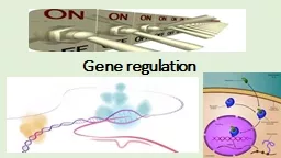
/
Characterisation of the Warm Acclimated Protein gene (wap65) in the An Characterisation of the Warm Acclimated Protein gene (wap65) in the An

Characterisation of the Warm Acclimated Protein gene (wap65) in the An - PDF document

trish-goza
trish-goza . @trish-goza
Follow
406 views
Uploaded On 2015-09-26

Characterisation of the Warm Acclimated Protein gene (wap65) in the An - PPT Presentation

WAP65 Melody S Clark and Gavin Burns British Antarctic Survey Natural Environment Research Council High Cross Madingley Road Cambridge CB3 0ET UK 1 Author for correspondenceM S Clark Brit ID: 141789

WAP65 Melody Clark and

Share:

Link:

Embed:

Download Presentation from below link

Download Pdf The PPT/PDF document "Characterisation of the Warm Acclimated ..." is the property of its rightful owner. Permission is granted to download and print the materials on this web site for personal, non-commercial use only, and to display it on your personal computer provided you do not modify the materials and that you retain all copyright notices contained in the materials. By downloading content from our website, you accept the terms of this agreement.


Presentation Transcript

WAP65 Characterisation of the Warm Acclimated Protein gene (wap65) in the Antarctic plunderfish (Harpagifer antarcticus Melody S Clark and Gavin Burns British Antarctic Survey, Natural Environment Research Council, High Cross, Madingley Road, Cambridge, CB3 0ET, UK 1 Author for correspondence:M. S. Clark, British Antarctic Survey, Natural Environment Research Council, High Cross, Madingley Road, Cambridge, CB3 0ET, UK. Email: mscl@bas.ac.uk Key Words: Harpagifer, wap65, acclimation, climate change 1 WAP65 experimental tank at time zero and maintained at 6.0 ± 0.08 o C. Five animals were 24 and 48 hours using Home Office approved procedures. Cloning and sequencing Approximately 1,000 clones were sequenced from a directionally cloned non- liver cDNA library. All clones were vector and quality clipped before subjecting to BLAST sequence similarity searching. Analysis of BLAST results identified 10 ESTs, which showed high sequence similarity to the warm-temperature-acclimation-related-65 protein (accession number Q4W7I1). These 10 EST clones were concatenated and edited using the phred, Phrap and consed packages (Ewing and Green 1998; Gordon et al, 1998). Sequence analysis was performed using the EMBOSS suite of open source software urceforge.net . Alignments were exported into Boxshade (http://www.ch.embnet.org/software/BOX_form.html) for annotation. The antarcticus Wap65 sequence was submitted to the EMBL database with the accession number AM408054. RNA Isolation and Q-PCR Total RNA was extracted from the tissue samples using TRI Reagent (Sigma) according to the manufacturer’s instructions. 1g of total RNA was DNase treated and reverse transcribed using a first strand synthesis kit (Promega). Profiling of tissue-specific expression and Q-PCR was carried out using the following primers: Acclim2F (TAGAGCACTACTACTGTTTCCA) and Acclim2Rev (AGGCCGTCACGCTTGGTGT); (ActinF: ACAGACTACCTCATGAAGATCCT; ActinR: GAGGCCAGGATGGAGCCTCC). Actin was used as the housekeeping sequence for both RT and Q-PCR experiments as it had previously been shown not to change under the experimental conditions used (data not shown). For Q-PCR, both primer sets were checked over a four fold 10x dilution series with RSq values and PCR efficiency values (1.00 and 100.8% respectively for Wap65) calculated using the MxPro - MX3000P v 3.00 Build 311 Schema 74 software. Wap65 and actin sequences were amplified from each time point using Brilliant SYBR ® Green QPCR Master Mix (Stratagene) and an MX3000P (Stratagene). PCR conditions were as follows: 95°C 10 minutes, 40 cycles of 95°C 30 seconds, 60°C 1 minute and 72°C for 1 minute with a final dissociation curve step. The plate set-up for each Q-PCR experiment consisted of 5 control individuals and 5 experimental individuals (both in triplicate). Analysis was performed using the MxPro - MX3000P v 3.00 Build 311 Schema 74 software and data exported into the Relative Expression Software tool (REST) (http://www.gene-quantification.info/), which incorporates the Pfaffl method of compensating for the PCR efficiency and also uses a Pair Wise Fixed Reallocation Randomisation Test (Pfaffl et al, 2002). sample t-test using MINITAB v14 to deteconfidence range. Results Concatenation of EST data (10 clones) produced a 1367nt consensus sequence of high quality reads. BLAST sequence similarity searching of this sequence revealed 72% sequence identity (Expect = 1.9 e-174 , Score = 1711) with the warm-temperature-acclimation-related-65 protein (accession number Q4W7I1) from the Medaka fish . Sequence comparisons identified that the full-length wap-65 coding sequence of 431 amino acids was present with 23 nts and 51nts of 5’ and 3’ UTR respectively. Sequence alignments using Clustal W (Figure 1) indicate H. antarcticus gene shares greater sequence similarity to the fish wap65-2 4 WAP65 isoform (72.2% sequence identity to O. latipes wap65-2, but only 49% sequence identity to O. latipes wap65-1). No library clones weresimilarity to wap65-1. Examination of the H. antarcticus gene with regard to hemopexin motifs indicates a similar protein structure with 10 cysteines conserved (out of 12 in mammals) to produce disulphide bridges. This gene also contains 6 (Trp196, Tyr201, Phe208, Tyr222, tyr229 and Phe230) out of the 7 aromatic residues plus Pro 294 that have been defined as important for the structure and stability of the haem pocket and also both histidine (His213 anwhich form the bis-histidyl Fe(III) complex involved in haem axial ligand binding (Paoli et al, 1999). However the H. antarcticus gene only contains a single N-glycosylation site and N-glycosylation has been shown to be important in haem binding. Tissue distribution of H. antarcticus wap65-2 was examined by RT-PCR over a range of 15 tissues in control animals. There was a very limited distribution with strong expression in the liver and much lower expression in the posterior kidney only (Figure 2). Q-PCR revealed that H. antarcticus wap65-2 was not induced in response to an increased environmental temperature of 6ºC. In fact a sigmodal-shape response was produced when the data is plotted as log fold expression change verses time, with considerable initial down regulation (approximately 40 fold) for the first 8 hours followed by re-equilibration to base-line levels around zero (Figure 3). The smallest p value for this dataset is level, but the p values are supportive of the general trend outlined above. In this experiment, variation in gene expression was high due to a limited data set and high inter-individual variation, as would be expected from a wild population study. This wide genetic variation clearly affects significance testing and the resultant p values. Discussion The designation of wap65, isoform 2 via sequence similarity analyses was validated by the tissue distribution. Wap65-2 has a much more restricted tissue distribution (mainly liver), compared to wap65-1, which is present in multiple tissues O. latipes (Hirayama et al, 2003; 2004). In spite of the presence of the two conserved histidine residues proposed to be essential for haem binding, other fish orthologues of Wap65-2, which also contain these residues, do not show an affinity for haem (Hirayama et al, 2004). There is sufficient conservation of gene H. antarcticus wap65-2 and O. latipes wap65-2 to suggest that the two genes are functional orthologues. Although true acclimation experiments for periods of several weeks were not carried out on H. antarcticus, the 6ºC time course assay was carried out for a 48 hour period, during which, based on previously published findings, wap65 should have been significantly induced. In contrast, in antarcticus expression of wap65-2 was down regulated for a period of between 8-12 hours, after which levels returned to those of the control base line. The consequences of which are that the wap65-2 is not involved in temperature acclimation. The initial drop in expression may well be due to a primary “shock” response. This is mirrored in other genes that we have surveyed from the same animals in this time course assay (unpublished data), with the fish subsequently adjusting, at least in terms of gene transcription processes, to the elevated water temperature. If this is subsequently proved to be the situation, then the speed of the initial drop in expression levels and lag to return to a steady state is probably a function of mRNA stability and gene regulation respectively. Several experiments have been successfully carried out to acclimate Antarctic fish to 4°C (Carpenter and Hofmann, 2000; Lowe et al, 2005; Jin et al, 2006). Therefore, although these fish survive in a stable environment of –1.8ºC to +1.0°C almost year-round, they do potentially have the ability to acclimate to higher water temperatures 5 WAP65 than they experience in the natural environment. However, this acclimation process is unlikely to involve wap65-2. This finding adds further data to the complex regulation of wap65 isoforms in fish. This gene set has also been shown to be induced in response to immunological stimulus using LPS and hypoxia in C. auratus (Kikuchi et al, 1997; Gracey et al, 2001). However, there is no elevation in the expression of either of the two wap65 isoforms to induction by LPS in O. latipes (Hirayama et al, 2004) or to environmental temperature increases in O. latipes and T. rubripes (Hirayama et al, 2003). Indeed, so far, the correlation of increased expression levels of wap65 with increased environmental temperature have only been identified in the Cypriniformes, specifically the wap65-1 isoform. and O. latipes belong to the orders: Perciformes and Atheriniformes, so potentially this acclimation function of wap65 is phylogenetically constrained. The biochemical processes, by which Antarctic fish acclimate to warmer sea temperatures remains unknown. Experiments are on-going in our laboratory using cDNA microarrays and long term acclimation experiments to decipher this process. Acknowledgements This paper was produced within the BAS The authors would like to thank the Rothera dive team for sample collection, Pete Rothery for statistics advice and Keiron Fraser for help in performing the heat shock experiments. 6 WAP65 References Amores A, Force A, Yan YL, et al. 1998. Zebrafish hox clusters and vertebrate genome evolution. Science 282:1711-4. Carpenter CM, Hofmann GE. 2000. Expression of 70 kDa heat shock proteins in antarctic and New Zealand notothenioid fish. Comp Biochem Physiol A Mol Integr Physiol 125:229-38. Ewing B, Hillier L, Wendl MC, Green P. 1998. Base-calling of automated sequencer traces using phred. I. Accuracy assessment. Genome Res 8:175-85. Gordon D, Abajian C, Green P. 1998. Consed: a graphical tool for sequence finishing. Genome Res 8:195-202. Gracey AY, Troll, JV, Somero GN. 2001. Hypoxia-induced gene expression profiling in the euryoxic fish . Proc Natl Acad Sci U S A, 98:1993-8. Hazel JR, Prosser CL. 1974. Molecular mechanisms of temperature compensation in poikilotherms, Physiol Rev 54:620-77. Hirayama, M, Kobiyama, A, Kinoshita, S, Watabe S. 2004. The occurance of two types of hemopexin-like protein in medaka and differences in their affinity to heme, J. Expt. Biol. 207:1387-1398. Hirayama M, Nakaniwa M, Ikeda D, et al. 2003. Primary structures and gene organizations of two types of Wap65 from the pufferfish Takifugu rubripes. Fish 29:211-224. Jin Y, DeVries AL. 2006. Antifreeze glycoprotein levels in Antarctic notothenioid fishes inhabiting different thermal environments and the effect of warm acclimation. Comp Biochem Physiol B Biochem Mol Biol 144:290-300. Kikuchi K, Watabe S, Aida K. 1997. The Wap65 gene expression of goldfish ) in association with warm temperature as well as bacterial 17:423-432. Kikuchi K, Watabe S, Suzuki, Y, Aida K, Nakajima H. 1993. The 65-kDa cytosolic protein associated with warm temperature acclimation in goldfish, Carassius auratus. J. Comp. Physiol. B 163:349-354. Kinoshita S, Itoi S, Watabe S. 2001. cDNA cloning and characterization of the warm-temperature-acclimation-associated protein Wap65 from carp, Cyprinus carpio, Fish. 24:125-134. molarity, glucose concentration and erythrocyte responses of two Antarctichronic thermal change. J. Fish Biol. 67:752-766. Nakaniwa M, Hirayama M, Shimizu A, et al. 2005. Genomic sequences encoding two types of medaka hemopexin-like protein Wap65 and their gene expression profiles in embryos, J. Expt. Biol208:1915-1925. Paoli M, Anderson BF, Baker HM, Morgan WT, Smith A, Baker EN. 1999. Crystal structure of hemopexin reveals a novel high-affinity heme site formed by two beta-propeller domains, Nature. Struct. Biol.6:926-931. Peck LS, Conway LZ. 2000. The myth of metabolic cold adaptation: oxygen consumption in stenothermal Antarctic bivalve molluscs. In: Harper, E, Crame, A.J (eds) Evolutionary Biology of the bivalvia Geological Society of London Special publication 177:441-450. Cambridge University Press, Cambridge. Pfaffl MW, Horgan GW, Dempfle L. 2002. Relative expression software tool (REST) for group-wise comparison and statistical analysis of relative expression results in real-time PCR. Nucleic Acids Res 30:e36. Somero GN, DeVries AL. 1967. Temperature tolerance of some Antarctic fishes. Science 156:257-8. 7 WAP65 Watabe S, Kikuchi K, Aida K. 1993. Cold- and warm-temperaturespecific cytosolic protein in goldfish and carp. Nippon Suisan Gakkaishi.59:151-156. 8 WAP65 Figure Legends Figure 1 ClustalW alignment of translated wap-65 genes from a number of fish species. Species ID and acceTakifugu rubripes (Q75UL8, Q75UL9), Ola: (Q8JIP8, Q8JIP9), Han: Harpagifer antarcticus (AM408054), Tni: (Q4STQ5), Xhe: Xiphophorus helleri (Q2EF31) and Cca: Cyprinus carpio (Q90WF7). Annotation above the sequence: hemopexin-like repeats are shown by lines, conserved cysteine residues are denoted by a “*”, N-glycosylation site by a line ended with diamonds, residues important for the structure and stability of the haem pocket are denoted with a “+” and the two histidine residues which form the bis-histidyl Fe(III) complex involved in haem axial ligand binding are indicated by a “#”. Figure 2 RT- defined tissue distribution of wap65-2 in H. antarcticus control (non-treated) animals. Actin RT-PCR was used as a quantification control for the different tissues. Figure 3 Q-PCR results using liver tissue for H. antarcticus wap65-2 gene over a 48 hour time course series with a 6ºC temperature heat shock. B: Graphical representation of log expression fold changes (with error bars) in the H. antarcticus 9 Ola_WAP65_1 1 MKLLFLCLALLAWARR--------------KGAVDRCGIEDAVANEGIYFFKDHLFKGFHGQ Cca_WAP65 1 MRLILSFAADT--AGHKPELHHEAKLVNEGIYFFKGDHLFKGFHGK consensus 1 *. .... ..*........... . .. . ...**..*...**.. ...*. .**........*.* * Xhe_WAP65 73 AQLSKELGPINAAFRMHNTENPNLFQDDKVYSYFNQTQIQEFPGVPHLDAAVECP Tru_WAP65_2 80 AQLVSEIDDSISAAFRMHNKANPDIYFLEDKVFSYYEQVHINEFPGVPHLDAAVECP Ola_WAP65_2 70 AQPSQYKELGHVDAAFRMHNPENQGLFLDDKVFSYFEHTLEEGYPKEIQEFPGVPHLDAAVECP Han_WAP65 69 AQLSNESFQQHNIGHVDAAFRMHNIEHLDLFLDDKVFSYYEQAEIQHLDAAVECP Tru_WAP65_2 160 KGECMDSVLFFKGQDVHMYDLTKTVHLPCTSAFRWLEHYYCFHGHNFTRFNPISGEVNGTYPKDARHYFMR Ola_WAP65_2 145 KGECVDSVLFFKGPDVHYDITKTVHLPCTSFRWLEHYYCFHGHNFTRFQPVTGEVTGNYPKDARRYFMR Han_WAP65 149 KGECMDSVLFFKGQDVHVYDITKTVHLPCTSARWLEHYYCFHGNNFTKFPVSGEVSGVYPKDARSYFMK Tni_WAP65 141 HPECEDSVIFFKGKEIHYVRTKVDEEFKMPNCTSAFRFMEHFYCFHGHMFSKFDPKTGEVGKYPKEARDYFMR Tru_WAP65_1 141 HPECEDSVIFFKGDEIHYVRTDEEFKMPNCTSAFRFMEHFYCFHGHMFSKFDPKTGEVGKYPKEARDYFMR Ola_WAP65_1 146 KPECVEDSVIFFKKNEIHFVKKTVDFRMPNCTSAFRFMEHYYCFHGHKFSKFDPKTGEVGKYPKDARKFFMR Tru_WAP65_1 221 CAKSEES--DPVNAGHFLEANTLKADTEN Ola_WAP65_1 226 CSKDEDN--DHENIYAFRGHHYIRDEGNTLKADTESAFKELHSEVDAVFSYNSHLYM Cca_WAP65 235 CPFGHICSRVHLDAITSDDDGSIYAFRGYHFVDKFSDTESAFKELHSEVDAVFSYEG consensus 241 *. ..... . ....**.....*.*.*..*..*.*.* .........*.... . ...**....*******....*. * Xhe_WAP65 305 IKDQVYIYKADAHTLIEGYPKTVKEELGIEGTVDAAFVCPTENGNMRDVDLTATPRVISREFPLDID Tru_WAP65_2 317 IKYTLVEGYPKTLEEELGVEGPVDAAFVCPGQTVGERFLLTATPRVVARNLFV-LDID Xhe_WAP65 384 AGLCGSQYESPILMGRIAPVASDMGCE Tru_WAP65_2 396 AACDAKGSKYASVTLSKIAEMGC Ola_WAP65_2 381 AACSSQGSNSRIAPVGCE Han_WAP65 385 AALCVFGATTLAMSRIAPEMGCE Tni_WAP65 378 AAMCFASPMIPEQRRVSLEMFGCDH Tru_WAP65_1 379 AAICFIAGRLELFGCDH Ola_WAP65_1 384 GAMCGNHYYHFESPKTFAARLELFGCDH Cca_WAP65 393 AAMCYDSPIMMMAKIMPEQHRVSQG consensus 401 ...* .*.... *...*...*................. ...**.. Fi g ure 1 WAP65 Actin Intestine Liver Spleen White muscle Ovary Skin Cartilage tail ray Cartilage gill Brain Heart Stomach Adipose tissue Posterior kidney Head kidney Figure 2 Time (hours) REST P-value Fold change Range A 8 0.069 0.045 0.010-0.144 22.20 down regulated 12 0.996 0.690 0.310-1.530 .43 down regulated Time (hours) 0102030405060