
Acid-Fast Stain Author: Multiple Authors Citation:
LV
Published · 27 slides · 0 views

1 / 1

Description
Acid-Fast Stain Author: Multiple Authors Citation: Multiple Authors. 2008. Acid-fast stain. Publication Date: September 2008 Bacillus cereusacid fast (Enlarged view)FIG. 1. Bacillus cereus, non-acid-fast. (Jerry Keplinger, East Tennessee
Related Topics
Share
Embed code
Download this presentation From Below
"Acid-Fast Stain Author: Multiple Authors Citation:" is the property of its rightful owner. Permission is granted to download and print the materials on this website for personal, non-commercial use only, and to display it on your personal computer provided you do not modify the materials and that you retain all copyright notices contained in the materials. By downloading content from our website, you accept the terms of this agreement.
Presentation Transcript
01
Acid-Fast Stain Author: Multiple Authors
Citation: Multiple Authors. 2008. Acid-fast stain.
Publication Date: September 2008<br>
Citation: Multiple Authors. 2008. Acid-fast stain.
Publication Date: September 2008<br>
02
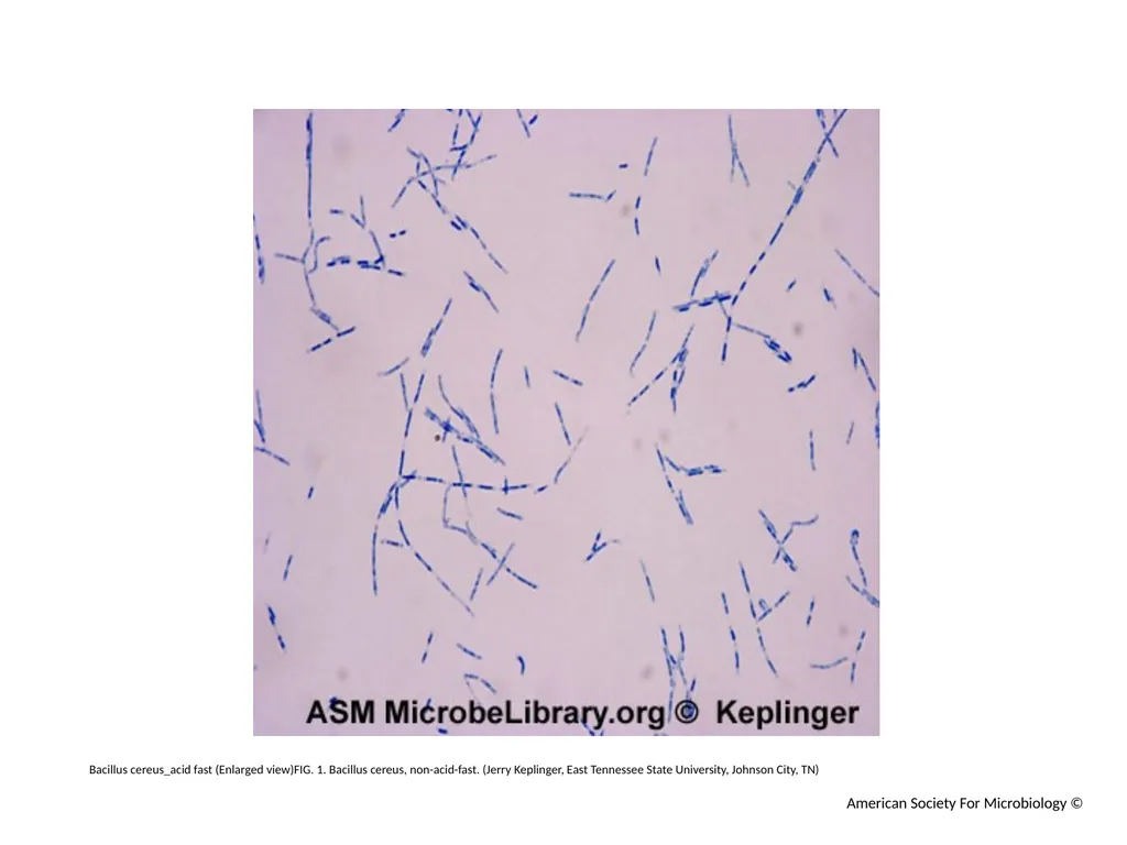
Bacillus cereus_acid fast (Enlarged view)FIG. 1. Bacillus cereus, non-acid-fast. (Jerry Keplinger, East Tennessee State University, Johnson City, TN) American Society For Microbiology ©<br>
03
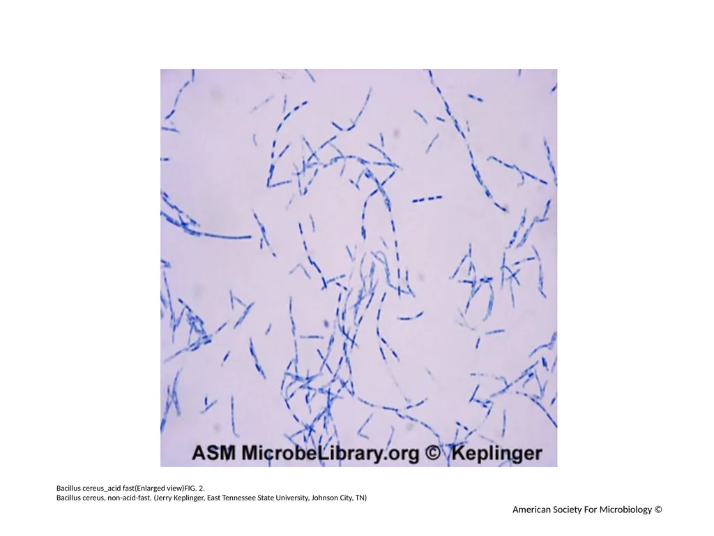
Bacillus cereus_acid fast(Enlarged view)FIG. 2.
Bacillus cereus, non-acid-fast. (Jerry Keplinger, East Tennessee State University, Johnson City, TN) American Society For Microbiology ©<br>
Bacillus cereus, non-acid-fast. (Jerry Keplinger, East Tennessee State University, Johnson City, TN) American Society For Microbiology ©<br>
04
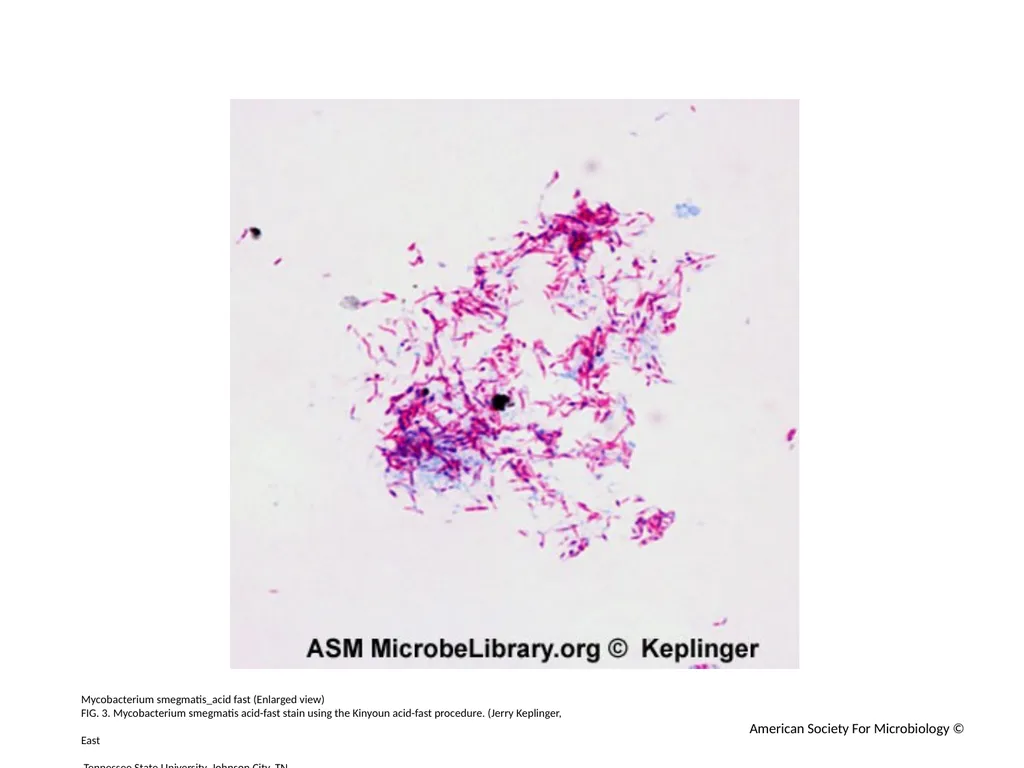
Mycobacterium smegmatis_acid fast (Enlarged view)
FIG. 3. Mycobacterium smegmatis acid-fast stain using the Kinyoun acid-fast procedure. (Jerry Keplinger,
East
Tennessee State University, Johnson City, TN
) American Society For Microbiology ©<br>
FIG. 3. Mycobacterium smegmatis acid-fast stain using the Kinyoun acid-fast procedure. (Jerry Keplinger,
East
Tennessee State University, Johnson City, TN
) American Society For Microbiology ©<br>
05
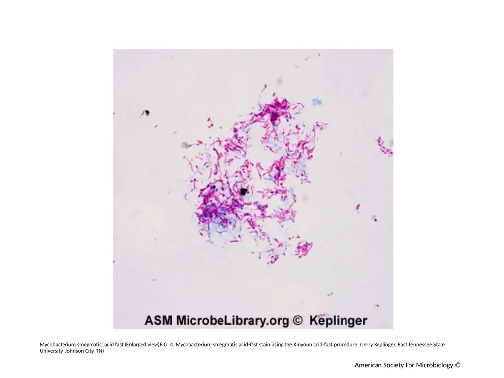
Mycobacterium smegmatis_acid fast (Enlarged view)FIG. 4. Mycobacterium smegmatis acid-fast stain using the Kinyoun acid-fast procedure. (Jerry Keplinger, East Tennessee State University, Johnson City, TN) American Society For Microbiology ©<br>
06
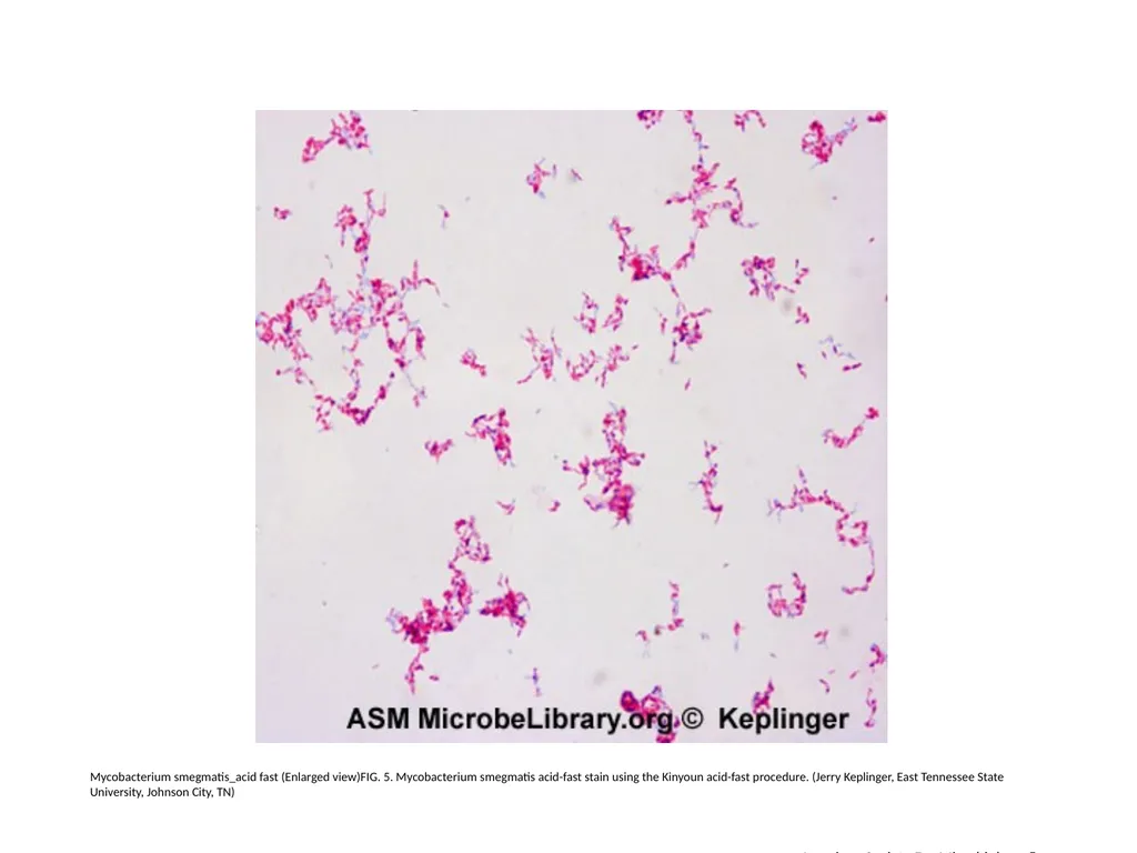
Mycobacterium smegmatis_acid fast (Enlarged view)FIG. 5. Mycobacterium smegmatis acid-fast stain using the Kinyoun acid-fast procedure. (Jerry Keplinger, East Tennessee State University, Johnson City, TN) American Society For Microbiology ©<br>
07
Mycobacterium tuberculosis_acid fast (Enlarged view)
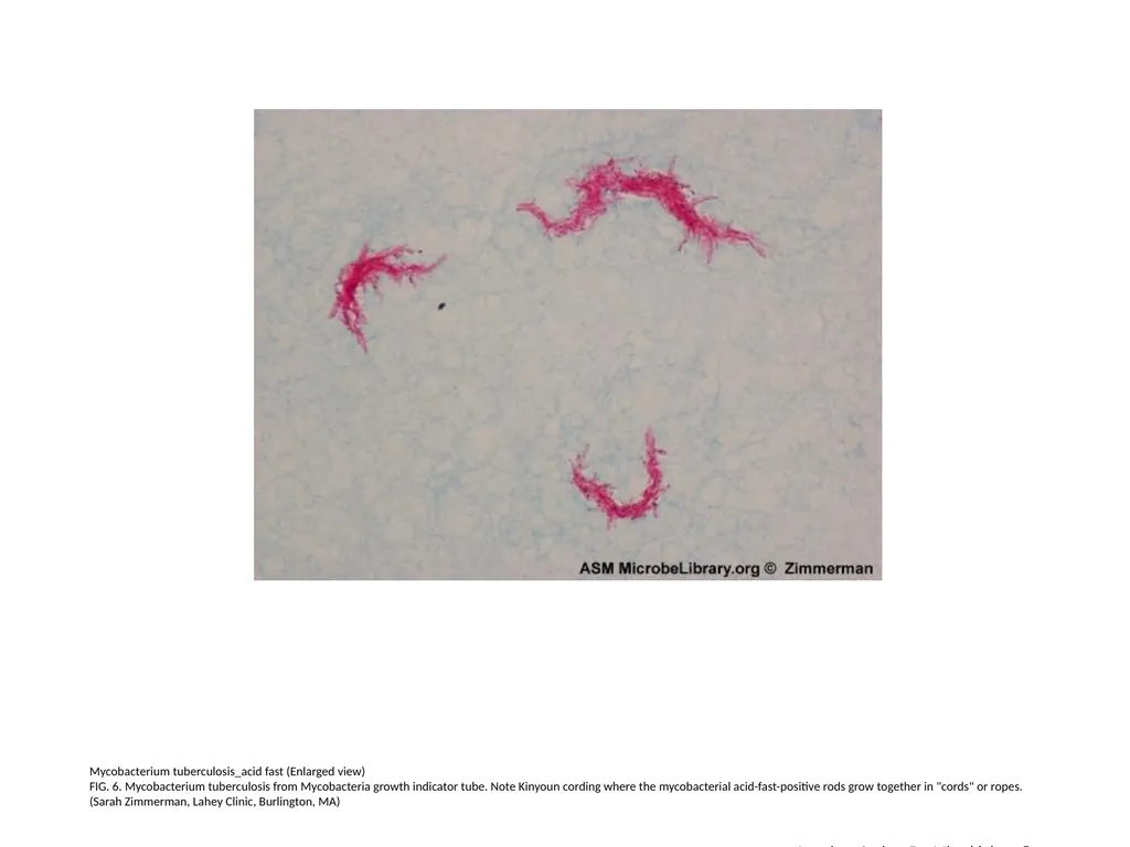
FIG. 6. Mycobacterium tuberculosis from Mycobacteria growth indicator tube. Note Kinyoun cording where the mycobacterial acid-fast-positive rods grow together in "cords" or ropes. (Sarah Zimmerman, Lahey Clinic, Burlington, MA) American Society For Microbiology ©<br>
FIG. 6. Mycobacterium tuberculosis from Mycobacteria growth indicator tube. Note Kinyoun cording where the mycobacterial acid-fast-positive rods grow together in "cords" or ropes. (Sarah Zimmerman, Lahey Clinic, Burlington, MA) American Society For Microbiology ©<br>
08
Mycobacterium tuberculosis_acid fast (Enlarged view)
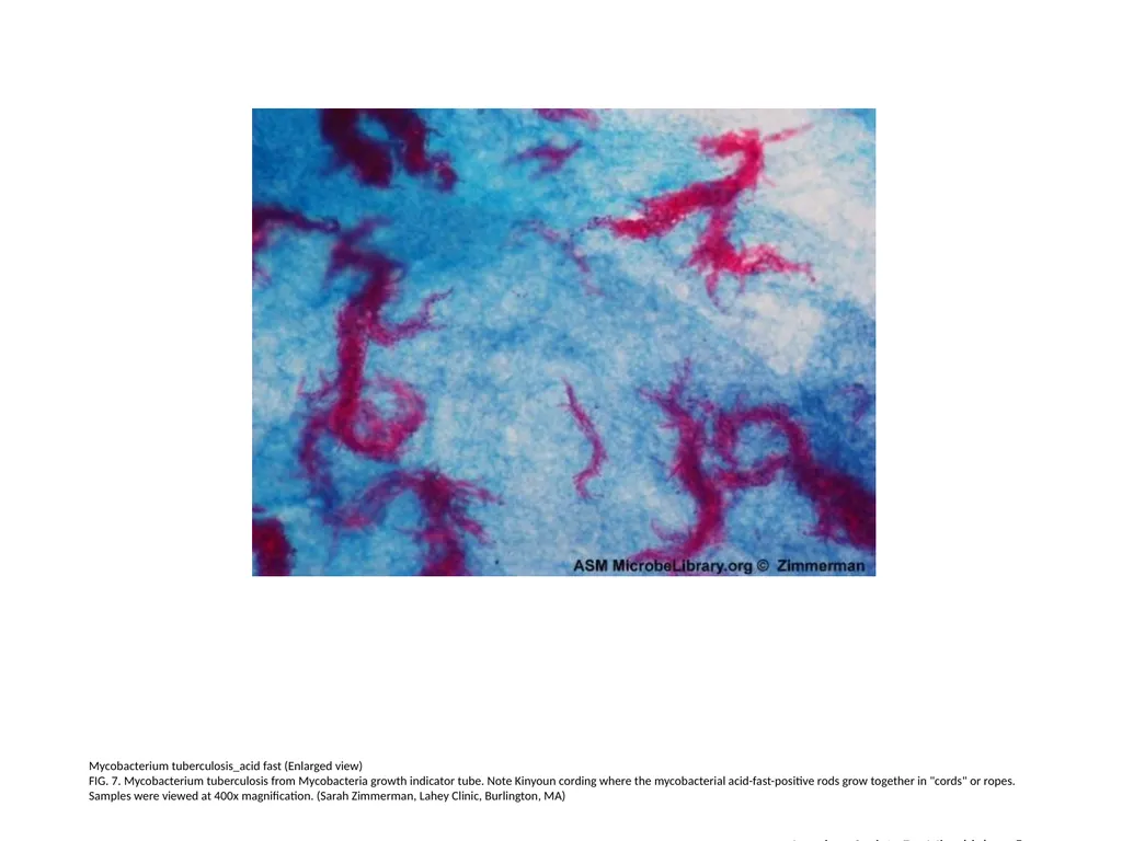
FIG. 7. Mycobacterium tuberculosis from Mycobacteria growth indicator tube. Note Kinyoun cording where the mycobacterial acid-fast-positive rods grow together in "cords" or ropes. Samples were viewed at 400x magnification. (Sarah Zimmerman, Lahey Clinic, Burlington, MA) American Society For Microbiology ©<br>
FIG. 7. Mycobacterium tuberculosis from Mycobacteria growth indicator tube. Note Kinyoun cording where the mycobacterial acid-fast-positive rods grow together in "cords" or ropes. Samples were viewed at 400x magnification. (Sarah Zimmerman, Lahey Clinic, Burlington, MA) American Society For Microbiology ©<br>
09
Mycobacterium tuberculosis_acid fast (Enlarged view)
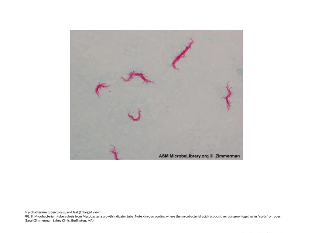
FIG. 8. Mycobacterium tuberculosis from Mycobacteria growth indicator tube. Note Kinyoun cording where the mycobacterial acid-fast-positive rods grow together in "cords" or ropes. (Sarah Zimmerman, Lahey Clinic, Burlington, MA) American Society For Microbiology ©<br>
FIG. 8. Mycobacterium tuberculosis from Mycobacteria growth indicator tube. Note Kinyoun cording where the mycobacterial acid-fast-positive rods grow together in "cords" or ropes. (Sarah Zimmerman, Lahey Clinic, Burlington, MA) American Society For Microbiology ©<br>
10
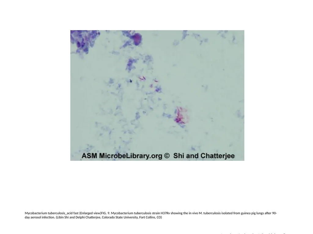
Mycobacterium tuberculosis_acid fast (Enlarged view)FIG. 9. Mycobacterium tuberculosis strain H37Rv showing the in vivo M. tuberculosis isolated from guinea pig lungs after 90-day aerosol infection. (Libin Shi and Delphi Chatterjee, Colorado State University, Fort Collins, CO) American Society For Microbiology ©<br>
11
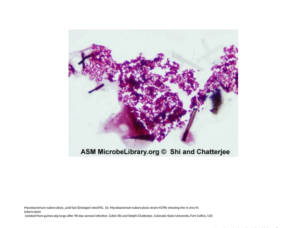
Mycobacterium tuberculosis_acid fast (Enlarged view)FIG. 10. Mycobacterium tuberculosis strain H37Rv showing the in vivo M.
tuberculosis
isolated from guinea pig lungs after 90-day aerosol infection. (Libin Shi and Delphi Chatterjee, Colorado State University, Fort Collins, CO) American Society For Microbiology ©<br>
tuberculosis
isolated from guinea pig lungs after 90-day aerosol infection. (Libin Shi and Delphi Chatterjee, Colorado State University, Fort Collins, CO) American Society For Microbiology ©<br>
12
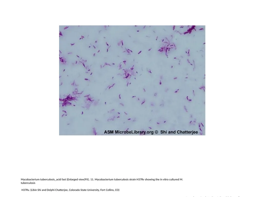
Mycobacterium tuberculosis_acid fast (Enlarged view)FIG. 11. Mycobacterium tuberculosis strain H37Rv showing the in vitro cultured M.
tuberculosis
H37Rv. (Libin Shi and Delphi Chatterjee, Colorado State University, Fort Collins, CO) American Society For Microbiology ©<br>
tuberculosis
H37Rv. (Libin Shi and Delphi Chatterjee, Colorado State University, Fort Collins, CO) American Society For Microbiology ©<br>
13
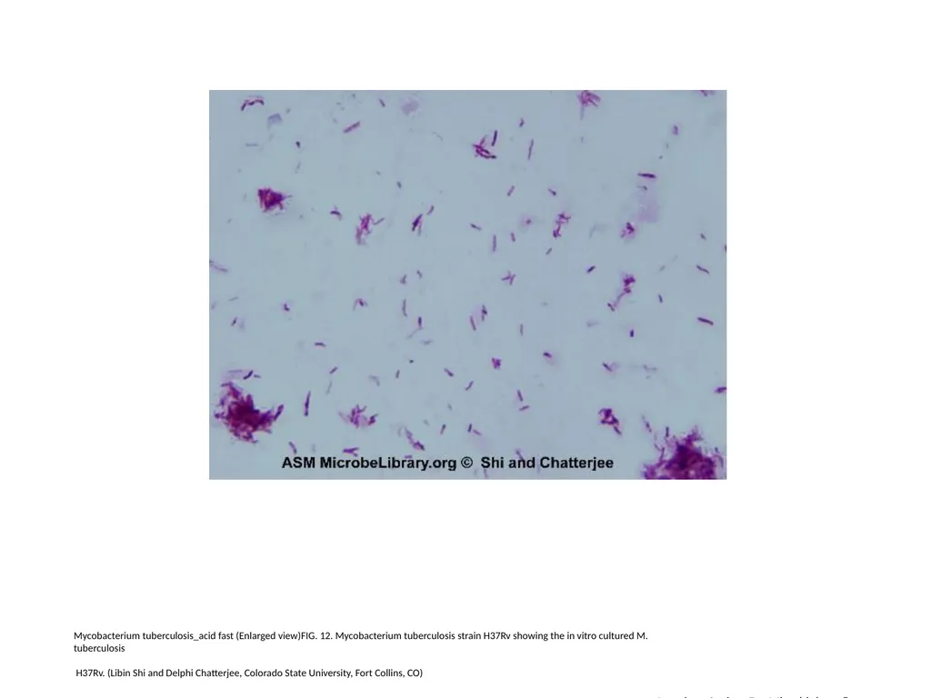
Mycobacterium tuberculosis_acid fast (Enlarged view)FIG. 12. Mycobacterium tuberculosis strain H37Rv showing the in vitro cultured M.
tuberculosis
H37Rv. (Libin Shi and Delphi Chatterjee, Colorado State University, Fort Collins, CO) American Society For Microbiology ©<br>
tuberculosis
H37Rv. (Libin Shi and Delphi Chatterjee, Colorado State University, Fort Collins, CO) American Society For Microbiology ©<br>
14
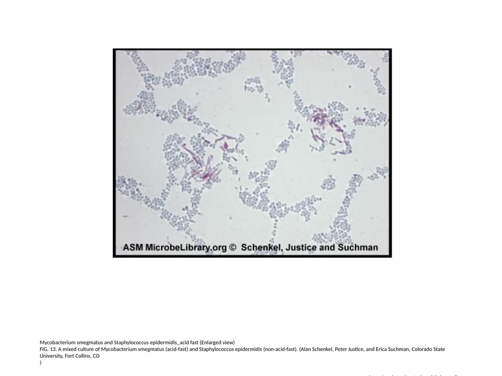
Mycobacterium smegmatus and Staphylococcus epidermidis_acid fast (Enlarged view)
FIG. 13. A mixed culture of Mycobacterium smegmatus (acid-fast) and Staphylococcus epidermidis (non-acid-fast). (Alan Schenkel, Peter Justice, and Erica Suchman, Colorado State University, Fort Collins, CO
) American Society For Microbiology ©<br>
FIG. 13. A mixed culture of Mycobacterium smegmatus (acid-fast) and Staphylococcus epidermidis (non-acid-fast). (Alan Schenkel, Peter Justice, and Erica Suchman, Colorado State University, Fort Collins, CO
) American Society For Microbiology ©<br>
15
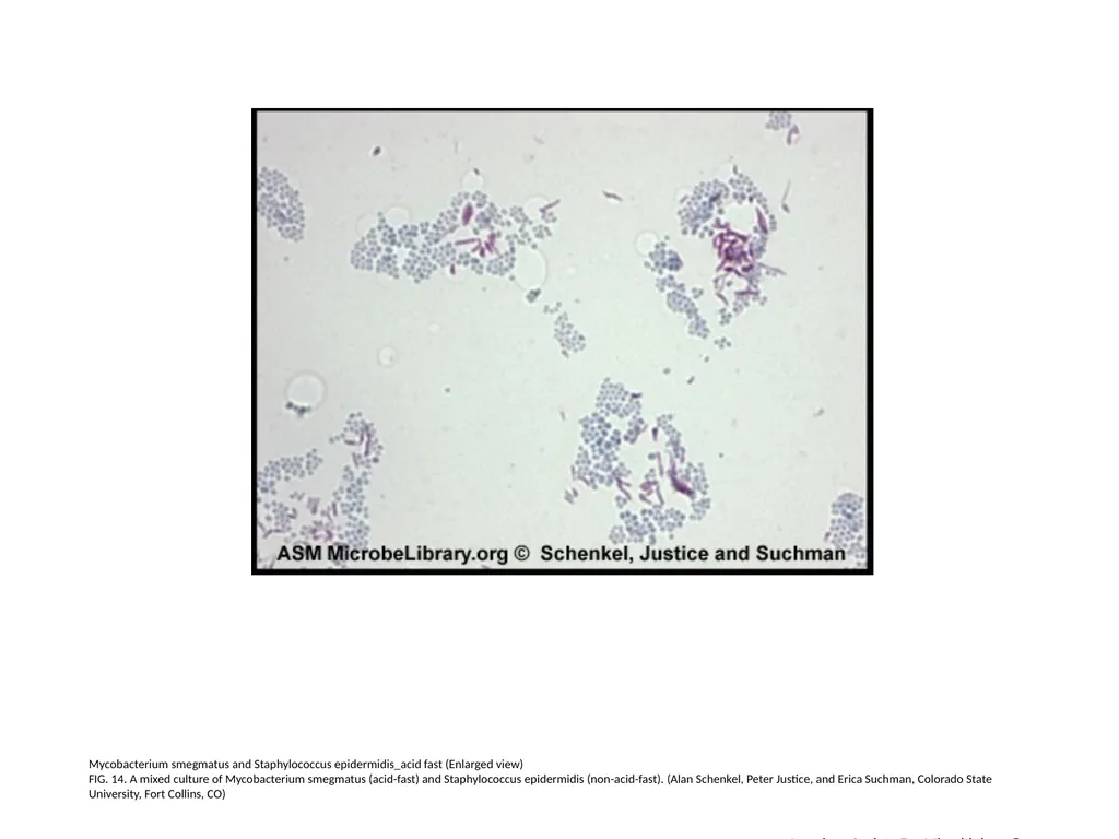
Mycobacterium smegmatus and Staphylococcus epidermidis_acid fast (Enlarged view)
FIG. 14. A mixed culture of Mycobacterium smegmatus (acid-fast) and Staphylococcus epidermidis (non-acid-fast). (Alan Schenkel, Peter Justice, and Erica Suchman, Colorado State University, Fort Collins, CO) American Society For Microbiology ©<br>
FIG. 14. A mixed culture of Mycobacterium smegmatus (acid-fast) and Staphylococcus epidermidis (non-acid-fast). (Alan Schenkel, Peter Justice, and Erica Suchman, Colorado State University, Fort Collins, CO) American Society For Microbiology ©<br>
16
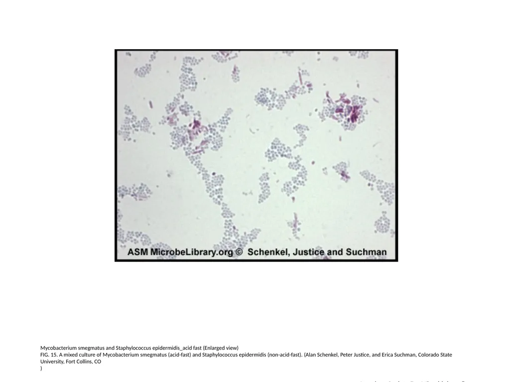
Mycobacterium smegmatus and Staphylococcus epidermidis_acid fast (Enlarged view)
FIG. 15. A mixed culture of Mycobacterium smegmatus (acid-fast) and Staphylococcus epidermidis (non-acid-fast). (Alan Schenkel, Peter Justice, and Erica Suchman, Colorado State University, Fort Collins, CO
) American Society For Microbiology ©<br>
FIG. 15. A mixed culture of Mycobacterium smegmatus (acid-fast) and Staphylococcus epidermidis (non-acid-fast). (Alan Schenkel, Peter Justice, and Erica Suchman, Colorado State University, Fort Collins, CO
) American Society For Microbiology ©<br>
17
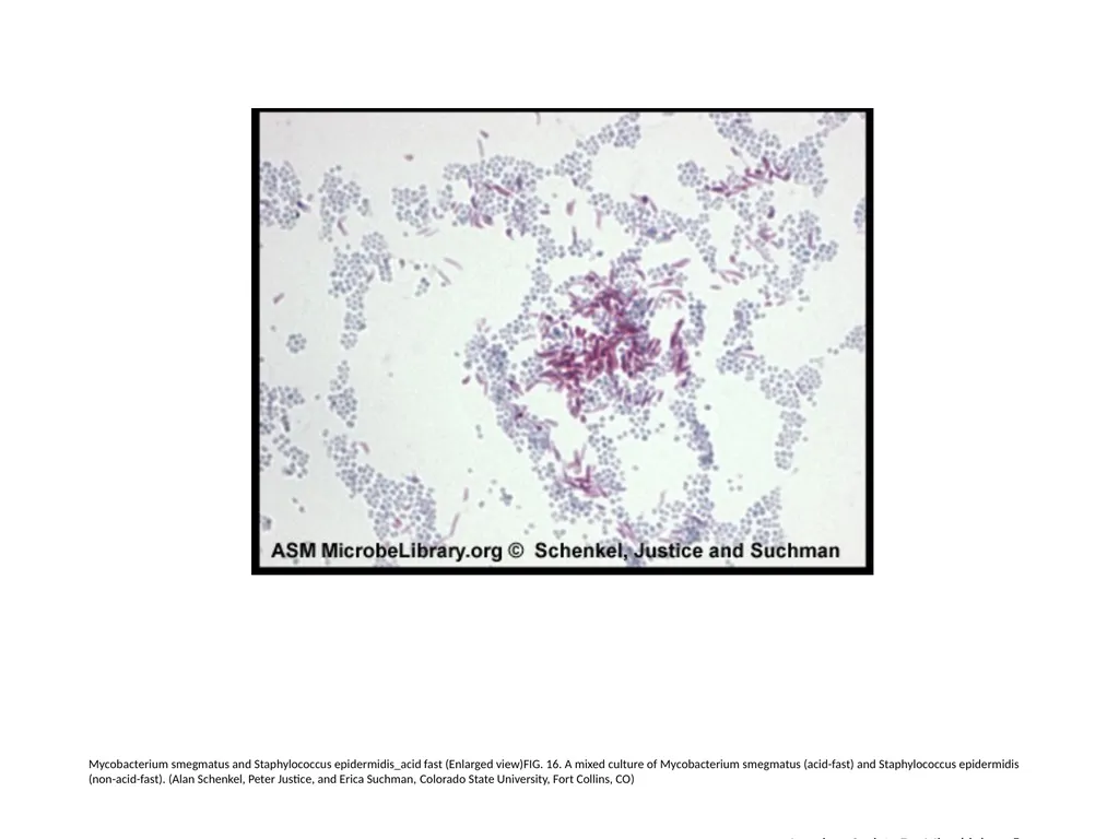
Mycobacterium smegmatus and Staphylococcus epidermidis_acid fast (Enlarged view)FIG. 16. A mixed culture of Mycobacterium smegmatus (acid-fast) and Staphylococcus epidermidis (non-acid-fast). (Alan Schenkel, Peter Justice, and Erica Suchman, Colorado State University, Fort Collins, CO) American Society For Microbiology ©<br>
18
Mycobacterium smegmatus and Staphylococcus epidermidis_acid fast (Enlarged view)
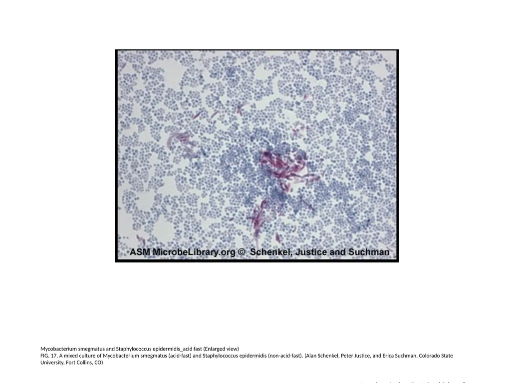
FIG. 17. A mixed culture of Mycobacterium smegmatus (acid-fast) and Staphylococcus epidermidis (non-acid-fast). (Alan Schenkel, Peter Justice, and Erica Suchman, Colorado State University, Fort Collins, CO) American Society For Microbiology ©<br>
FIG. 17. A mixed culture of Mycobacterium smegmatus (acid-fast) and Staphylococcus epidermidis (non-acid-fast). (Alan Schenkel, Peter Justice, and Erica Suchman, Colorado State University, Fort Collins, CO) American Society For Microbiology ©<br>
19
Mesenteric lymph node_acid fast (Enlarged view)
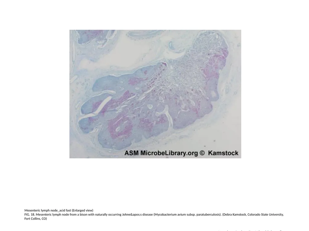
FIG. 18. Mesenteric lymph node from a bison with naturally occurring Johne's disease (Mycobacterium avium subsp. paratuberculosis). (Debra Kamstock, Colorado State University, Fort Collins, CO) American Society For Microbiology ©<br>
FIG. 18. Mesenteric lymph node from a bison with naturally occurring Johne's disease (Mycobacterium avium subsp. paratuberculosis). (Debra Kamstock, Colorado State University, Fort Collins, CO) American Society For Microbiology ©<br>
20
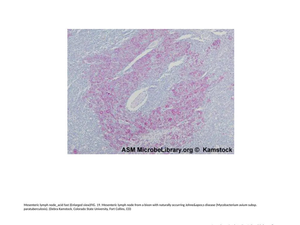
Mesenteric lymph node_acid fast (Enlarged view)FIG. 19. Mesenteric lymph node from a bison with naturally occurring Johne's disease (Mycobacterium avium subsp. paratuberculosis). (Debra Kamstock, Colorado State University, Fort Collins, CO) American Society For Microbiology ©<br>
21
Mesenteric lymph node_acid fast (Enlarged view)
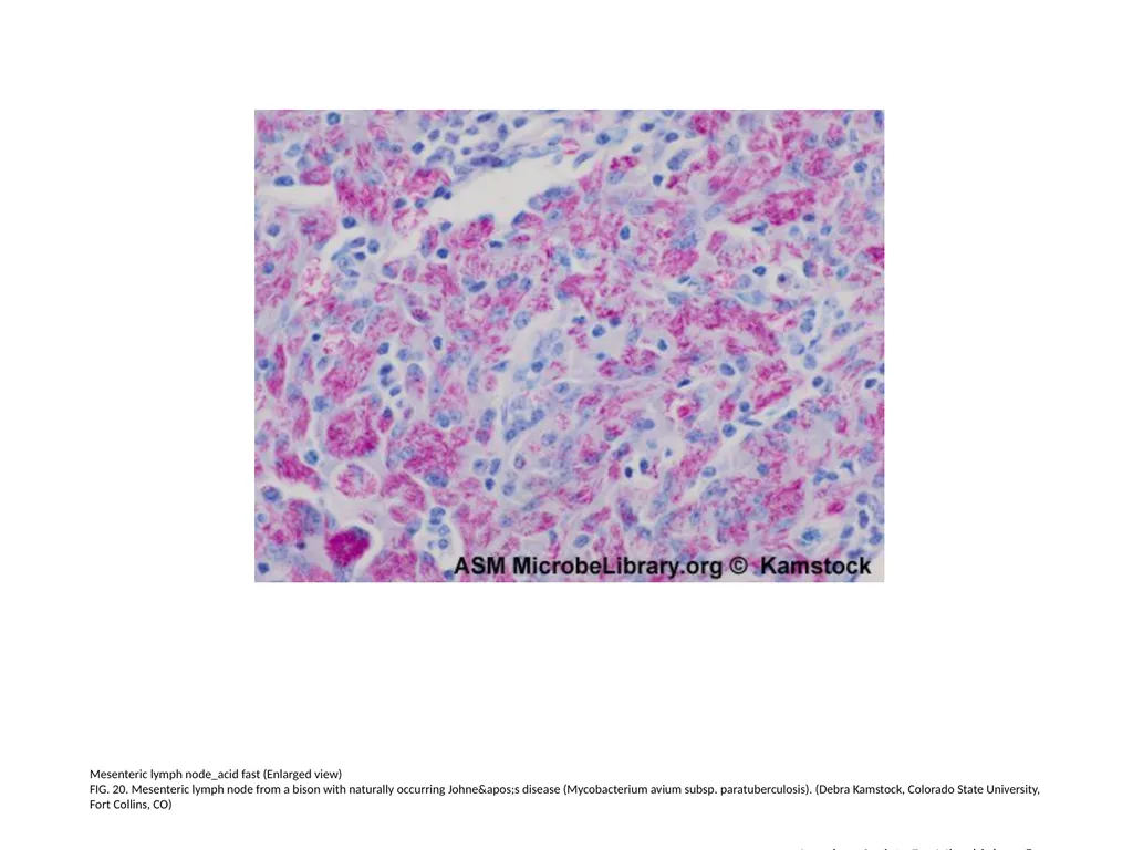
FIG. 20. Mesenteric lymph node from a bison with naturally occurring Johne's disease (Mycobacterium avium subsp. paratuberculosis). (Debra Kamstock, Colorado State University, Fort Collins, CO) American Society For Microbiology ©<br>
FIG. 20. Mesenteric lymph node from a bison with naturally occurring Johne's disease (Mycobacterium avium subsp. paratuberculosis). (Debra Kamstock, Colorado State University, Fort Collins, CO) American Society For Microbiology ©<br>
22
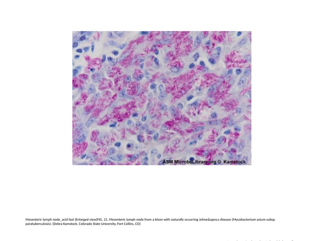
Mesenteric lymph node_acid fast (Enlarged view)FIG. 21. Mesenteric lymph node from a bison with naturally occurring Johne's disease (Mycobacterium avium subsp. paratuberculosis). (Debra Kamstock, Colorado State University, Fort Collins, CO) American Society For Microbiology ©<br>
23
Mesenteric lymph node_acid fast (Enlarged view)
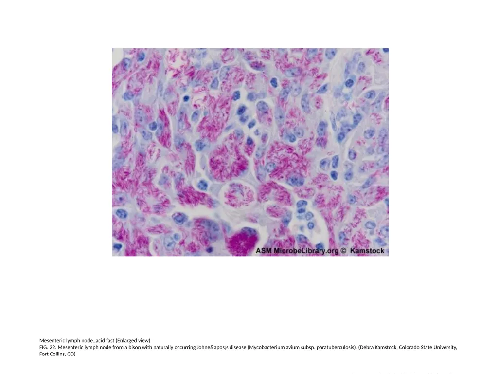
FIG. 22. Mesenteric lymph node from a bison with naturally occurring Johne's disease (Mycobacterium avium subsp. paratuberculosis). (Debra Kamstock, Colorado State University, Fort Collins, CO) American Society For Microbiology ©<br>
FIG. 22. Mesenteric lymph node from a bison with naturally occurring Johne's disease (Mycobacterium avium subsp. paratuberculosis). (Debra Kamstock, Colorado State University, Fort Collins, CO) American Society For Microbiology ©<br>
24
American Society For Microbiology © Mycobacterium tuberculosis_acid fast (Enlarged view)FIG. 23.
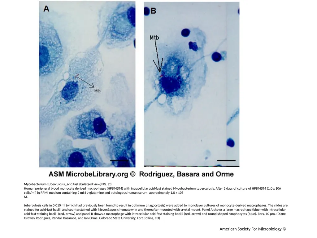
Human peripheral blood monocyte derived macrophages (HPBMDM) with intracellular acid-fast stained Mycobacterium tuberculosis. After 5 days of culture of HPBMDM (1.0 x 10⁶ cells/ml) in RPMI medium containing 2 mM L-glutamine and autologous human serum, approximately 1.0 x 10⁵
M.
tuberculosis cells in 0.010 ml (which had previously been found to result in optimum phagocytosis) were added to monolayer cultures of monocyte-derived macrophages. The slides are stained for acid-fast bacilli and counterstained with Meyer's hematoxylin and thereafter mounted with crystal mount. Panel A shows a large macrophage (blue) with intracellular acid-fast-staining bacilli (red, arrow) and panel B shows a macrophage with intracellular acid-fast-staining bacilli (red, arrow) and round shaped lymphocytes (blue). Bars, 10 µm. (Diane Ordway Rodriguez, Randall Basaraba, and Ian Orme, Colorado State University, Fort Collins, CO)<br>
Human peripheral blood monocyte derived macrophages (HPBMDM) with intracellular acid-fast stained Mycobacterium tuberculosis. After 5 days of culture of HPBMDM (1.0 x 10⁶ cells/ml) in RPMI medium containing 2 mM L-glutamine and autologous human serum, approximately 1.0 x 10⁵
M.
tuberculosis cells in 0.010 ml (which had previously been found to result in optimum phagocytosis) were added to monolayer cultures of monocyte-derived macrophages. The slides are stained for acid-fast bacilli and counterstained with Meyer's hematoxylin and thereafter mounted with crystal mount. Panel A shows a large macrophage (blue) with intracellular acid-fast-staining bacilli (red, arrow) and panel B shows a macrophage with intracellular acid-fast-staining bacilli (red, arrow) and round shaped lymphocytes (blue). Bars, 10 µm. (Diane Ordway Rodriguez, Randall Basaraba, and Ian Orme, Colorado State University, Fort Collins, CO)<br>
25
American Society For Microbiology © Mycobacterium tuberculosis_acid fast (Enlarged view)
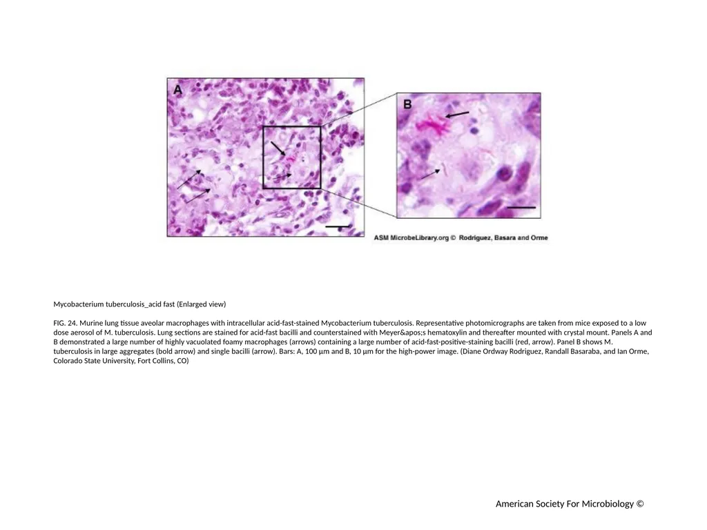
FIG. 24. Murine lung tissue aveolar macrophages with intracellular acid-fast-stained Mycobacterium tuberculosis. Representative photomicrographs are taken from mice exposed to a low dose aerosol of M. tuberculosis. Lung sections are stained for acid-fast bacilli and counterstained with Meyer's hematoxylin and thereafter mounted with crystal mount. Panels A and B demonstrated a large number of highly vacuolated foamy macrophages (arrows) containing a large number of acid-fast-positive-staining bacilli (red, arrow). Panel B shows M. tuberculosis in large aggregates (bold arrow) and single bacilli (arrow). Bars: A, 100 µm and B, 10 µm for the high-power image. (Diane Ordway Rodriguez, Randall Basaraba, and Ian Orme, Colorado State University, Fort Collins, CO)<br>
FIG. 24. Murine lung tissue aveolar macrophages with intracellular acid-fast-stained Mycobacterium tuberculosis. Representative photomicrographs are taken from mice exposed to a low dose aerosol of M. tuberculosis. Lung sections are stained for acid-fast bacilli and counterstained with Meyer's hematoxylin and thereafter mounted with crystal mount. Panels A and B demonstrated a large number of highly vacuolated foamy macrophages (arrows) containing a large number of acid-fast-positive-staining bacilli (red, arrow). Panel B shows M. tuberculosis in large aggregates (bold arrow) and single bacilli (arrow). Bars: A, 100 µm and B, 10 µm for the high-power image. (Diane Ordway Rodriguez, Randall Basaraba, and Ian Orme, Colorado State University, Fort Collins, CO)<br>
26
American Society For Microbiology © Mycobacterium tuberculosis_acid fast (Enlarged view)
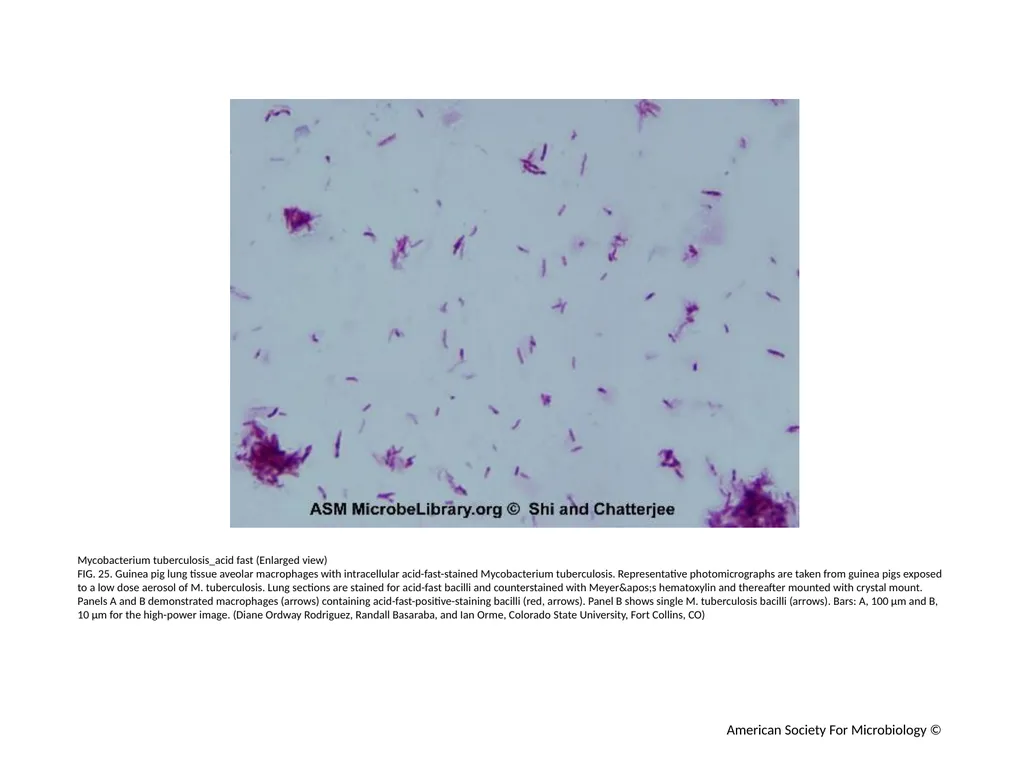
FIG. 25. Guinea pig lung tissue aveolar macrophages with intracellular acid-fast-stained Mycobacterium tuberculosis. Representative photomicrographs are taken from guinea pigs exposed to a low dose aerosol of M. tuberculosis. Lung sections are stained for acid-fast bacilli and counterstained with Meyer's hematoxylin and thereafter mounted with crystal mount. Panels A and B demonstrated macrophages (arrows) containing acid-fast-positive-staining bacilli (red, arrows). Panel B shows single M. tuberculosis bacilli (arrows). Bars: A, 100 µm and B, 10 µm for the high-power image. (Diane Ordway Rodriguez, Randall Basaraba, and Ian Orme, Colorado State University, Fort Collins, CO)<br>
FIG. 25. Guinea pig lung tissue aveolar macrophages with intracellular acid-fast-stained Mycobacterium tuberculosis. Representative photomicrographs are taken from guinea pigs exposed to a low dose aerosol of M. tuberculosis. Lung sections are stained for acid-fast bacilli and counterstained with Meyer's hematoxylin and thereafter mounted with crystal mount. Panels A and B demonstrated macrophages (arrows) containing acid-fast-positive-staining bacilli (red, arrows). Panel B shows single M. tuberculosis bacilli (arrows). Bars: A, 100 µm and B, 10 µm for the high-power image. (Diane Ordway Rodriguez, Randall Basaraba, and Ian Orme, Colorado State University, Fort Collins, CO)<br>
27
Acid-fast stain of Mycobacterium smegmatis and Staphylococcus aureus
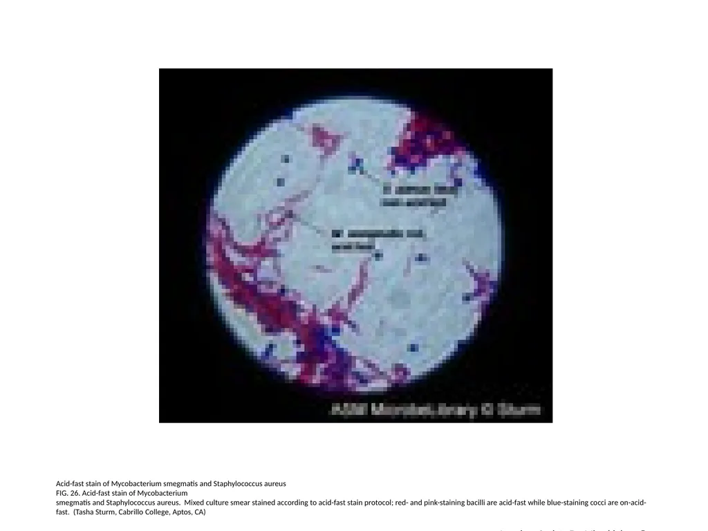
FIG. 26. Acid-fast stain of Mycobacterium
smegmatis and Staphylococcus aureus. Mixed culture smear stained according to acid-fast stain protocol; red- and pink-staining bacilli are acid-fast while blue-staining cocci are on-acid-fast. (Tasha Sturm, Cabrillo College, Aptos, CA) American Society For Microbiology ©<br>
FIG. 26. Acid-fast stain of Mycobacterium
smegmatis and Staphylococcus aureus. Mixed culture smear stained according to acid-fast stain protocol; red- and pink-staining bacilli are acid-fast while blue-staining cocci are on-acid-fast. (Tasha Sturm, Cabrillo College, Aptos, CA) American Society For Microbiology ©<br>