04
CLINICAL ASSESSMENT History.
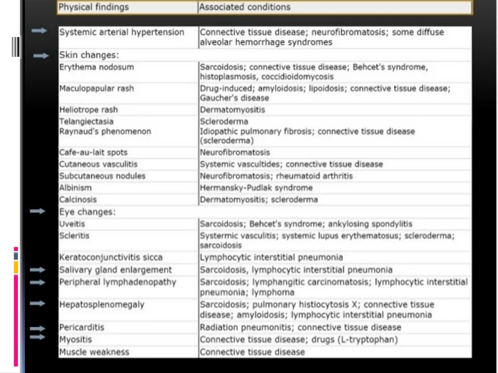
Physical Exam.
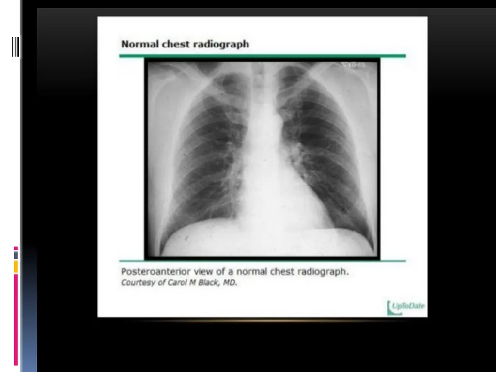
Chest Radiograph.
Pulmonary Function Testing.
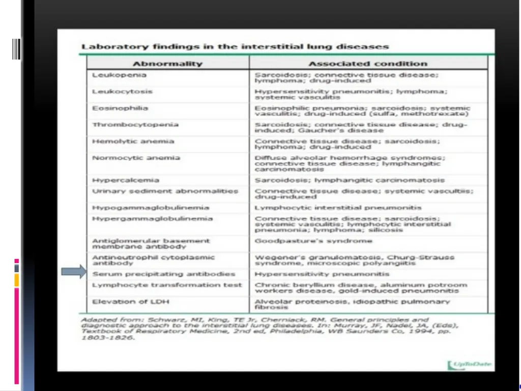
Laboratory Studies.
Tissue examination<br>
05
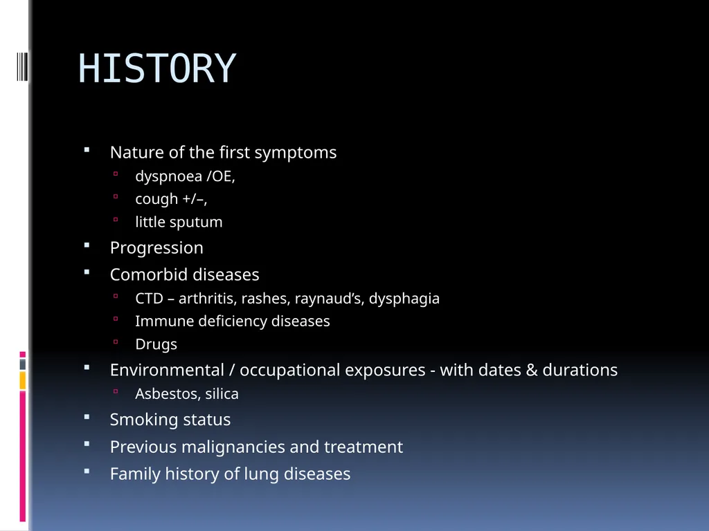
HISTORY Nature of the first symptoms
dyspnoea /OE,
cough +/–,
little sputum
Progression
Comorbid diseases
CTD – arthritis, rashes, raynaud’s, dysphagia
Immune deficiency diseases
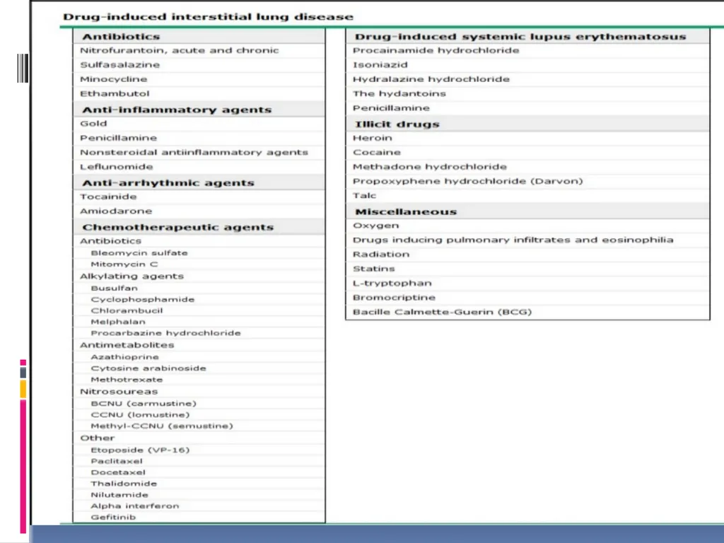
Drugs
Environmental / occupational exposures - with dates & durations
Asbestos, silica
Smoking status
Previous malignancies and treatment
Family history of lung diseases<br>
06
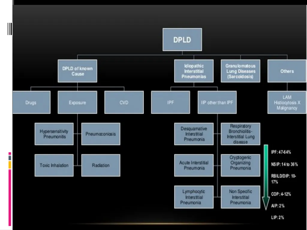
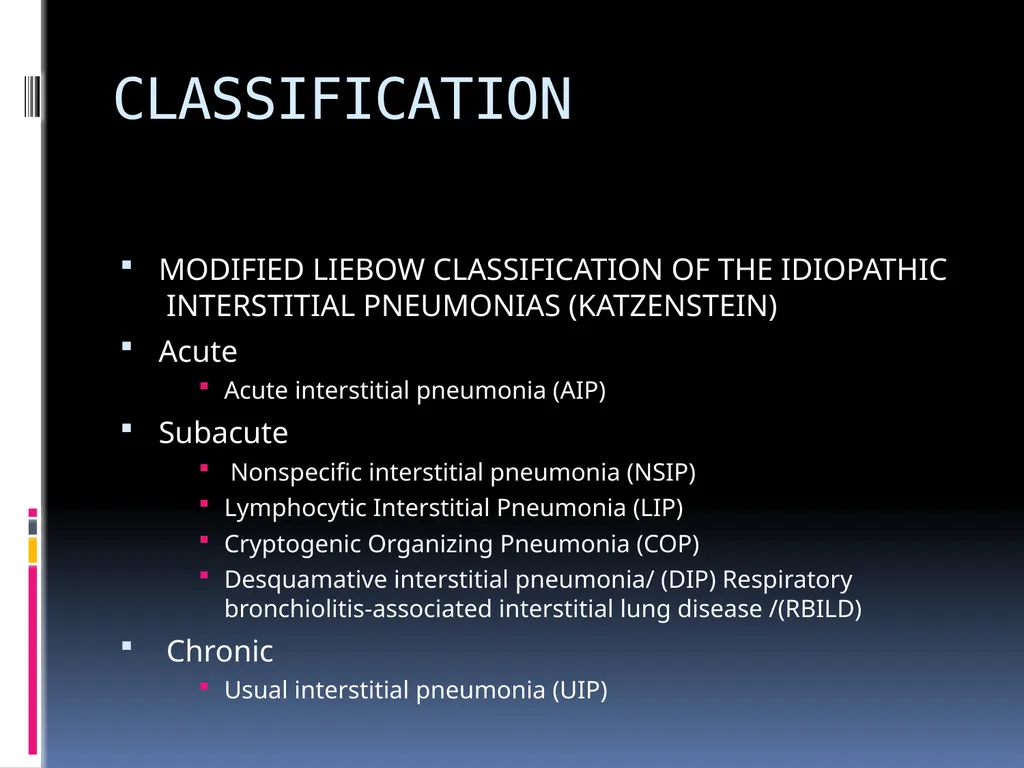
CLASSIFICATION MODIFIED LIEBOW CLASSIFICATION OF THE IDIOPATHIC INTERSTITIAL PNEUMONIAS (KATZENSTEIN)
Acute
Acute interstitial pneumonia (AIP)
Subacute
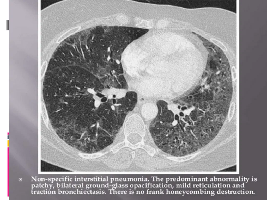
Nonspecific interstitial pneumonia (NSIP)
Lymphocytic Interstitial Pneumonia (LIP)
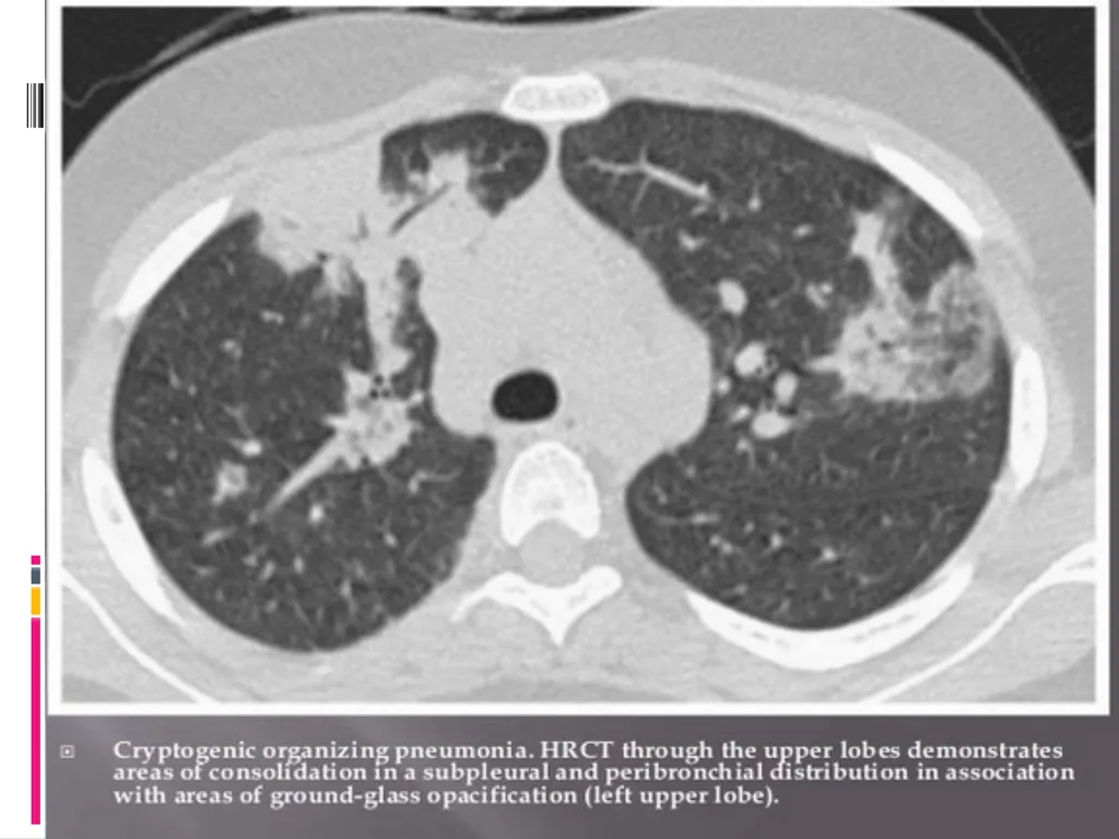
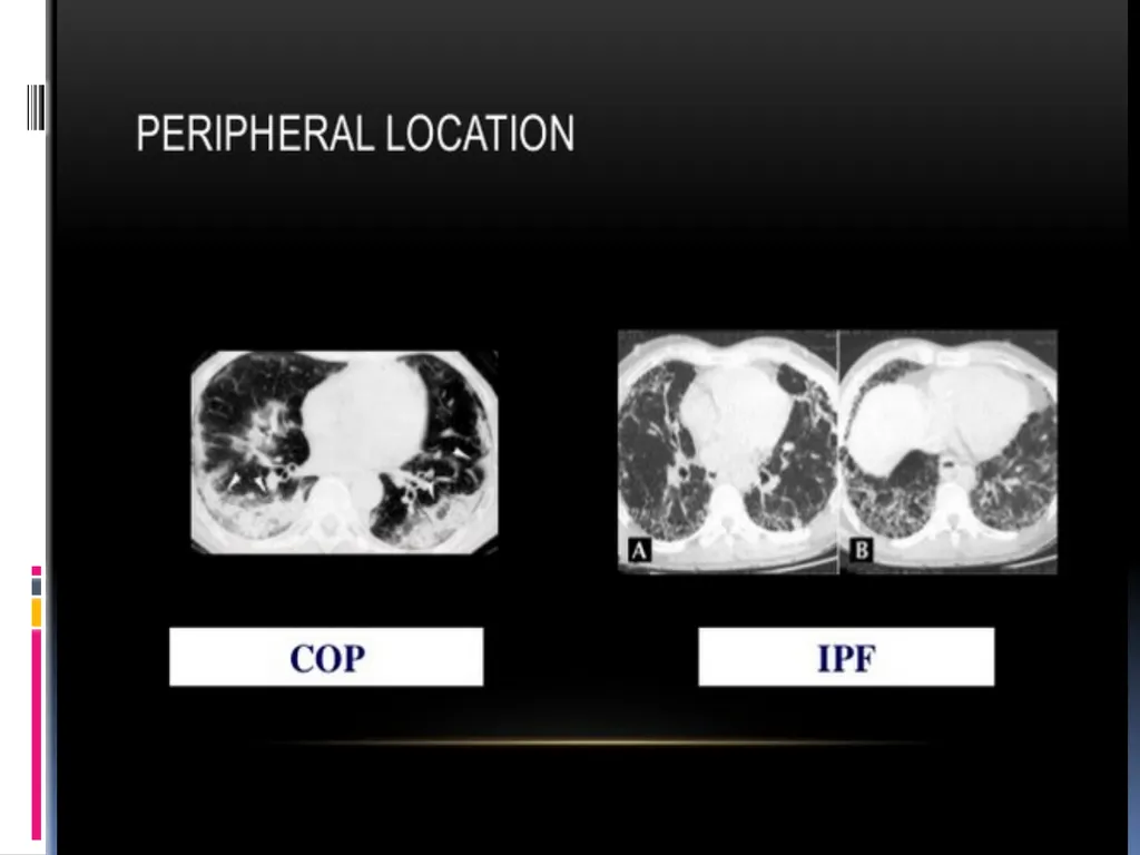
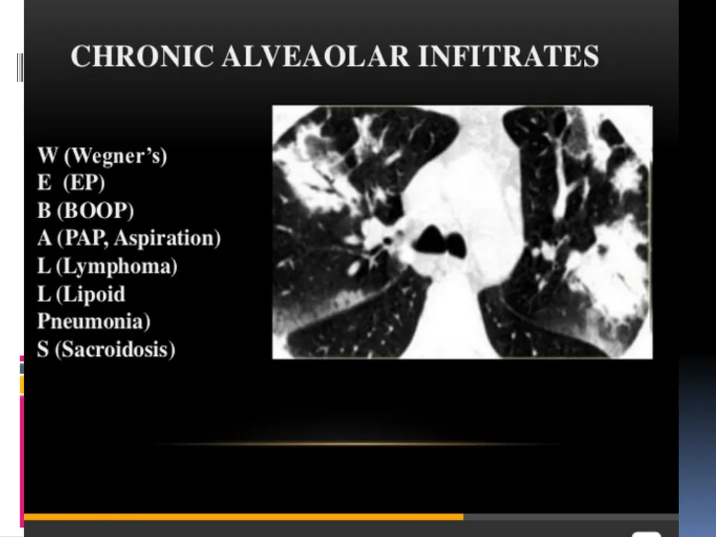
Cryptogenic Organizing Pneumonia (COP)
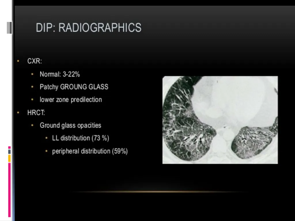
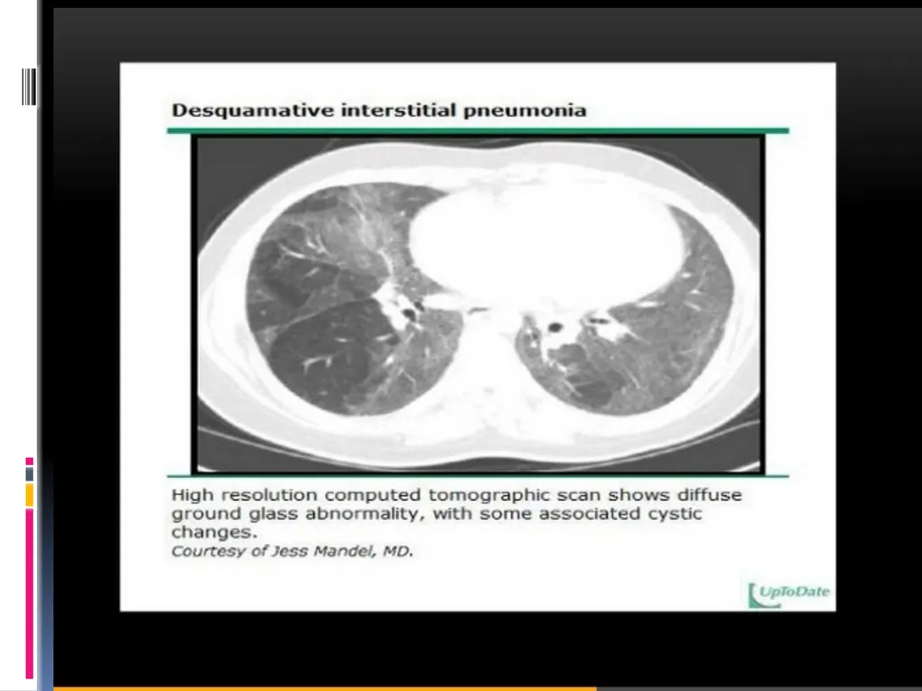
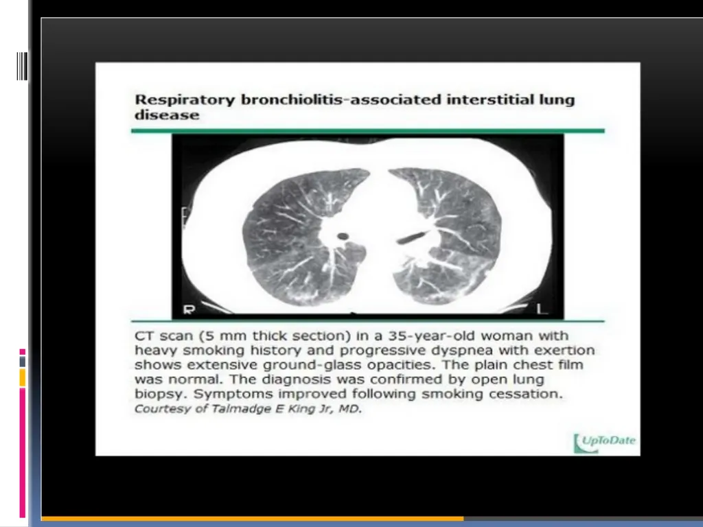
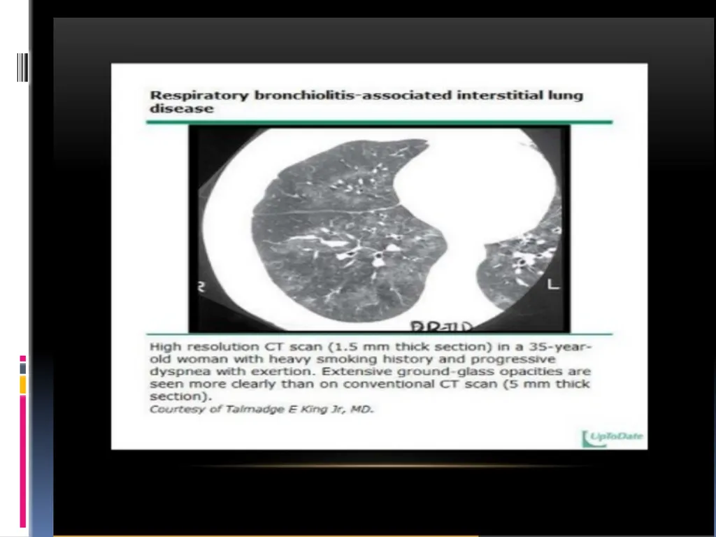
Desquamative interstitial pneumonia/ (DIP) Respiratory bronchiolitis-associated interstitial lung disease /(RBILD)
Chronic
Usual interstitial pneumonia (UIP)<br>
07
PHYSICAL EXAMINATION • Lung examination
• Cardiac examination
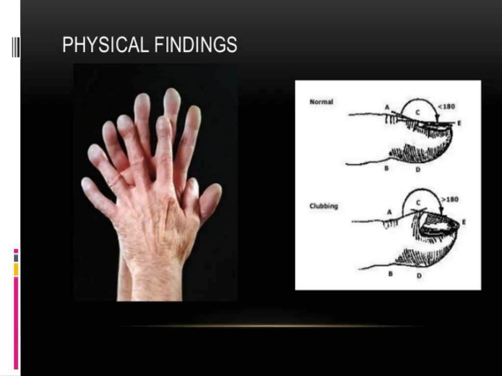
• Clubbing
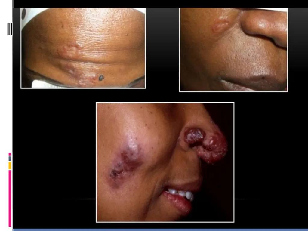
• Extrapulmonary findings of systemic disease<br>
08
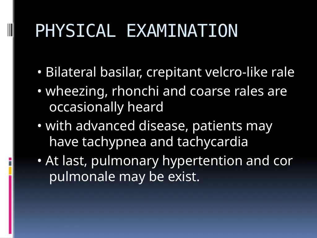
PHYSICAL EXAMINATION • Bilateral basilar, crepitant velcro-like rale
• wheezing, rhonchi and coarse rales are occasionally heard
• with advanced disease, patients may have tachypnea and tachycardia
• At last, pulmonary hypertention and cor pulmonale may be exist.<br>
10
LABORTARY FINDINGS<br>
11
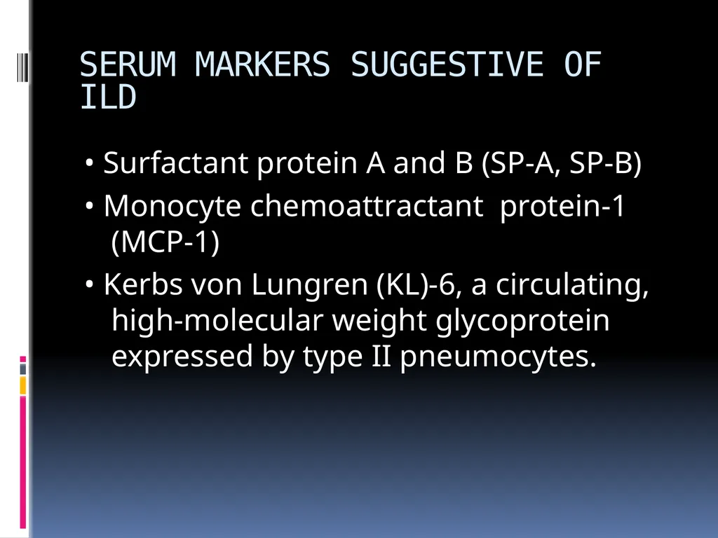
SERUM MARKERS SUGGESTIVE OF ILD • Surfactant protein A and B (SP-A, SP-B)
• Monocyte chemoattractant protein-1 (MCP-1)
• Kerbs von Lungren (KL)-6, a circulating, high-molecular weight glycoprotein expressed by type II pneumocytes.<br>
12
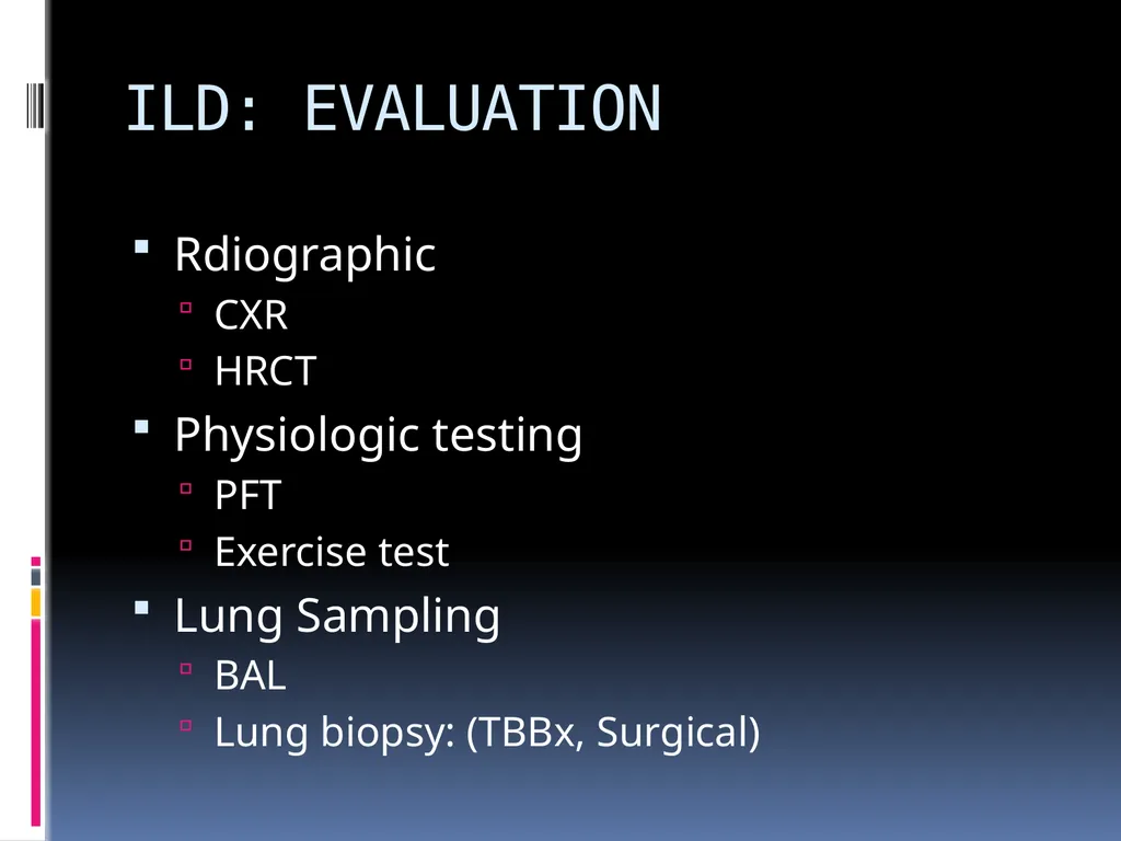
ILD: EVALUATION Rdiographic
CXR
HRCT
Physiologic testing
PFT
Exercise test
Lung Sampling
BAL
Lung biopsy: (TBBx, Surgical)<br>
13
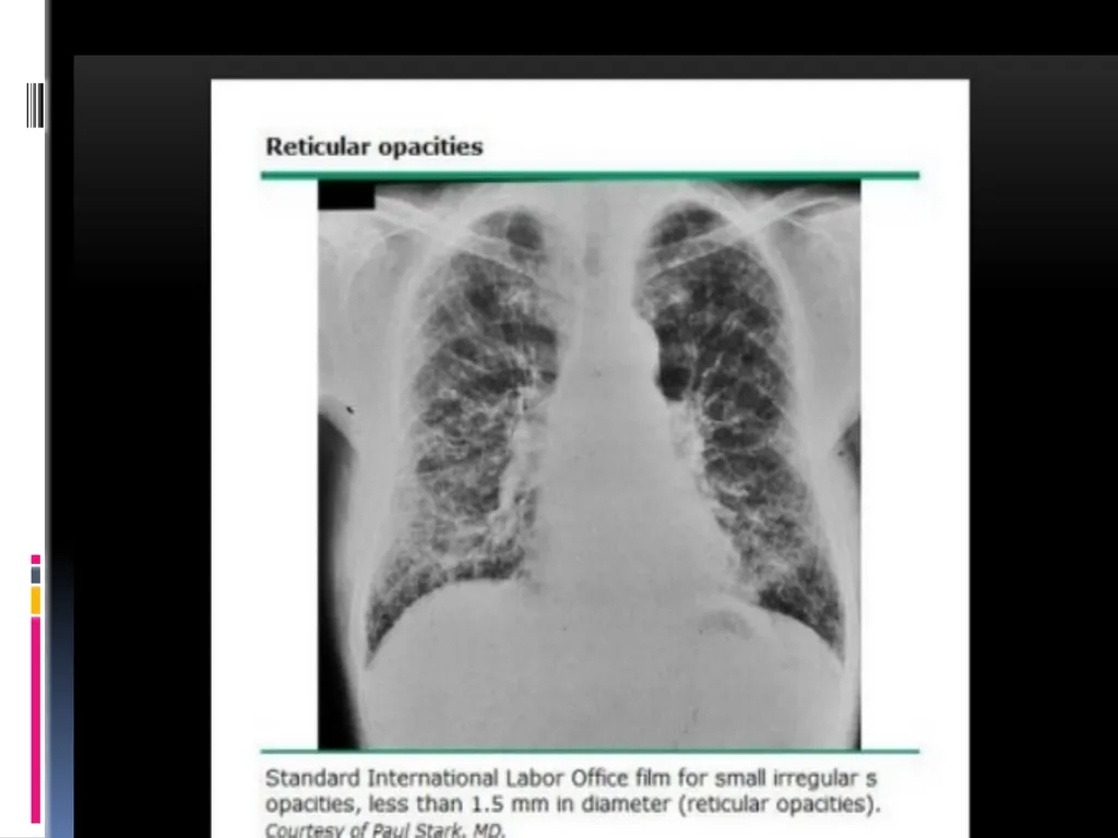
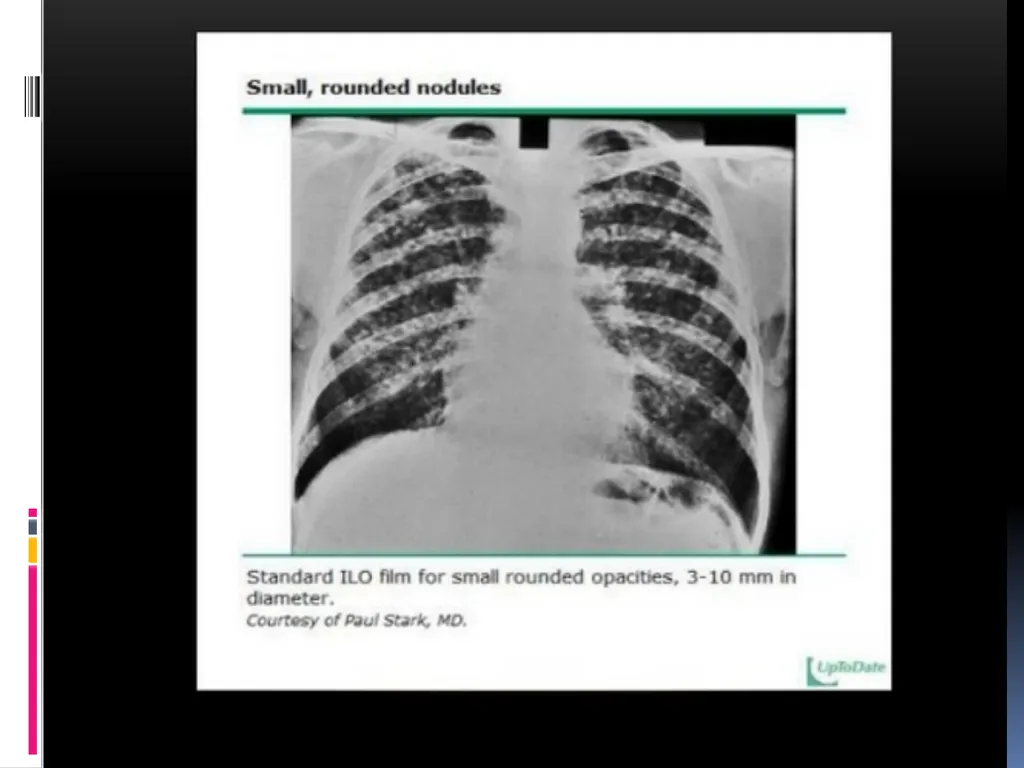
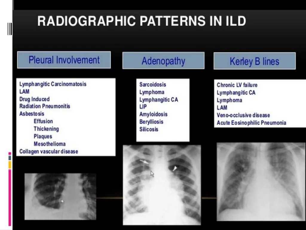
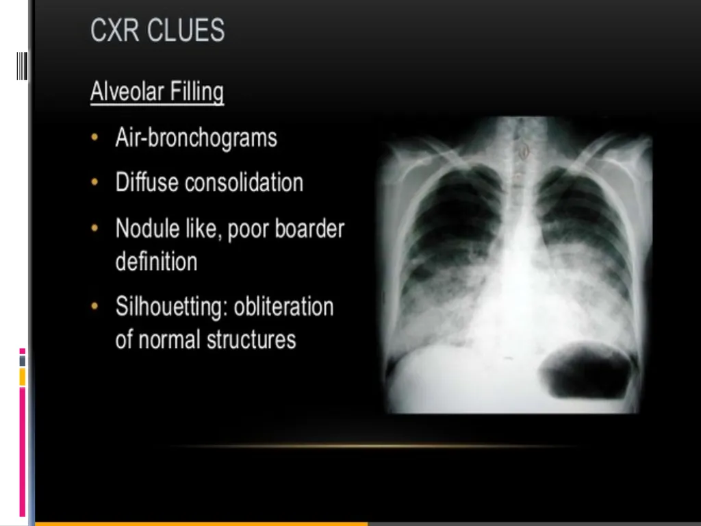
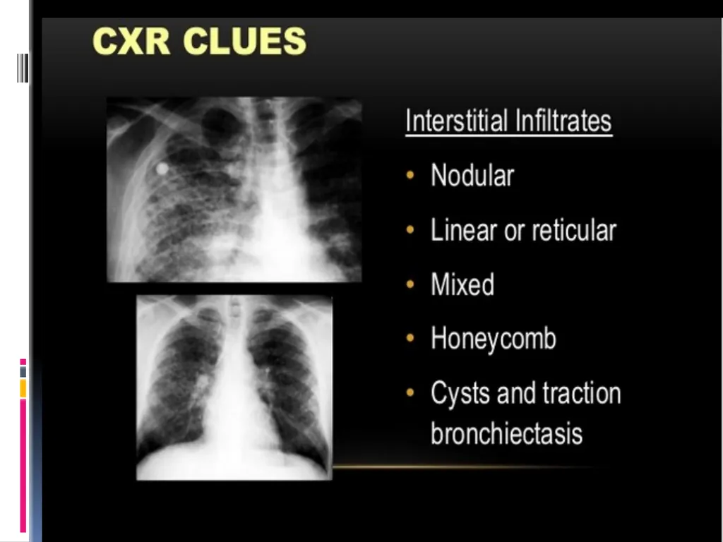
CHEST RADIOGRAPHY It is the important method to diagnose the ILDs.<br>
14
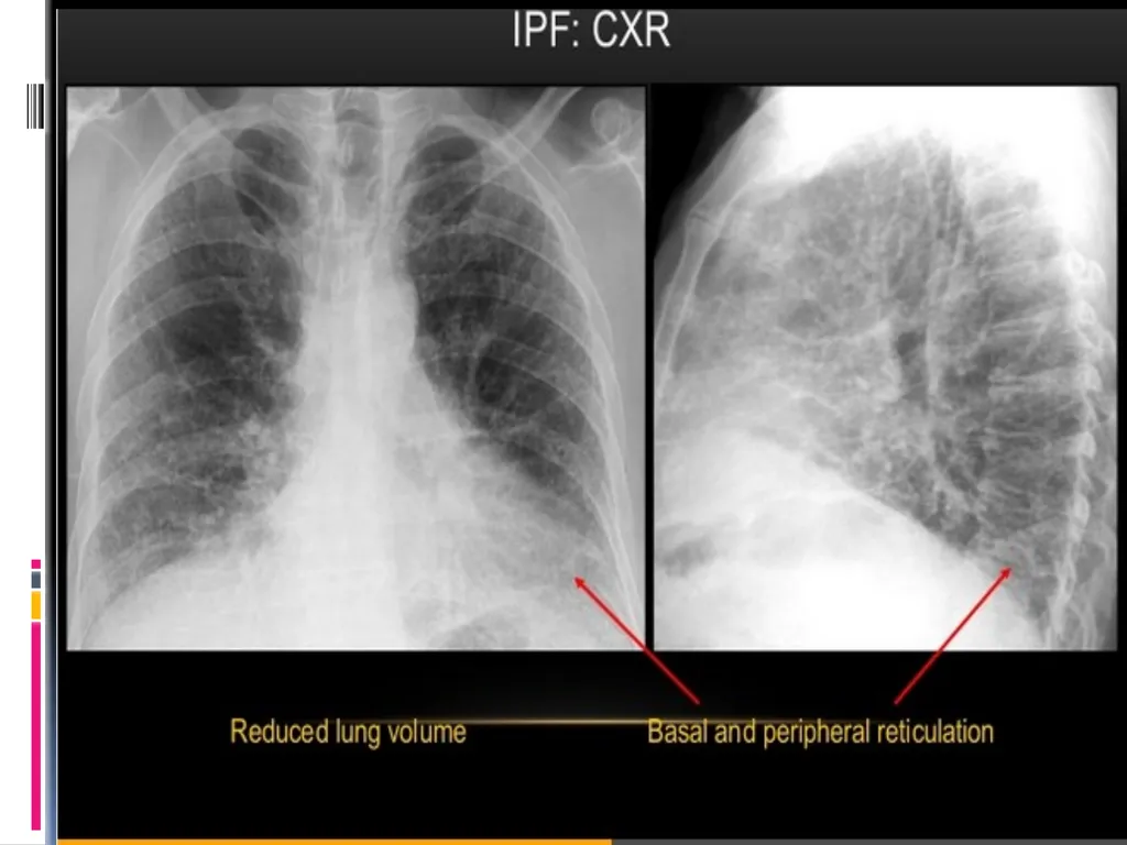
IMAGING -CXR The correlation between the radiographic pattern and the stage of disease (clinical or histopathologic) is generally poor
Honeycombing correlates a poor prognosis.
Review all previous chest films to assess the rate of change in disease activity.<br>
15
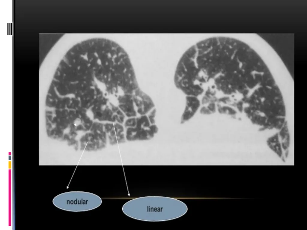
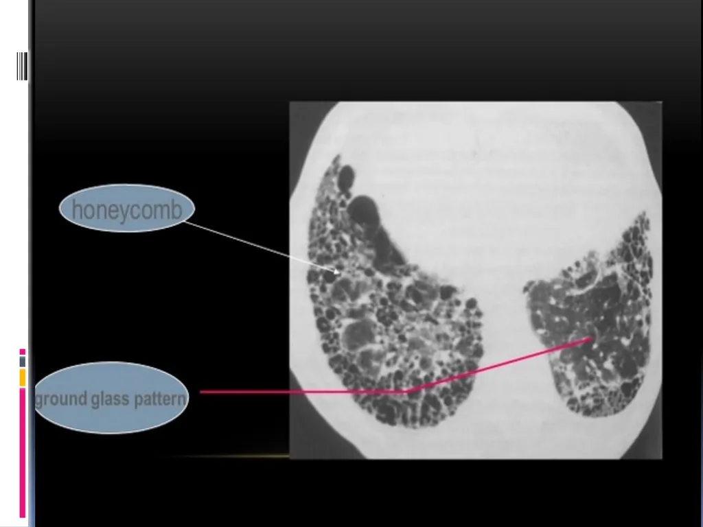
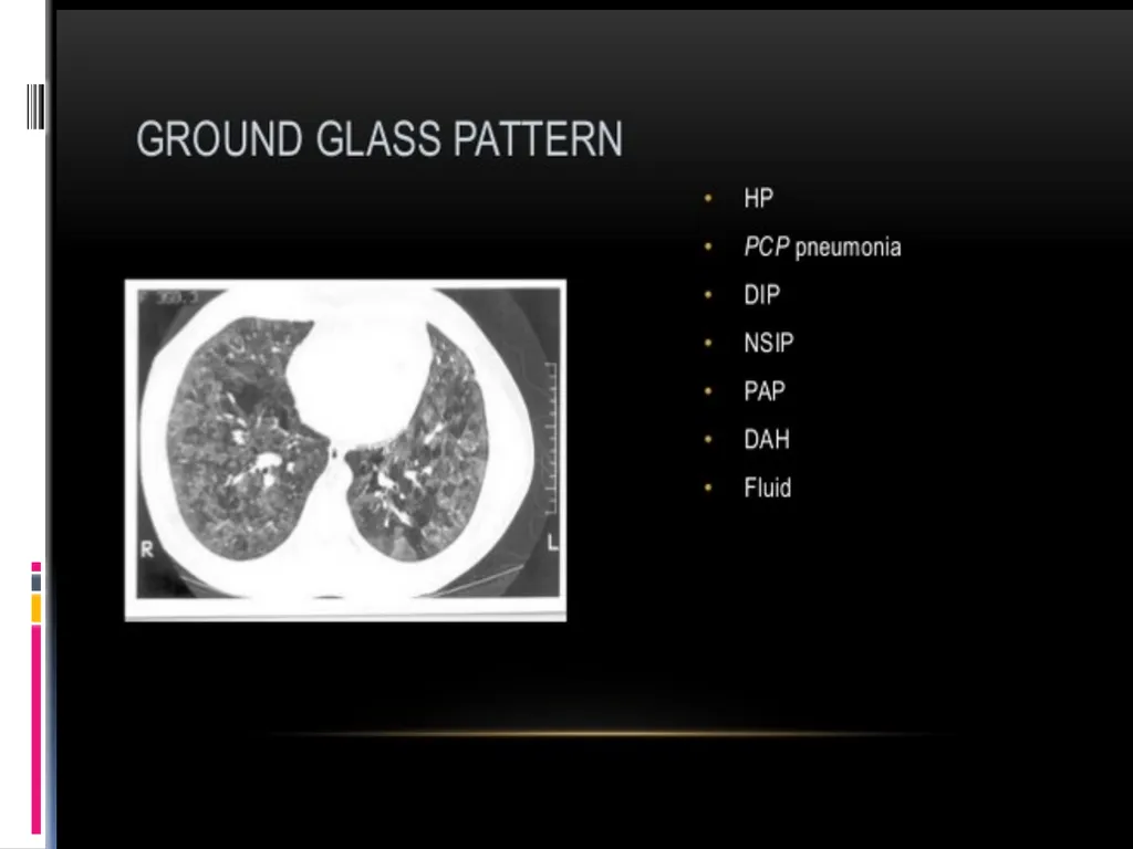
A diffuse ground glass pattern ->early in the disease.
Progresses -> nodules, linear(reticular) infiltrates, or a combination.
infiltrates become coarser and lung volume is lost-> honeycomb pattern<br>
16
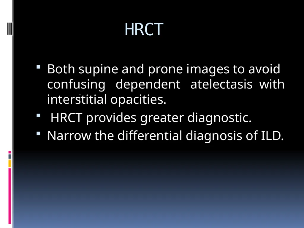
HRCT Both supine and prone images to avoid confusing dependent atelectasis with interstitial opacities.
HRCT provides greater diagnostic.
Narrow the differential diagnosis of ILD. ;<br>
17
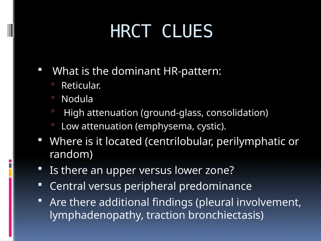
HRCT CLUES What is the dominant HR-pattern:
Reticular.
Nodula
High attenuation (ground-glass, consolidation)
Low attenuation (emphysema, cystic).
Where is it located (centrilobular, perilymphatic or random)
Is there an upper versus lower zone?
Central versus peripheral predominance
Are there additional findings (pleural involvement, lymphadenopathy, traction bronchiectasis)<br>
18
HRCT FINDINGS Bilateral symmetric hilar adenopathy and upper lung zone reticular opacities - > sarcoidosis.
Pleural plaques with linear calcification -> Asbstosis
Centrilobular nodules that spare the subpleural
region ->hypersensitivity pneumonitis, sarcoidosis,Langerhans cell histiocytosis;respiratory, follicular, and cellular bronchiolitis.<br>
19
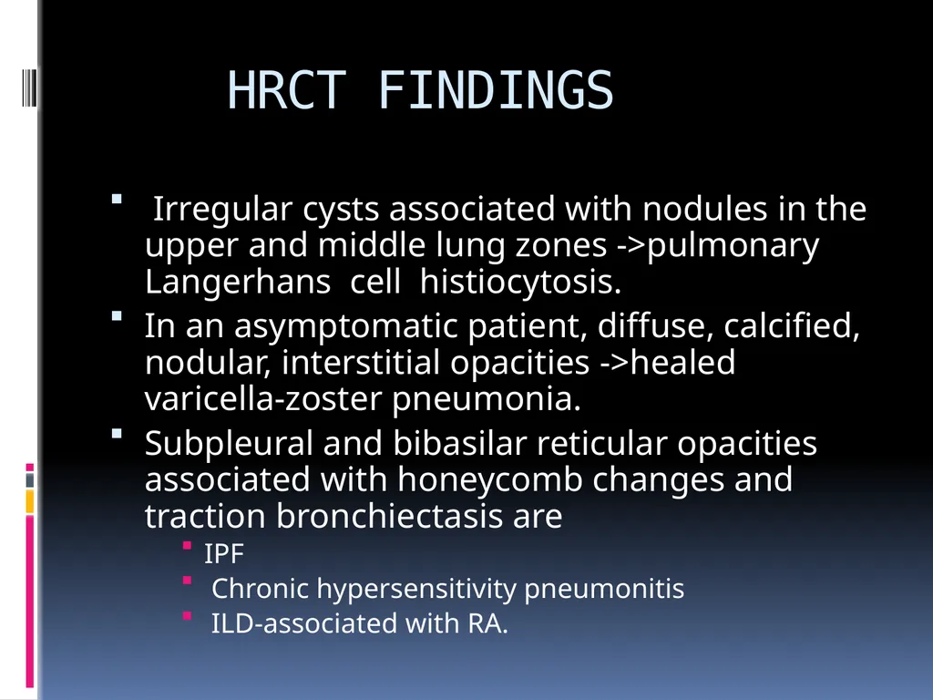
HRCT FINDINGS Irregular cysts associated with nodules in the upper and middle lung zones ->pulmonary Langerhans cell histiocytosis.
In an asymptomatic patient, diffuse, calcified, nodular, interstitial opacities ->healed varicella-zoster pneumonia.
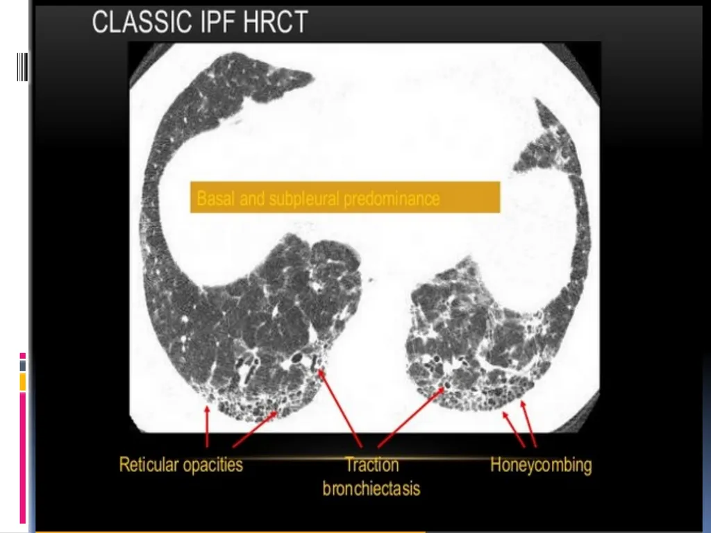
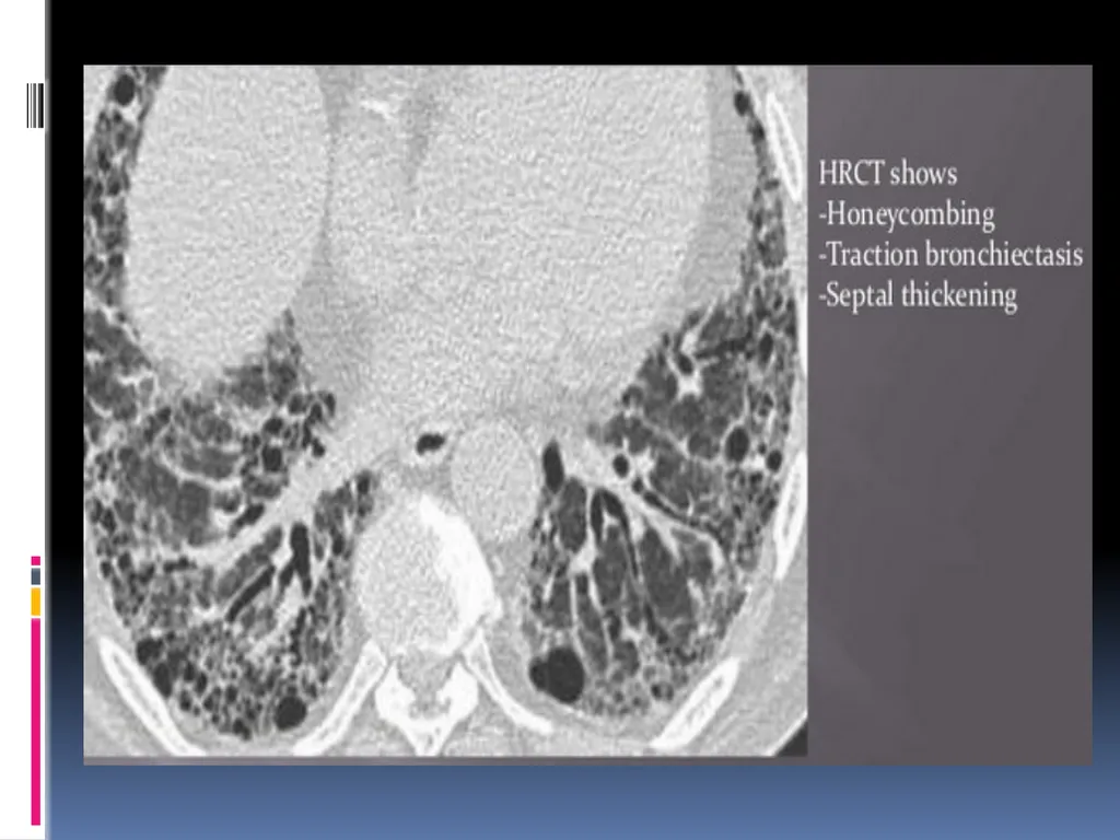
Subpleural and bibasilar reticular opacities associated with honeycomb changes and traction bronchiectasis are
IPF
Chronic hypersensitivity pneumonitis
ILD-associated with RA.<br>
20
NSIP may be distinguished from UIP on CT by a more prominent component of ground- glass attenuation and a finer reticular pattern in the absence of honeycombing.<br>
21
FDG-PET SCANNING The role of (18)F-2-deoxy-2-fluoro-D-glucose (FDG) (PET) in the evaluation of ILD is unclear.
Positive FDG uptake can be seen in
Sarcoidosis
Pulmonary
Langerhans cell
histiocytosis
Lymphangitic carcinomatosis
Not typically obtained in the evaluation of ILD.<br>
22
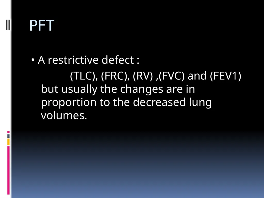
PFT • A restrictive defect :
(TLC), (FRC), (RV) ,(FVC) and (FEV1) but usually the changes are in proportion to the decreased lung volumes.<br>
23
AN INTERSTITIAL PATTERN ON CXR ACCOMPANIED BY OBSTRUCTIVE AIRFLOW SUGGESTIVE OF :
Sarcoidosis
Lymphangioleiomyomatosis
Hypersensitivity pneumonitis
Pulmonary Langerhans cell histiocytosis
Tuberous sclerosis and pulmonary lymphangioleiomyomatosis
Combined COPD and ILD
Constrictive bronchiolitis<br>
24
PFT A reduction (DLCO) is a common, but nonspecific finding in ILD- , the severity of the DLCO reduction does not correlate well with disease prognosis, unless the DLCO is less than 35 % of predicted.
Due to effacement of the alveolar capillary units but more importantly, to the extent of mismatching of V/Q of the alveoli.<br>
25
MODERATE TO SEVERE REDUCTION OF DLCO IN THEPRESENCE OF NORMAL LUNG VOLUMES IN A PATIENTWITH ILD SUGGESTS ONE OF THE FOLLOWING:
1. Combined emphysema and ILD
2. Combined ILD and PVD
3. PLCH
4. LAM.<br>
26
GAS EXCHANGE AT REST AND ON EXERTION • Resting ABG may be normal in early ILD or may reveal hypoxemia (secondary to V/Q mismatch) CO2 retention is rare and usually a manifestation of end-stage disease.<br>
27
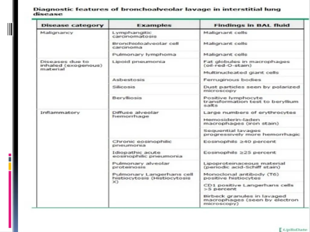
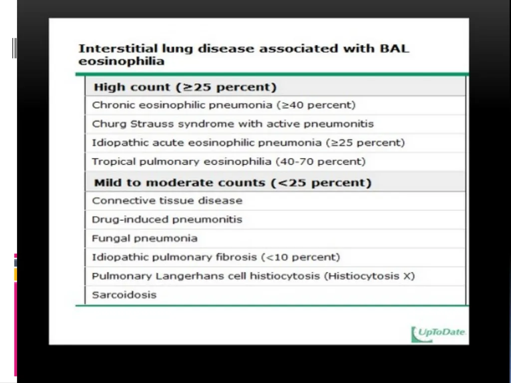
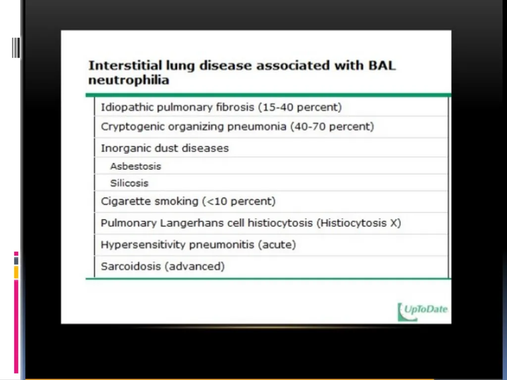
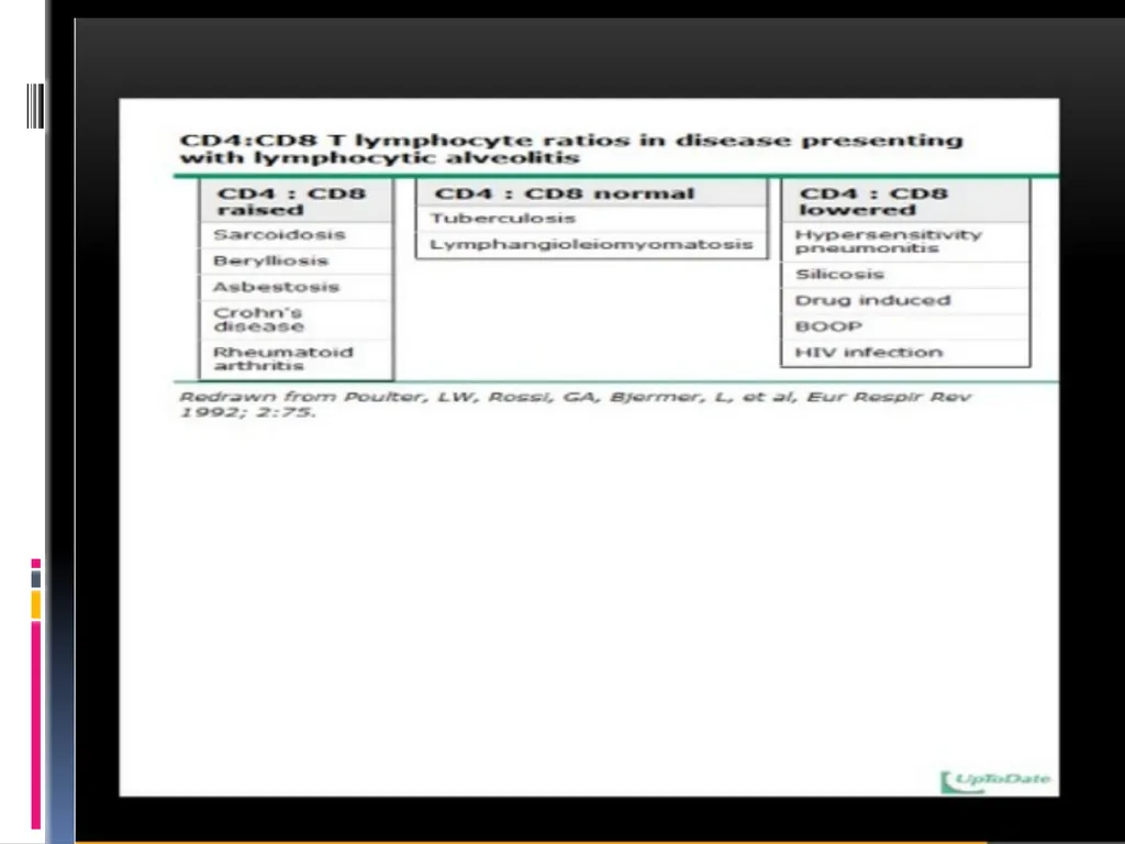
ROLE OF BRONCHOALVEOLAR LAVAGE • The lavage fluid is sent for cell counts, cultures for mycobacterial, viral and fungal pathogens, and cytologic analysis
• Virtually all patients presenting with hemoptysis and radiographic ILD should undergo BAL -> to confirm an alveolar source of bleeding and identify any infectious etiologies.<br>
28
BAL is less likely to be helpful in patients with a radiographic pattern suggestive of IPF.
BAL does not have an established role in the assessment of ILD progression or response to therapy<br>
29
LUNG BIOPSY • Obtained by flexible fiberoptic bronchoscopy, video-assisted thoracoscopic (VATS) biopsy, or open lung biopsy.
• The histopathologic pattern found on the lung biopsy specimen is evaluated in combination with the clinical information to determine the diagnosis.<br>
30
ROLE OF LUNG BIOPSY 1 Atypical or progressive symptoms and signs (age less than 50 years, fever, weight loss, hemoptysis, signs of vasculitis)
2. Atypical radiographic features
3. Unexplained extrapulmonary manifestations
4. Rapid clinical deterioration
5. Sudden change in radiographic appearance.<br>
31
VIDEO ASSISTED THORACIC SURGERY VATS is the preferred procedure for obtaining a lung biopsy High diagnostic accuracy
Less morbidity and mortality than open lung biopsy
BAL and TBBx limited to excluding other IPF mimickers. Ideal biopsy
Two or more surgical wedge biopsies with areas of normal lung
Samples should measure 3- 5 cm in length and 2- 3 cm in depth .
Outpatient thoracoscopic lung biopsy can be a safe and effective procedure for patients with interstitial or focal lung disease .<br>
32
TREATMENT Treatments for interstitial lung disease vary according to the type of interstitial lung disease and its cause.
Antibiotics . These are effective treatments for most interstitial pneumonias.
Azithromycin (Zithromax) and levofloxacin (Levaquin) eliminate the bacteria that cause most interstitial pneumonias.
Viral pneumonias usually resolve on their own.
Fungal pneumonias are rare, but can be treated with antifungal drugs.<br>
33
CORTICOSTEROIDS In some forms of interstitial lung disease, ongoing inflammation in the lungs causes damage and scarring. Corticosteroids like prednisone and methylprednisolone reduce the activity of the immune system. This reduces the amount of inflammation in the lungs and the rest of the body.<br>
34
The most evidence is for
either the combination of corticosteroids with azathioprine
NAC(N Acetyl cysteine) or the novel antifibrotic (pirfenidone).
( The first option is criticized, as it has only been shown in one study and there was no placebo group. Therefore, a conformational study is under the way (prednisone, azathioprine, and NAC: A Study That Evaluates Response in IPF: PANTHER trial). For pirfenidone, more studies have been done, but not all of them are convincing)<br>
35
OXYGEN THERAPY Oxygen is required for some people with ILD because of low level of oxygen in the blood.
Some may need oxygen therapy all of the time while others may need it only during sleep ando exercise.
Improving the level of oxygen in the blood through the use of supplemental oxygen can help relieve the strain on the heart and lungs and improve symptoms of shortness of breath and fatigue.<br>
36
PULMONARY REHABILITATION A pulmonary rehabilitation program is recommended to help you achieve your highest level of functioning.
Includes education, exercise conditioning, breathing techniques, and energy saving techniques, respiratory therapy evaluation, nutritional counseling and psychosocial support.
The specific goals of pulmonary rehabilitation are to improve quality of life.<br>
37
LUNG TRANSPLANTATION If other therapies fail to adequately treat ILD, lung transplant is an option for some advanced cases.
With improved surgical techniques and post-transplant care, this may offer improved quality of life and prolonged survival to selected patients.<br>
38
PROGNOSIS Even with treatment, many types of ILD progress naturally with a worsening of symptoms, x-ray, and physiologic findings. Sometimes worsening is due to a complication of the disease or therapy.
This may include conditions such as pulmonary hypertension or right heart failure. Some therapies may result in infection, muscle weakness and osteoporosis.<br>
39
KEY POINTS Suspect interstitial lung diseases with insidious dyspnea , even with minimal or no radiographic findings.
A thorough history with exposures and systemic ROS is key.
Firm diagnosis for IPF requires ruling out treatable causes and considering biopsy for atypical presentations<br>
40
Case 1 65 year old male with progressive breathlessness.
Chest Xray shows prominent bronchovascular markings<br>
42
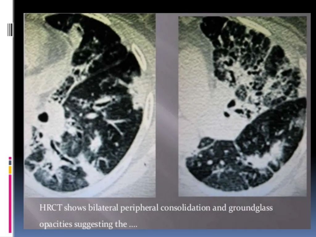
Case 2 50 year old male with history of dry cough , malaise , low grade fever.
Chest Xray- shows persistent pneumonitis patches.<br>
44
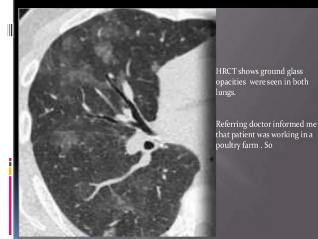
Case 3 North Indian worker brought by his employer. He complains of recent breathlessness on coming to work.
Normal chest Xray.<br>
45
Diagnosis Possible Subacute hypersensitivity pneumonitis.<br>