
د. خالد Arthrology Field of study concerned with






Description: د. خالد Arthrology Field of study concerned with the study of joints. Definition of joint Is the articulation between boney surfaces wich allow free or limited movement. The classification is according to the type of material by wich the
Related Topics
Download Presentation
"د. خالد Arthrology Field of study concerned with" is the property of its rightful owner. Permission is granted to download and print the materials on this website for personal, non-commercial use only, and to display it on your personal computer provided you do not modify the materials and that you retain all copyright notices contained in the materials. By downloading content from our website, you accept the terms of this agreement.
Presentation Transcript
slide1. د. خالد
Arthrology Field of study concerned with the study of joints.<br>
slide2. Definition of joint Is the articulation between boney surfaces wich allow free or limited movement.
The classification is according to the type of material by wich the articulating bones are united.<br>
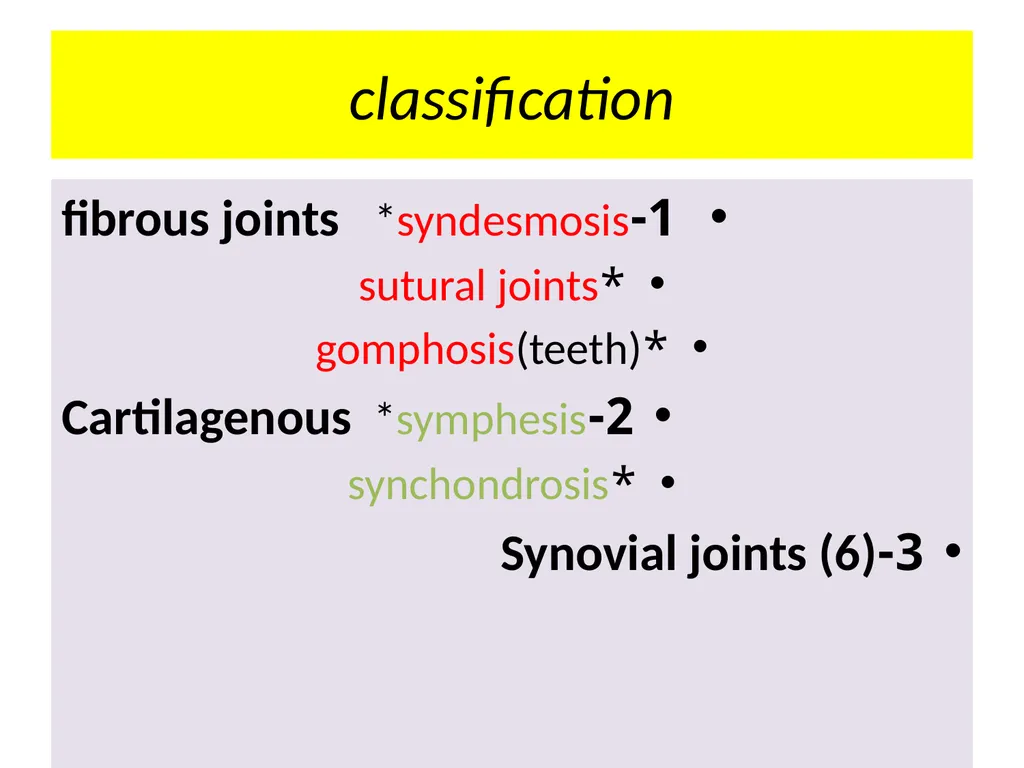
slide4. classification 1-fibrous joints *syndesmosis
*sutural joints
*gomphosis(teeth)
2-Cartilagenous *symphesis
*synchondrosis
3-Synovial joints (6)<br>
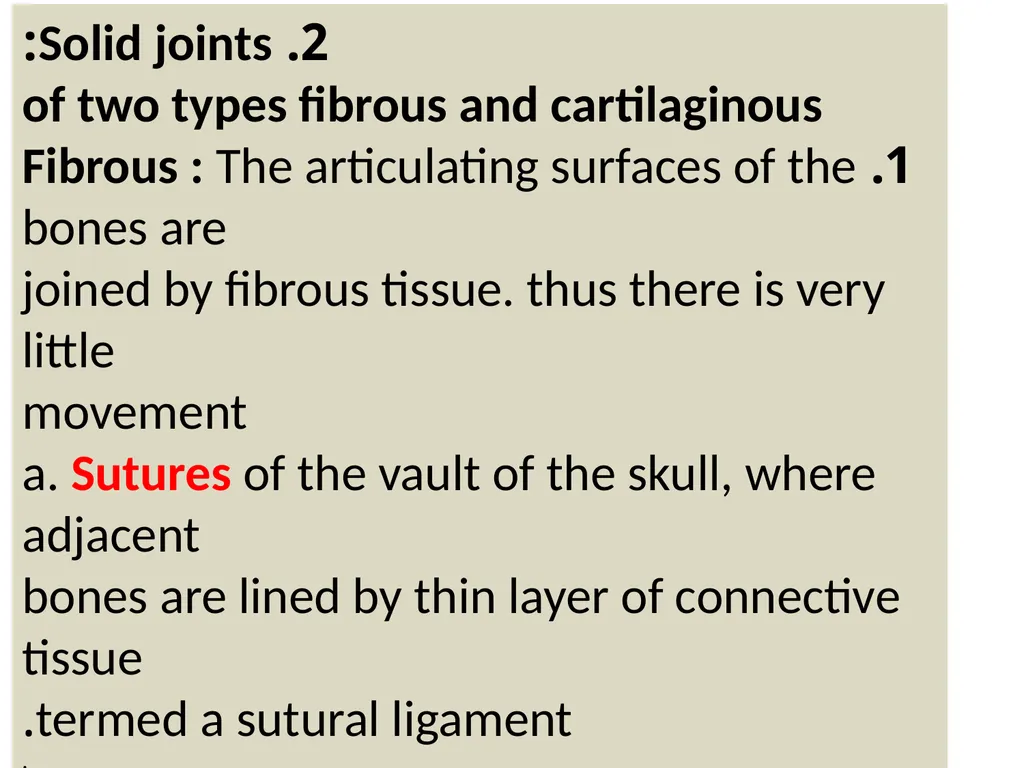
slide5. 2. Solid joints:
of two types fibrous and cartilaginous
1. Fibrous : The articulating surfaces of the bones are
joined by fibrous tissue. thus there is very little
movement
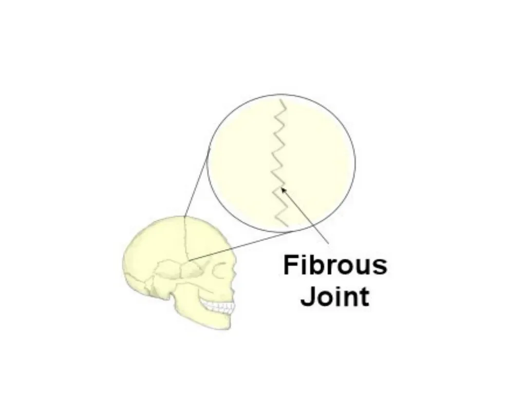
a. Sutures of the vault of the skull, where adjacent
bones are lined by thin layer of connective tissue
termed a sutural ligament.
.<br>
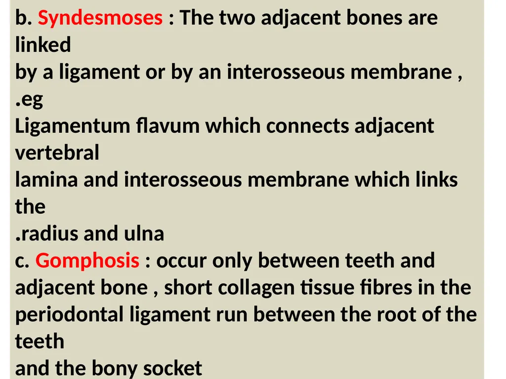
slide6. b. Syndesmoses : The two adjacent bones are linked
by a ligament or by an interosseous membrane , eg.
Ligamentum flavum which connects adjacent vertebral
lamina and interosseous membrane which links the
radius and ulna.
c. Gomphosis : occur only between teeth and
adjacent bone , short collagen tissue fibres in the
periodontal ligament run between the root of the teeth
and the bony socket<br>
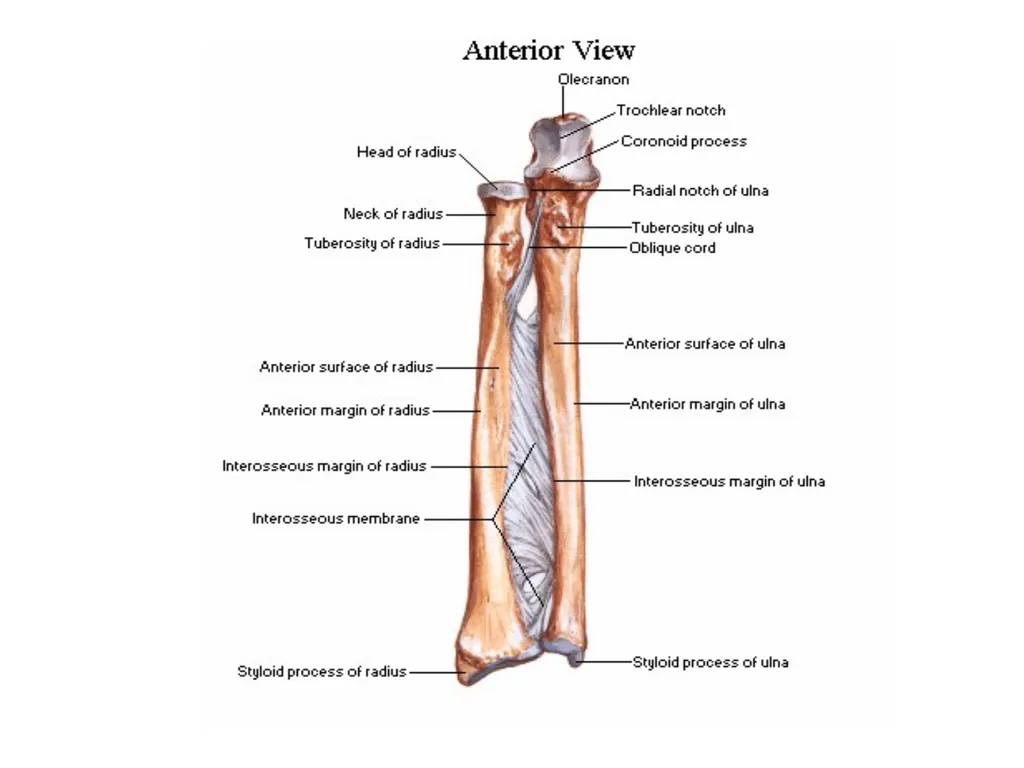
slide8. FIGURE The side-to-side articulation of the ulna and radius
forms a syndesmotic joint. An interosseous ligament tightly binds
these bones and permits only slight movement between them.<br>
slide10. 2 . Cartilaginous Joints : of two types
a. Synchondrosis or Primary cartilaginous j.
Bones are united by plate of hyaline cartilage
eg. Union between the epiphysis and the
diaphysis of a growing bone, eg.2 between first
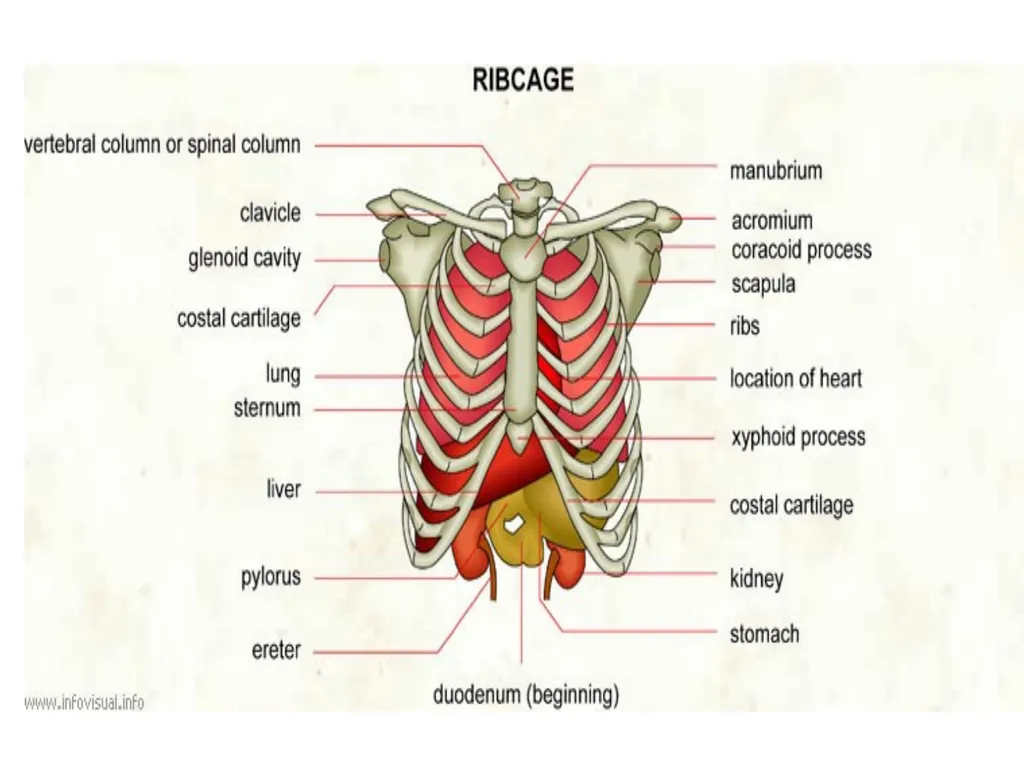
rib and manubrium sterni, no movement is
possible.<br>
slide11. A fracture of a long bone in a child may be extremely serious if it
involves the mitotically active epiphyseal plate of a synchondrotic
joint. If such an injury is left untreated, bone growth is usually
retarded or arrested, so that the appendage will be shorter
than normal.<br>
slide12. b. Symphyses or Secondary Cartilaginous J .
Bones are united by a plate of fibrocartilage,
and the articular surfaces are covered by thin
plate of hyaline cartilage eg. Joints between
vertebral bodies, eg . symphysis pubis, small
amount of movement is possible.<br>
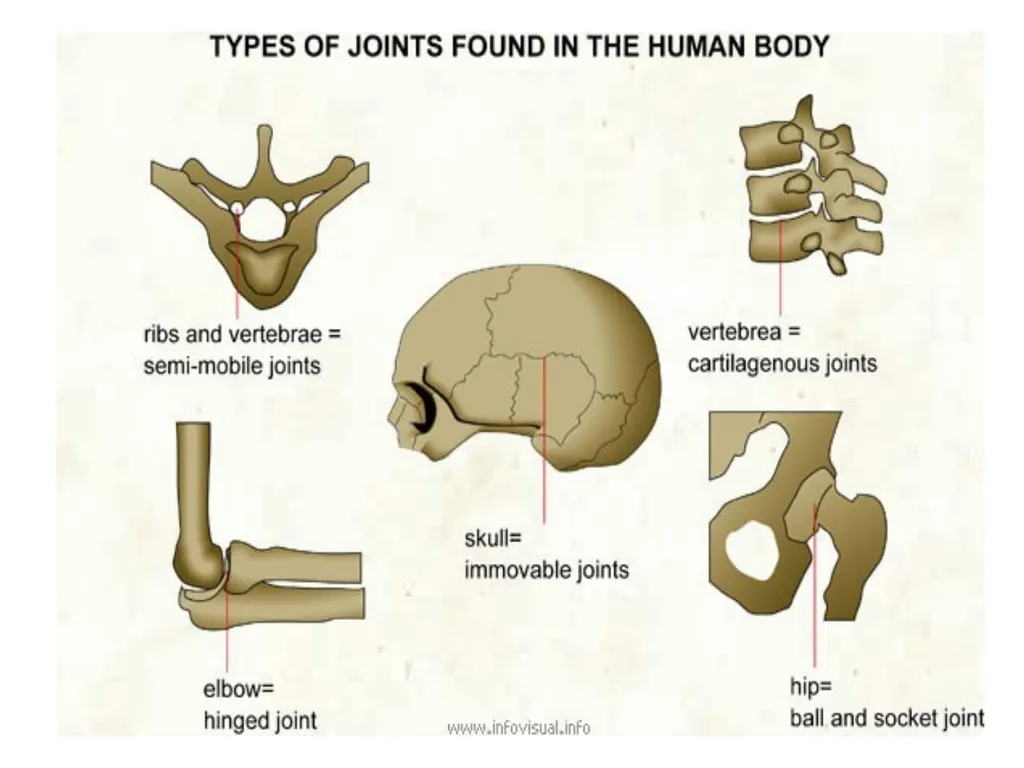
slide15. Types of Joint A joint is the point where two or more bones meet. There are three main types of joints; Fibrous (immoveable), Cartilaginous (partially moveable) and the Synovial (freely moveable) joint<br>
slide17. Fibrous joints
Fibrous (synarthrodial): This type of joint is held together by only a ligament. Examples are where the teeth are held to their bony sockets and at both the radioulnar and tibiofibular joints.sutural joints.
Cartilaginous
Cartilaginous (synchondroses and sympheses): These joints occur where the connection between the articulating bones is made up of cartilage for example between vertebrae in the spine.<br>
slide18. A cartilagenous joint between two vertebrae<br>
slide19. Synchondroses are temporary joints which are only present in children, up until the end of puberty. For example the epiphyseal plates in long bones. Symphesis joints are permanant cartilagenous joints, for example the pubic symphesis.<br>
slide20. Synovial Joints
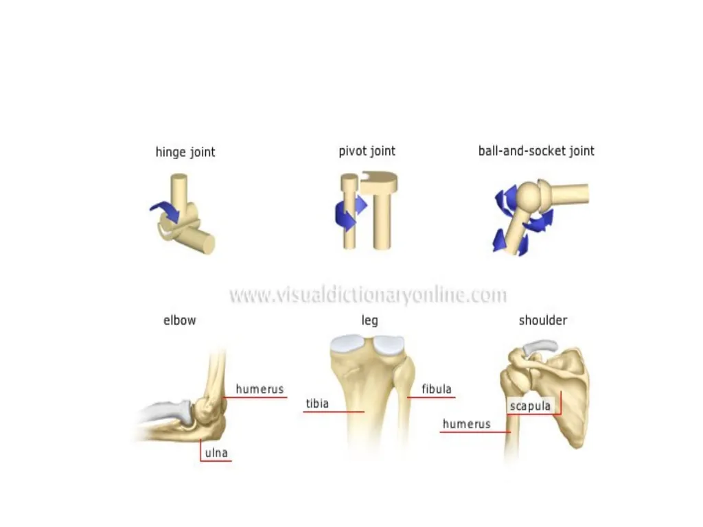
Synovial (diarthrosis): Synovial joints are by far the most common classification of joint within the human body. They are highly moveable and all have a synovial capsule (collagenous structure) surrounding the entire joint, a synovial membrane (the inner layer of the capsule) which secretes synovial fluid (a lubricating liquid) and cartilage known as hyaline cartilage which pads the ends of the articulating bones. There are 6 types of synovial joints which are classified by the shape of the joint and the movement available.<br>
slide21. Types of Synovial Joint<br>
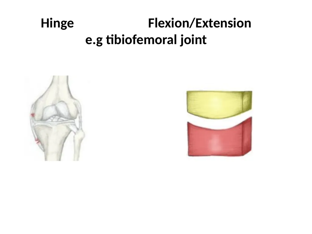
slide23. Hinge Flexion/Extension e.g tibiofemoral joint<br>
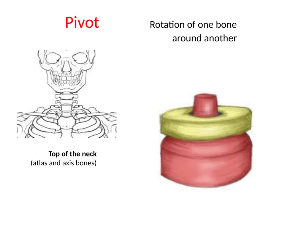
slide24. Pivot Rotation of one bone around another Top of the neck (atlas and axis bones)<br>
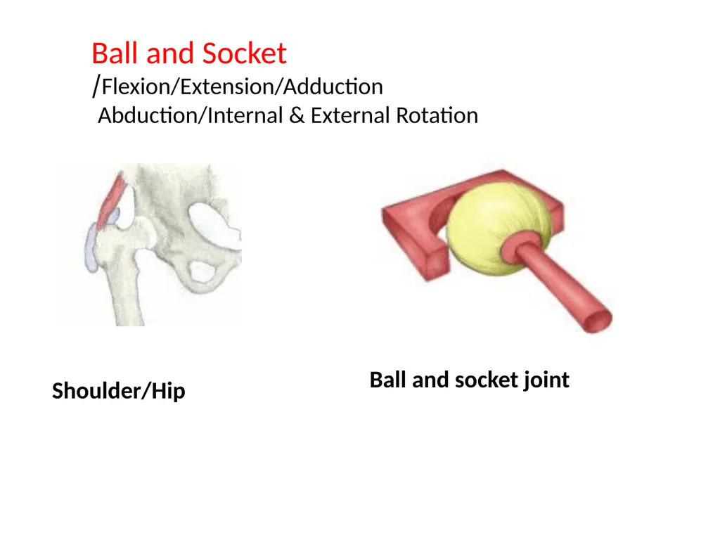
slide25. Ball and Socket Flexion/Extension/Adduction/
Abduction/Internal & External Rotation Shoulder/Hip Ball and socket joint<br>
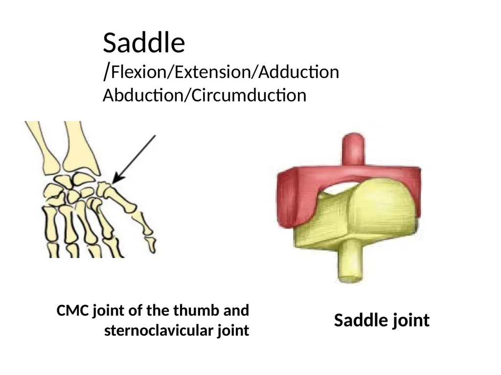
slide26. Saddle Flexion/Extension/Adduction/
Abduction/Circumduction CMC joint of the thumb and sternoclavicular joint Saddle joint<br>
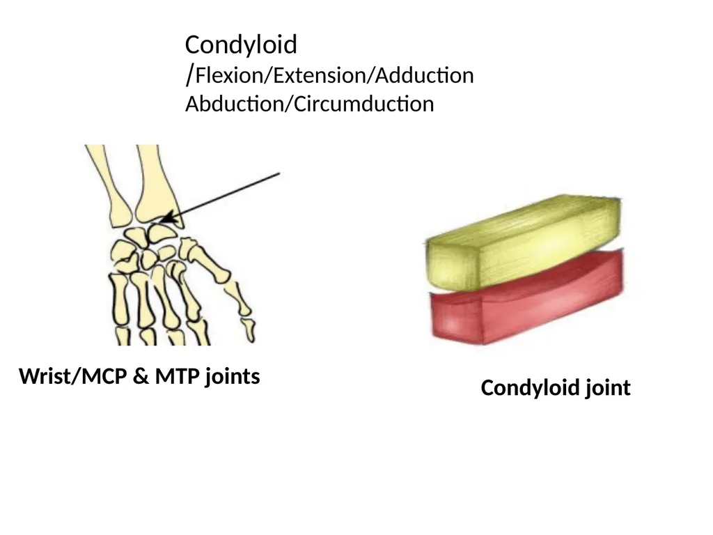
slide27. Condyloid Flexion/Extension/Adduction/
Abduction/Circumduction Wrist/MCP & MTP joints Condyloid joint<br>
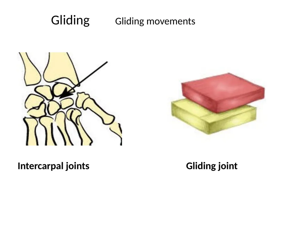
slide28. Gliding Gliding movements Intercarpal joints Gliding joint<br>
slide29. Gomphoses (socket joints) are immovable joints found between the teeth and jaws. They are held together by a periodontal ligament that acts as a shock absorber.<br>
slide31. Saddle Joints
Saddle joints are so named because the ends of each bone resemble a saddle, with concave and convex portions that fit together. Saddle joints allow angular movements similar to condyloid joints but with a greater range of motion. An example of a saddle joint is the thumb joint, which can move back and forth and up and down, but more freely than the wrist or fingers<br>
slide2. Definition of joint Is the articulation between boney surfaces wich allow free or limited movement.
The classification is according to the type of material by wich the articulating bones are united.<br>
slide4. classification 1-fibrous joints *syndesmosis
*sutural joints
*gomphosis(teeth)
2-Cartilagenous *symphesis
*synchondrosis
3-Synovial joints (6)<br>
slide5. 2. Solid joints:
of two types fibrous and cartilaginous
1. Fibrous : The articulating surfaces of the bones are
joined by fibrous tissue. thus there is very little
movement
a. Sutures of the vault of the skull, where adjacent
bones are lined by thin layer of connective tissue
termed a sutural ligament.
.<br>
slide6. b. Syndesmoses : The two adjacent bones are linked
by a ligament or by an interosseous membrane , eg.
Ligamentum flavum which connects adjacent vertebral
lamina and interosseous membrane which links the
radius and ulna.
c. Gomphosis : occur only between teeth and
adjacent bone , short collagen tissue fibres in the
periodontal ligament run between the root of the teeth
and the bony socket<br>
slide8. FIGURE The side-to-side articulation of the ulna and radius
forms a syndesmotic joint. An interosseous ligament tightly binds
these bones and permits only slight movement between them.<br>
slide10. 2 . Cartilaginous Joints : of two types
a. Synchondrosis or Primary cartilaginous j.
Bones are united by plate of hyaline cartilage
eg. Union between the epiphysis and the
diaphysis of a growing bone, eg.2 between first
rib and manubrium sterni, no movement is
possible.<br>
slide11. A fracture of a long bone in a child may be extremely serious if it
involves the mitotically active epiphyseal plate of a synchondrotic
joint. If such an injury is left untreated, bone growth is usually
retarded or arrested, so that the appendage will be shorter
than normal.<br>
slide12. b. Symphyses or Secondary Cartilaginous J .
Bones are united by a plate of fibrocartilage,
and the articular surfaces are covered by thin
plate of hyaline cartilage eg. Joints between
vertebral bodies, eg . symphysis pubis, small
amount of movement is possible.<br>
slide15. Types of Joint A joint is the point where two or more bones meet. There are three main types of joints; Fibrous (immoveable), Cartilaginous (partially moveable) and the Synovial (freely moveable) joint<br>
slide17. Fibrous joints
Fibrous (synarthrodial): This type of joint is held together by only a ligament. Examples are where the teeth are held to their bony sockets and at both the radioulnar and tibiofibular joints.sutural joints.
Cartilaginous
Cartilaginous (synchondroses and sympheses): These joints occur where the connection between the articulating bones is made up of cartilage for example between vertebrae in the spine.<br>
slide18. A cartilagenous joint between two vertebrae<br>
slide19. Synchondroses are temporary joints which are only present in children, up until the end of puberty. For example the epiphyseal plates in long bones. Symphesis joints are permanant cartilagenous joints, for example the pubic symphesis.<br>
slide20. Synovial Joints
Synovial (diarthrosis): Synovial joints are by far the most common classification of joint within the human body. They are highly moveable and all have a synovial capsule (collagenous structure) surrounding the entire joint, a synovial membrane (the inner layer of the capsule) which secretes synovial fluid (a lubricating liquid) and cartilage known as hyaline cartilage which pads the ends of the articulating bones. There are 6 types of synovial joints which are classified by the shape of the joint and the movement available.<br>
slide21. Types of Synovial Joint<br>
slide23. Hinge Flexion/Extension e.g tibiofemoral joint<br>
slide24. Pivot Rotation of one bone around another Top of the neck (atlas and axis bones)<br>
slide25. Ball and Socket Flexion/Extension/Adduction/
Abduction/Internal & External Rotation Shoulder/Hip Ball and socket joint<br>
slide26. Saddle Flexion/Extension/Adduction/
Abduction/Circumduction CMC joint of the thumb and sternoclavicular joint Saddle joint<br>
slide27. Condyloid Flexion/Extension/Adduction/
Abduction/Circumduction Wrist/MCP & MTP joints Condyloid joint<br>
slide28. Gliding Gliding movements Intercarpal joints Gliding joint<br>
slide29. Gomphoses (socket joints) are immovable joints found between the teeth and jaws. They are held together by a periodontal ligament that acts as a shock absorber.<br>
slide31. Saddle Joints
Saddle joints are so named because the ends of each bone resemble a saddle, with concave and convex portions that fit together. Saddle joints allow angular movements similar to condyloid joints but with a greater range of motion. An example of a saddle joint is the thumb joint, which can move back and forth and up and down, but more freely than the wrist or fingers<br>