
Blood Agar Plates and Hemolysis: Streptococcus
DP
Published · 44 slides · 0 views

1 / 1

Description
Blood Agar Plates and Hemolysis: Streptococcus Authors: Rebecca Buxton Citation: Rebecca Buxton. 2009. Blood agar plates and hemolysis: streptococcus. Publication Date: December 2009 Typticase soy agar w wo sheep blood (Enlarged View)
Related Topics
Share
Embed code
Download this presentation From Below
"Blood Agar Plates and Hemolysis: Streptococcus" is the property of its rightful owner. Permission is granted to download and print the materials on this website for personal, non-commercial use only, and to display it on your personal computer provided you do not modify the materials and that you retain all copyright notices contained in the materials. By downloading content from our website, you accept the terms of this agreement.
Presentation Transcript
01
Blood Agar Plates and Hemolysis: Streptococcus Authors: Rebecca Buxton
Citation: Rebecca Buxton. 2009. Blood agar plates and hemolysis: streptococcus.
Publication Date: December 2009<br>
Citation: Rebecca Buxton. 2009. Blood agar plates and hemolysis: streptococcus.
Publication Date: December 2009<br>
02
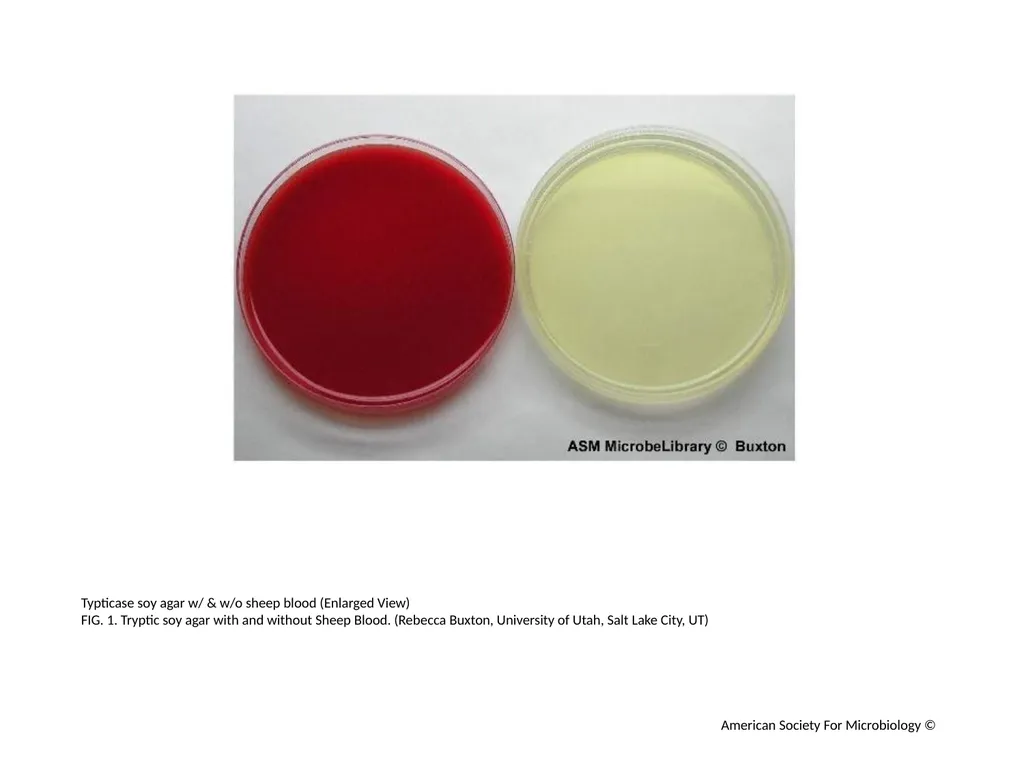
Typticase soy agar w/ & w/o sheep blood (Enlarged View)
FIG. 1. Tryptic soy agar with and without Sheep Blood. (Rebecca Buxton, University of Utah, Salt Lake City, UT) American Society For Microbiology ©<br>
FIG. 1. Tryptic soy agar with and without Sheep Blood. (Rebecca Buxton, University of Utah, Salt Lake City, UT) American Society For Microbiology ©<br>
03
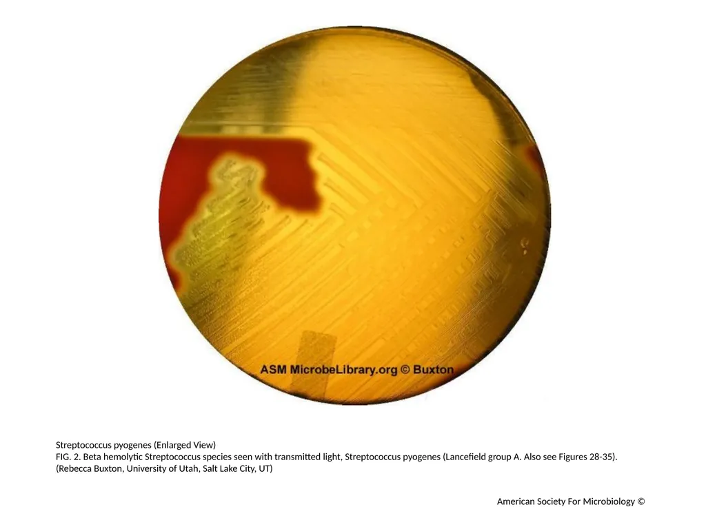
Streptococcus pyogenes (Enlarged View)
FIG. 2. Beta hemolytic Streptococcus species seen with transmitted light, Streptococcus pyogenes (Lancefield group A. Also see Figures 28-35). (Rebecca Buxton, University of Utah, Salt Lake City, UT) American Society For Microbiology ©<br>
FIG. 2. Beta hemolytic Streptococcus species seen with transmitted light, Streptococcus pyogenes (Lancefield group A. Also see Figures 28-35). (Rebecca Buxton, University of Utah, Salt Lake City, UT) American Society For Microbiology ©<br>
04
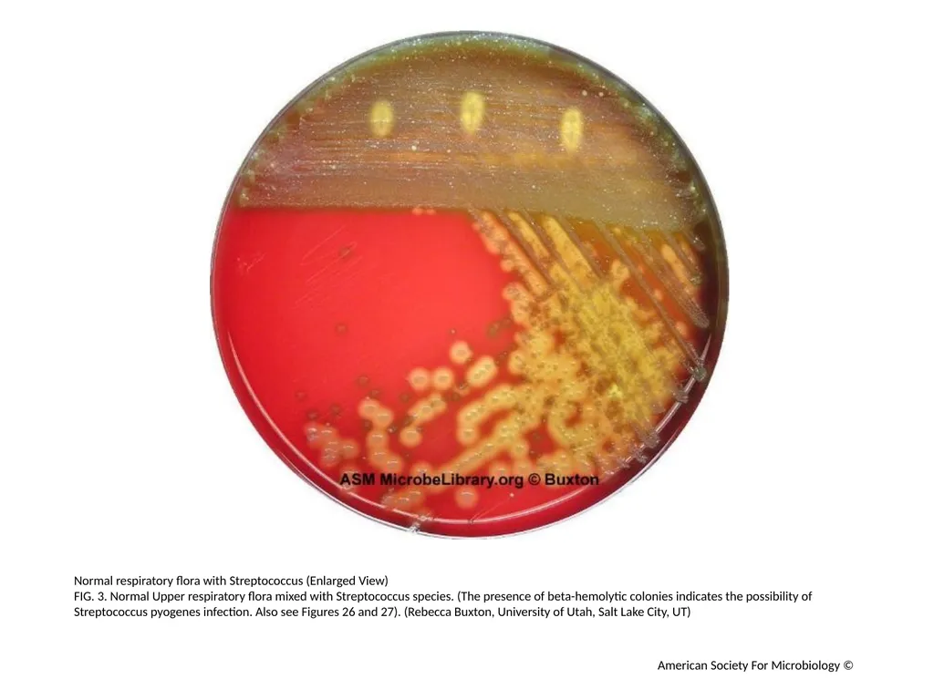
Normal respiratory flora with Streptococcus (Enlarged View)
FIG. 3. Normal Upper respiratory flora mixed with Streptococcus species. (The presence of beta-hemolytic colonies indicates the possibility of Streptococcus pyogenes infection. Also see Figures 26 and 27). (Rebecca Buxton, University of Utah, Salt Lake City, UT) American Society For Microbiology ©<br>
FIG. 3. Normal Upper respiratory flora mixed with Streptococcus species. (The presence of beta-hemolytic colonies indicates the possibility of Streptococcus pyogenes infection. Also see Figures 26 and 27). (Rebecca Buxton, University of Utah, Salt Lake City, UT) American Society For Microbiology ©<br>
05
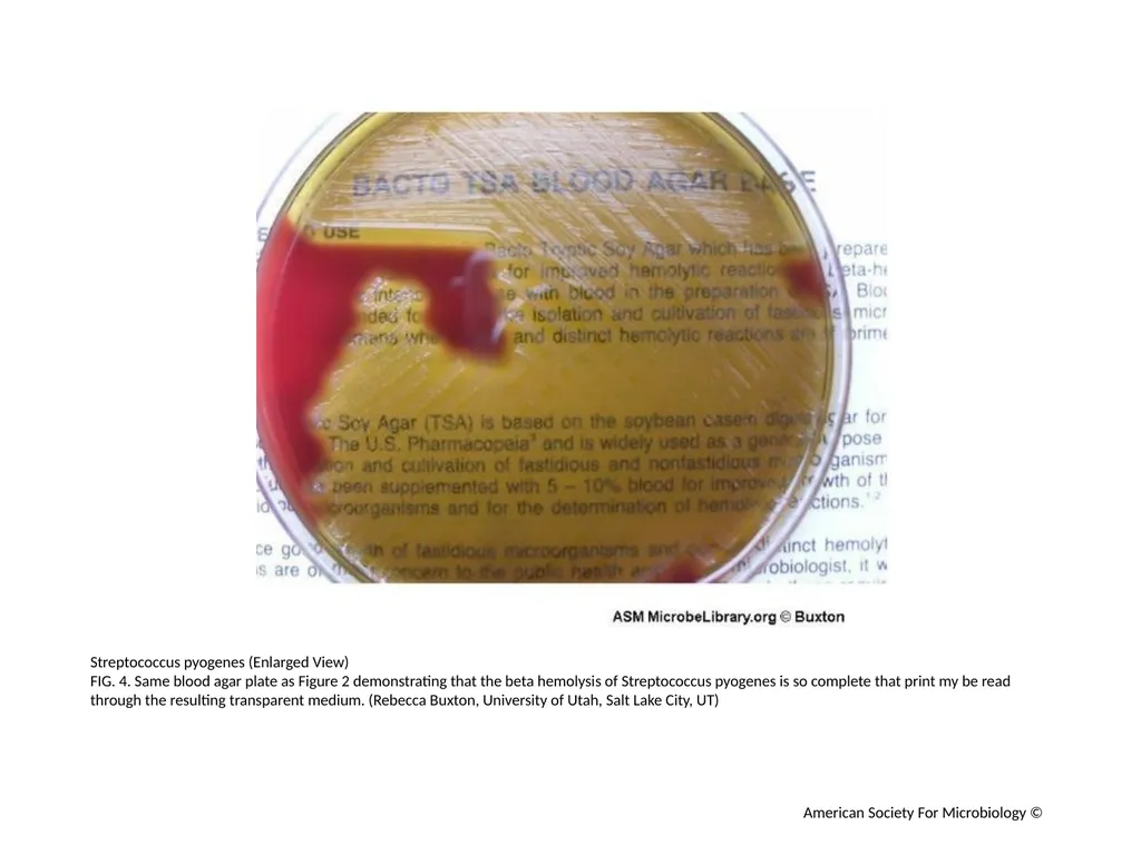
Streptococcus pyogenes (Enlarged View)
FIG. 4. Same blood agar plate as Figure 2 demonstrating that the beta hemolysis of Streptococcus pyogenes is so complete that print my be read through the resulting transparent medium. (Rebecca Buxton, University of Utah, Salt Lake City, UT) American Society For Microbiology ©<br>
FIG. 4. Same blood agar plate as Figure 2 demonstrating that the beta hemolysis of Streptococcus pyogenes is so complete that print my be read through the resulting transparent medium. (Rebecca Buxton, University of Utah, Salt Lake City, UT) American Society For Microbiology ©<br>
06
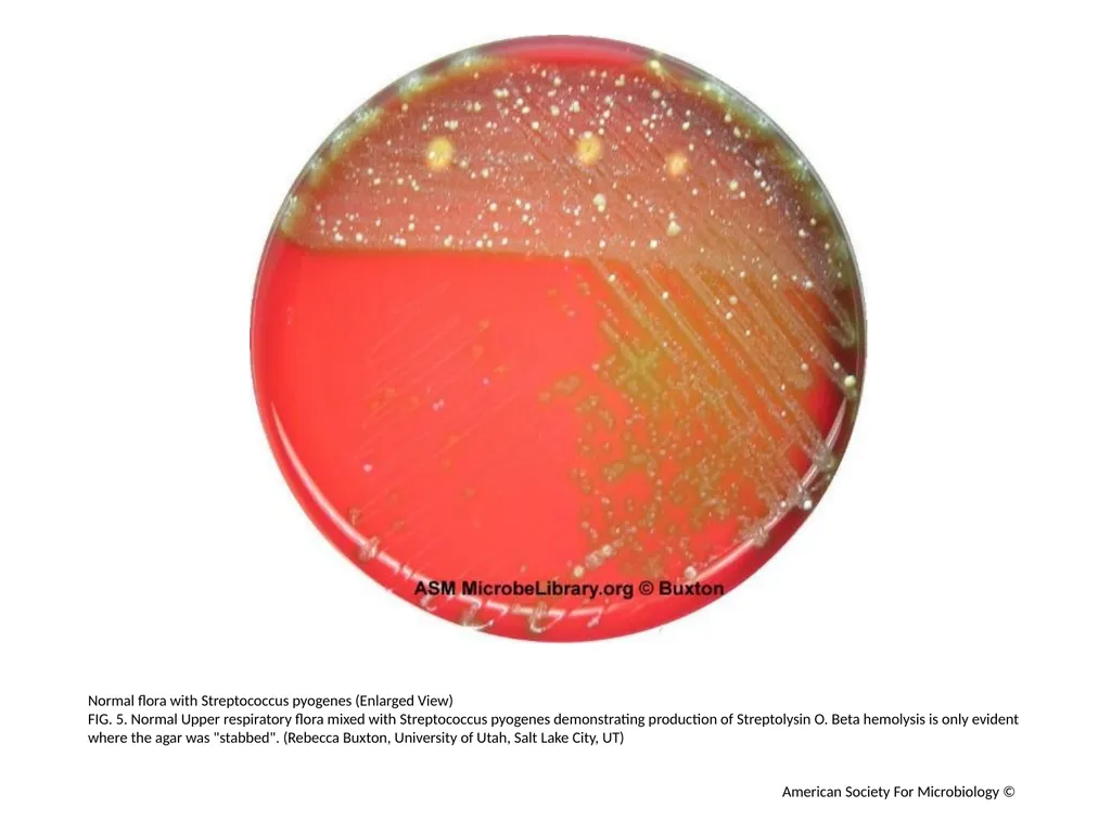
Normal flora with Streptococcus pyogenes (Enlarged View)
FIG. 5. Normal Upper respiratory flora mixed with Streptococcus pyogenes demonstrating production of Streptolysin O. Beta hemolysis is only evident where the agar was "stabbed". (Rebecca Buxton, University of Utah, Salt Lake City, UT) American Society For Microbiology ©<br>
FIG. 5. Normal Upper respiratory flora mixed with Streptococcus pyogenes demonstrating production of Streptolysin O. Beta hemolysis is only evident where the agar was "stabbed". (Rebecca Buxton, University of Utah, Salt Lake City, UT) American Society For Microbiology ©<br>
07
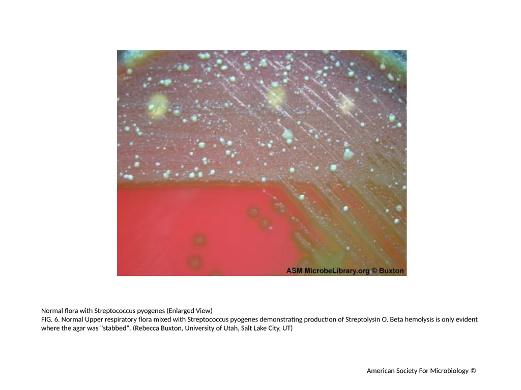
Normal flora with Streptococcus pyogenes (Enlarged View)
FIG. 6. Normal Upper respiratory flora mixed with Streptococcus pyogenes demonstrating production of Streptolysin O. Beta hemolysis is only evident where the agar was "stabbed". (Rebecca Buxton, University of Utah, Salt Lake City, UT) American Society For Microbiology ©<br>
FIG. 6. Normal Upper respiratory flora mixed with Streptococcus pyogenes demonstrating production of Streptolysin O. Beta hemolysis is only evident where the agar was "stabbed". (Rebecca Buxton, University of Utah, Salt Lake City, UT) American Society For Microbiology ©<br>
08
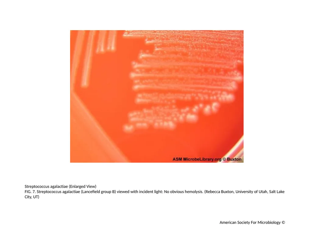
Streptococcus agalactiae (Enlarged View)
FIG. 7. Streptococcus agalactiae (Lancefield group B) viewed with incident light: No obvious hemolysis. (Rebecca Buxton, University of Utah, Salt Lake City, UT) American Society For Microbiology ©<br>
FIG. 7. Streptococcus agalactiae (Lancefield group B) viewed with incident light: No obvious hemolysis. (Rebecca Buxton, University of Utah, Salt Lake City, UT) American Society For Microbiology ©<br>
09
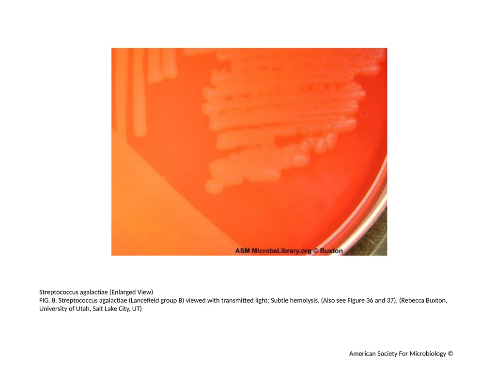
Streptococcus agalactiae (Enlarged View)
FIG. 8. Streptococcus agalactiae (Lancefield group B) viewed with transmitted light: Subtle hemolysis. (Also see Figure 36 and 37). (Rebecca Buxton, University of Utah, Salt Lake City, UT) American Society For Microbiology ©<br>
FIG. 8. Streptococcus agalactiae (Lancefield group B) viewed with transmitted light: Subtle hemolysis. (Also see Figure 36 and 37). (Rebecca Buxton, University of Utah, Salt Lake City, UT) American Society For Microbiology ©<br>
10
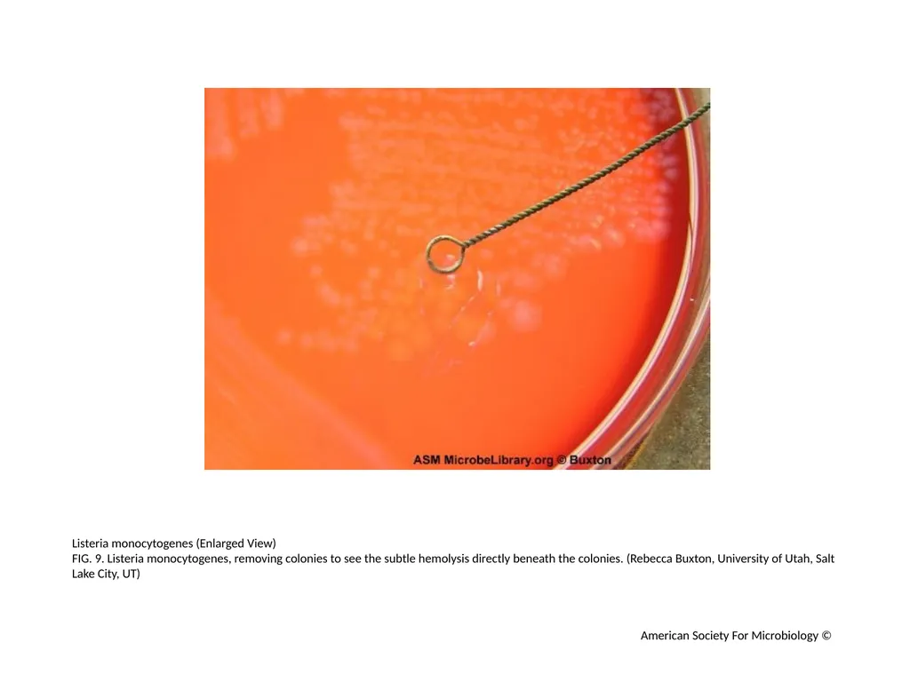
Listeria monocytogenes (Enlarged View)
FIG. 9. Listeria monocytogenes, removing colonies to see the subtle hemolysis directly beneath the colonies. (Rebecca Buxton, University of Utah, Salt Lake City, UT) American Society For Microbiology ©<br>
FIG. 9. Listeria monocytogenes, removing colonies to see the subtle hemolysis directly beneath the colonies. (Rebecca Buxton, University of Utah, Salt Lake City, UT) American Society For Microbiology ©<br>
11
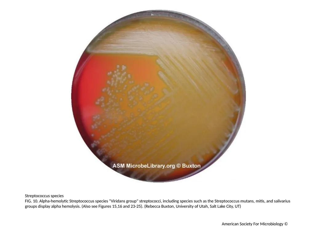
Streptococcus species
FIG. 10. Alpha-hemolytic Streptococcus species "Viridans group" streptococci, including species such as the Streptococcus mutans, mitis, and salivarius groups display alpha hemolysis. (Also see Figures 15,16 and 23-25). (Rebecca Buxton, University of Utah, Salt Lake City, UT) American Society For Microbiology ©<br>
FIG. 10. Alpha-hemolytic Streptococcus species "Viridans group" streptococci, including species such as the Streptococcus mutans, mitis, and salivarius groups display alpha hemolysis. (Also see Figures 15,16 and 23-25). (Rebecca Buxton, University of Utah, Salt Lake City, UT) American Society For Microbiology ©<br>
12
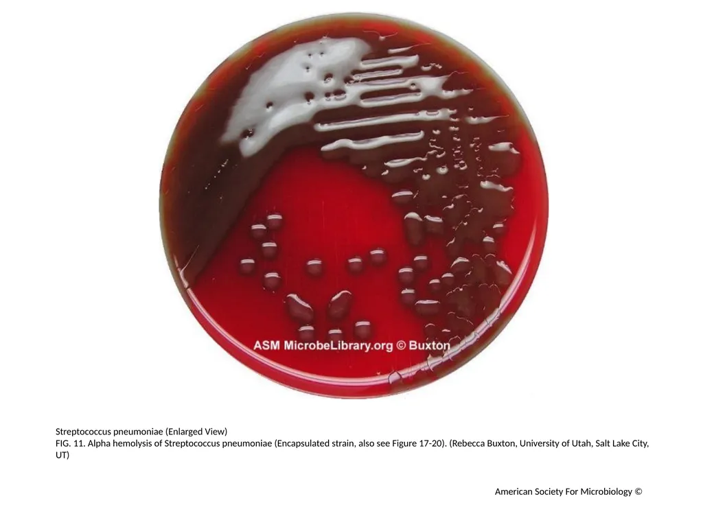
Streptococcus pneumoniae (Enlarged View)
FIG. 11. Alpha hemolysis of Streptococcus pneumoniae (Encapsulated strain, also see Figure 17-20). (Rebecca Buxton, University of Utah, Salt Lake City, UT) American Society For Microbiology ©<br>
FIG. 11. Alpha hemolysis of Streptococcus pneumoniae (Encapsulated strain, also see Figure 17-20). (Rebecca Buxton, University of Utah, Salt Lake City, UT) American Society For Microbiology ©<br>
13
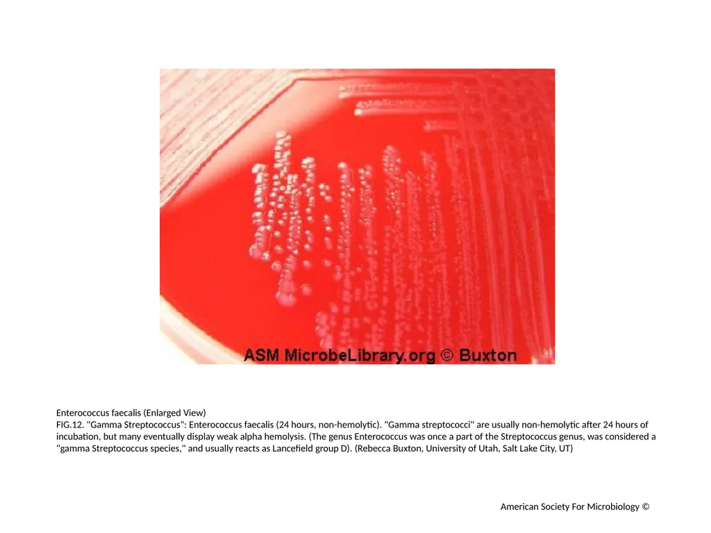
Enterococcus faecalis (Enlarged View)
FIG.12. "Gamma Streptococcus": Enterococcus faecalis (24 hours, non-hemolytic). "Gamma streptococci" are usually non-hemolytic after 24 hours of incubation, but many eventually display weak alpha hemolysis. (The genus Enterococcus was once a part of the Streptococcus genus, was considered a "gamma Streptococcus species," and usually reacts as Lancefield group D). (Rebecca Buxton, University of Utah, Salt Lake City, UT) American Society For Microbiology ©<br>
FIG.12. "Gamma Streptococcus": Enterococcus faecalis (24 hours, non-hemolytic). "Gamma streptococci" are usually non-hemolytic after 24 hours of incubation, but many eventually display weak alpha hemolysis. (The genus Enterococcus was once a part of the Streptococcus genus, was considered a "gamma Streptococcus species," and usually reacts as Lancefield group D). (Rebecca Buxton, University of Utah, Salt Lake City, UT) American Society For Microbiology ©<br>
14
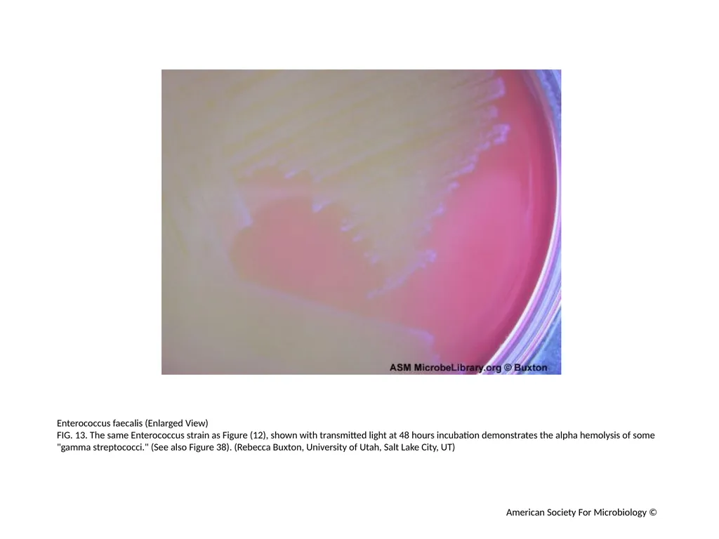
Enterococcus faecalis (Enlarged View)
FIG. 13. The same Enterococcus strain as Figure (12), shown with transmitted light at 48 hours incubation demonstrates the alpha hemolysis of some "gamma streptococci." (See also Figure 38). (Rebecca Buxton, University of Utah, Salt Lake City, UT) American Society For Microbiology ©<br>
FIG. 13. The same Enterococcus strain as Figure (12), shown with transmitted light at 48 hours incubation demonstrates the alpha hemolysis of some "gamma streptococci." (See also Figure 38). (Rebecca Buxton, University of Utah, Salt Lake City, UT) American Society For Microbiology ©<br>
15
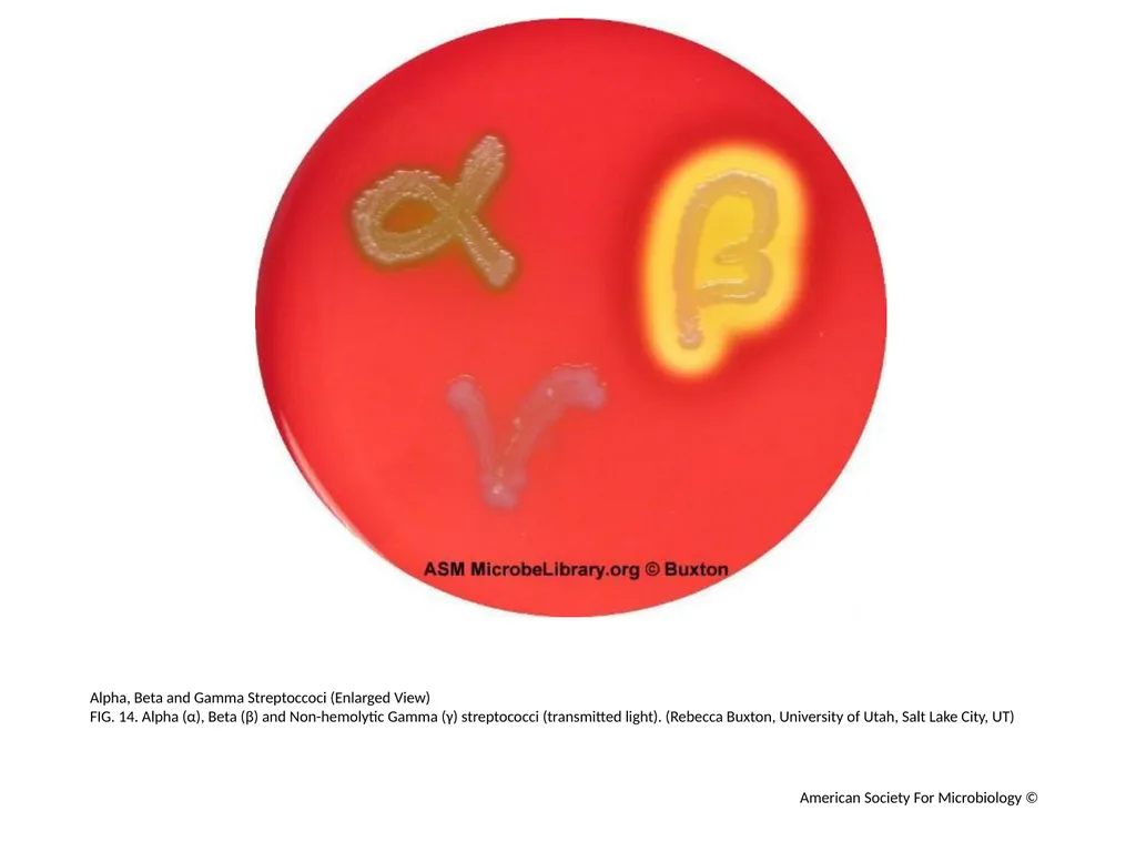
Alpha, Beta and Gamma Streptoccoci (Enlarged View)
FIG. 14. Alpha (α), Beta (β) and Non-hemolytic Gamma (γ) streptococci (transmitted light). (Rebecca Buxton, University of Utah, Salt Lake City, UT) American Society For Microbiology ©<br>
FIG. 14. Alpha (α), Beta (β) and Non-hemolytic Gamma (γ) streptococci (transmitted light). (Rebecca Buxton, University of Utah, Salt Lake City, UT) American Society For Microbiology ©<br>
16
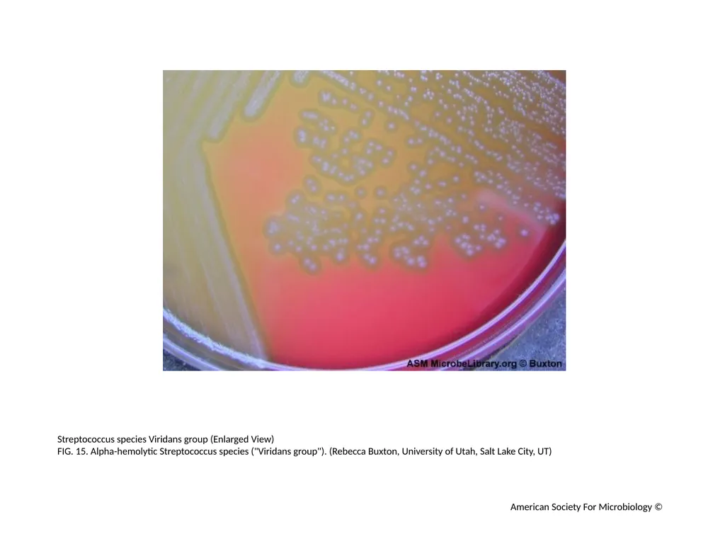
Streptococcus species Viridans group (Enlarged View)
FIG. 15. Alpha-hemolytic Streptococcus species ("Viridans group"). (Rebecca Buxton, University of Utah, Salt Lake City, UT) American Society For Microbiology ©<br>
FIG. 15. Alpha-hemolytic Streptococcus species ("Viridans group"). (Rebecca Buxton, University of Utah, Salt Lake City, UT) American Society For Microbiology ©<br>
17
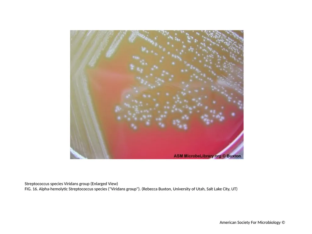
Streptococcus species Viridans group (Enlarged View)
FIG. 16. Alpha-hemolytic Streptococcus species ("Viridans group"). (Rebecca Buxton, University of Utah, Salt Lake City, UT) American Society For Microbiology ©<br>
FIG. 16. Alpha-hemolytic Streptococcus species ("Viridans group"). (Rebecca Buxton, University of Utah, Salt Lake City, UT) American Society For Microbiology ©<br>
18
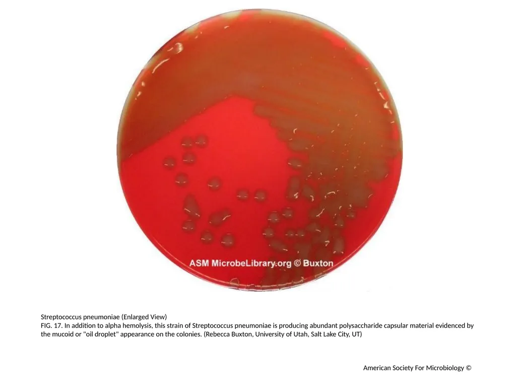
Streptococcus pneumoniae (Enlarged View)
FIG. 17. In addition to alpha hemolysis, this strain of Streptococcus pneumoniae is producing abundant polysaccharide capsular material evidenced by the mucoid or "oil droplet" appearance on the colonies. (Rebecca Buxton, University of Utah, Salt Lake City, UT) American Society For Microbiology ©<br>
FIG. 17. In addition to alpha hemolysis, this strain of Streptococcus pneumoniae is producing abundant polysaccharide capsular material evidenced by the mucoid or "oil droplet" appearance on the colonies. (Rebecca Buxton, University of Utah, Salt Lake City, UT) American Society For Microbiology ©<br>
19
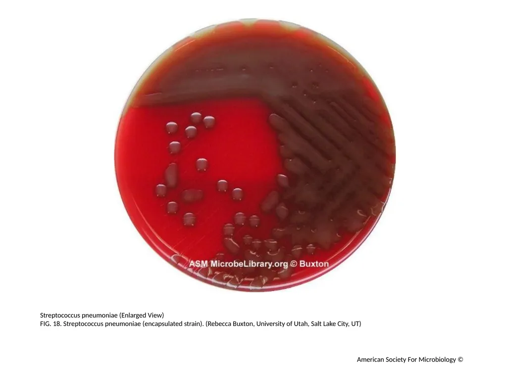
Streptococcus pneumoniae (Enlarged View)
FIG. 18. Streptococcus pneumoniae (encapsulated strain). (Rebecca Buxton, University of Utah, Salt Lake City, UT) American Society For Microbiology ©<br>
FIG. 18. Streptococcus pneumoniae (encapsulated strain). (Rebecca Buxton, University of Utah, Salt Lake City, UT) American Society For Microbiology ©<br>
20
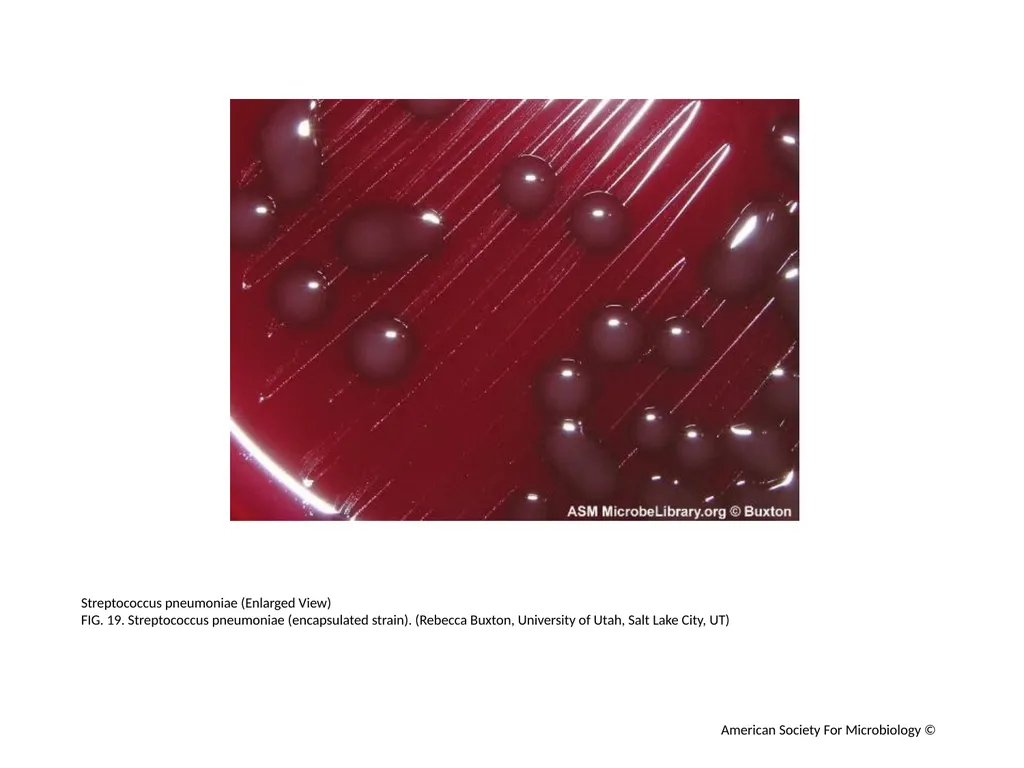
Streptococcus pneumoniae (Enlarged View)
FIG. 19. Streptococcus pneumoniae (encapsulated strain). (Rebecca Buxton, University of Utah, Salt Lake City, UT) American Society For Microbiology ©<br>
FIG. 19. Streptococcus pneumoniae (encapsulated strain). (Rebecca Buxton, University of Utah, Salt Lake City, UT) American Society For Microbiology ©<br>
21
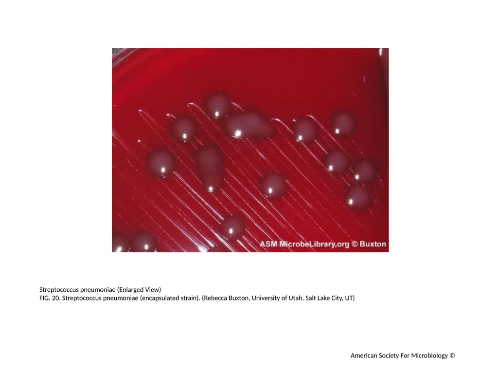
Streptococcus pneumoniae (Enlarged View)
FIG. 20. Streptococcus pneumoniae (encapsulated strain). (Rebecca Buxton, University of Utah, Salt Lake City, UT) American Society For Microbiology ©<br>
FIG. 20. Streptococcus pneumoniae (encapsulated strain). (Rebecca Buxton, University of Utah, Salt Lake City, UT) American Society For Microbiology ©<br>
22
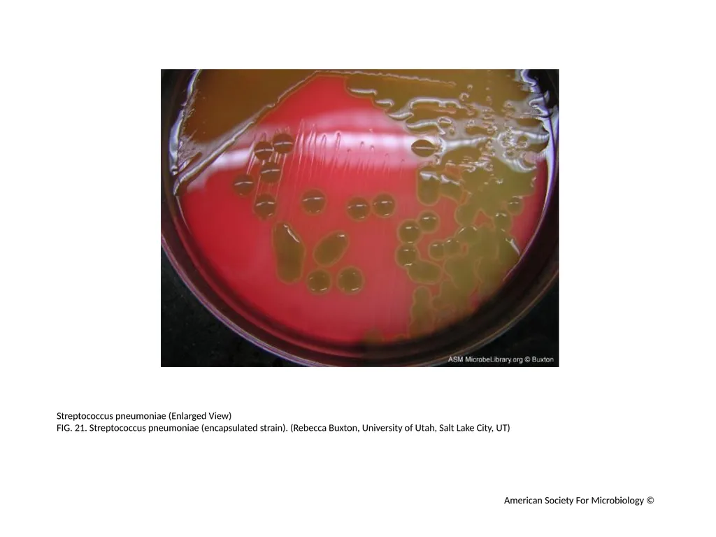
Streptococcus pneumoniae (Enlarged View)
FIG. 21. Streptococcus pneumoniae (encapsulated strain). (Rebecca Buxton, University of Utah, Salt Lake City, UT) American Society For Microbiology ©<br>
FIG. 21. Streptococcus pneumoniae (encapsulated strain). (Rebecca Buxton, University of Utah, Salt Lake City, UT) American Society For Microbiology ©<br>
23
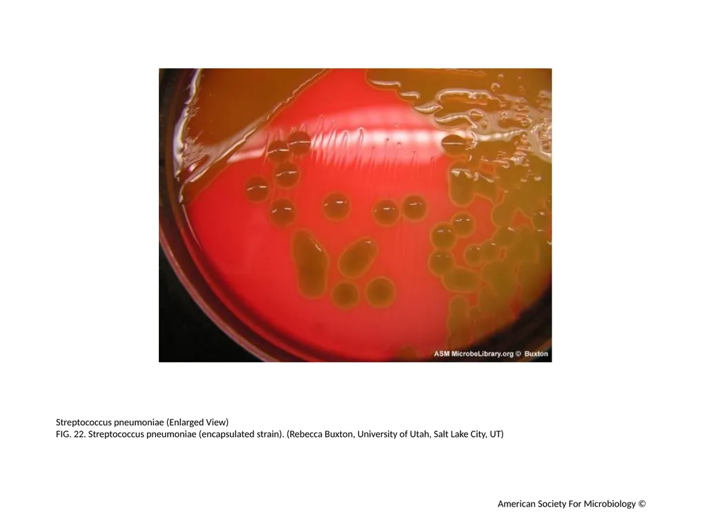
Streptococcus pneumoniae (Enlarged View)
FIG. 22. Streptococcus pneumoniae (encapsulated strain). (Rebecca Buxton, University of Utah, Salt Lake City, UT) American Society For Microbiology ©<br>
FIG. 22. Streptococcus pneumoniae (encapsulated strain). (Rebecca Buxton, University of Utah, Salt Lake City, UT) American Society For Microbiology ©<br>
24
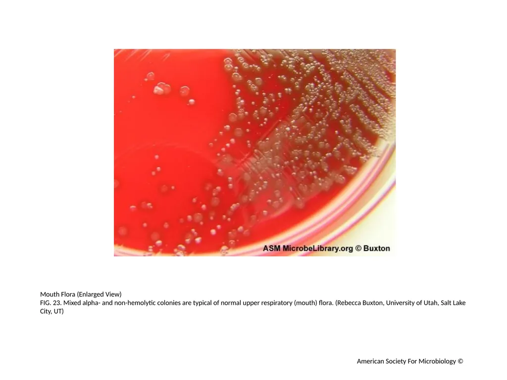
Mouth Flora (Enlarged View)
FIG. 23. Mixed alpha- and non-hemolytic colonies are typical of normal upper respiratory (mouth) flora. (Rebecca Buxton, University of Utah, Salt Lake City, UT) American Society For Microbiology ©<br>
FIG. 23. Mixed alpha- and non-hemolytic colonies are typical of normal upper respiratory (mouth) flora. (Rebecca Buxton, University of Utah, Salt Lake City, UT) American Society For Microbiology ©<br>
25
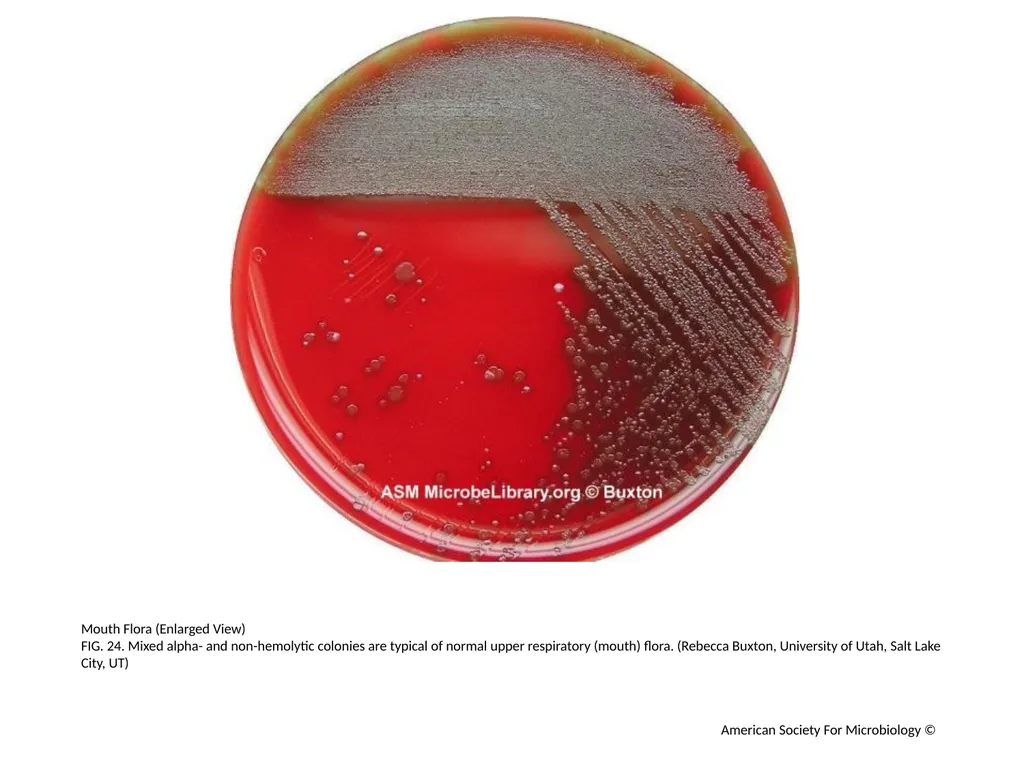
Mouth Flora (Enlarged View)
FIG. 24. Mixed alpha- and non-hemolytic colonies are typical of normal upper respiratory (mouth) flora. (Rebecca Buxton, University of Utah, Salt Lake City, UT) American Society For Microbiology ©<br>
FIG. 24. Mixed alpha- and non-hemolytic colonies are typical of normal upper respiratory (mouth) flora. (Rebecca Buxton, University of Utah, Salt Lake City, UT) American Society For Microbiology ©<br>
26
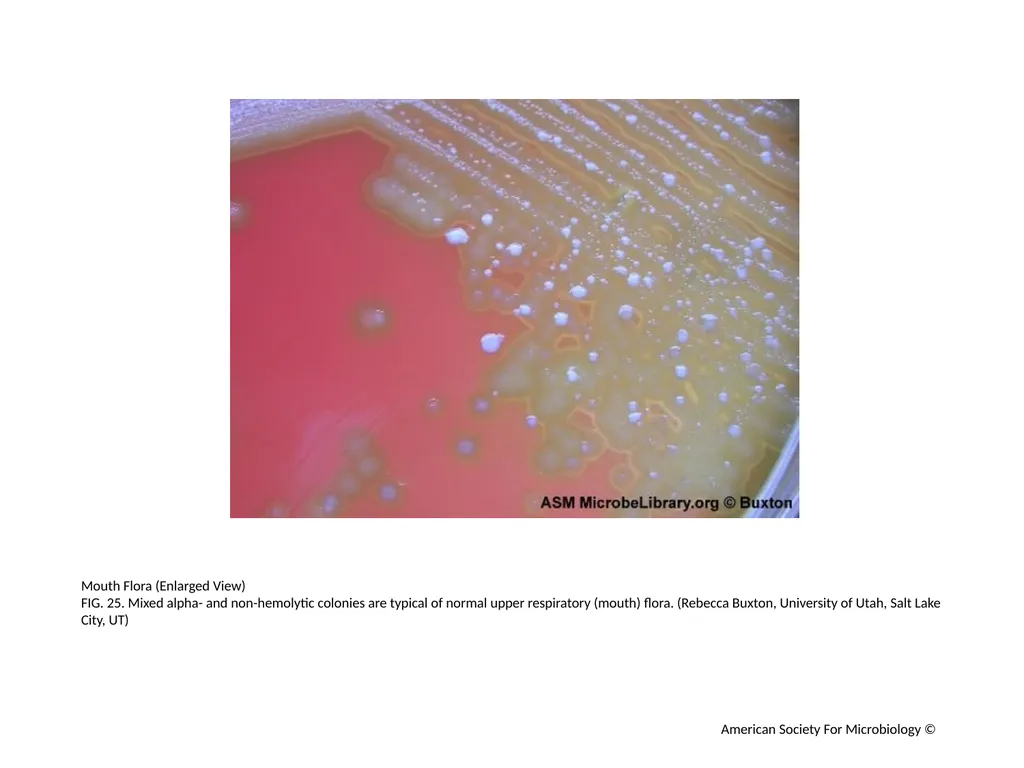
Mouth Flora (Enlarged View)
FIG. 25. Mixed alpha- and non-hemolytic colonies are typical of normal upper respiratory (mouth) flora. (Rebecca Buxton, University of Utah, Salt Lake City, UT) American Society For Microbiology ©<br>
FIG. 25. Mixed alpha- and non-hemolytic colonies are typical of normal upper respiratory (mouth) flora. (Rebecca Buxton, University of Utah, Salt Lake City, UT) American Society For Microbiology ©<br>
27
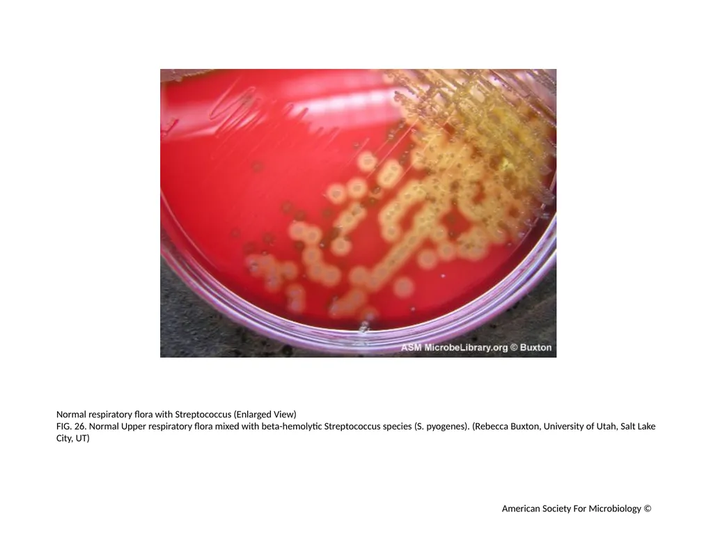
Normal respiratory flora with Streptococcus (Enlarged View)
FIG. 26. Normal Upper respiratory flora mixed with beta-hemolytic Streptococcus species (S. pyogenes). (Rebecca Buxton, University of Utah, Salt Lake City, UT) American Society For Microbiology ©<br>
FIG. 26. Normal Upper respiratory flora mixed with beta-hemolytic Streptococcus species (S. pyogenes). (Rebecca Buxton, University of Utah, Salt Lake City, UT) American Society For Microbiology ©<br>
28
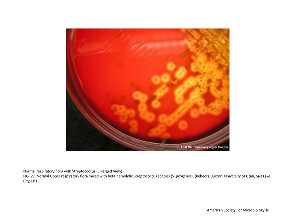
Normal respiratory flora with Streptococcus (Enlarged View)
FIG. 27. Normal Upper respiratory flora mixed with beta-hemolytic Streptococcus species (S. pyogenes). (Rebecca Buxton, University of Utah, Salt Lake City, UT) American Society For Microbiology ©<br>
FIG. 27. Normal Upper respiratory flora mixed with beta-hemolytic Streptococcus species (S. pyogenes). (Rebecca Buxton, University of Utah, Salt Lake City, UT) American Society For Microbiology ©<br>
29
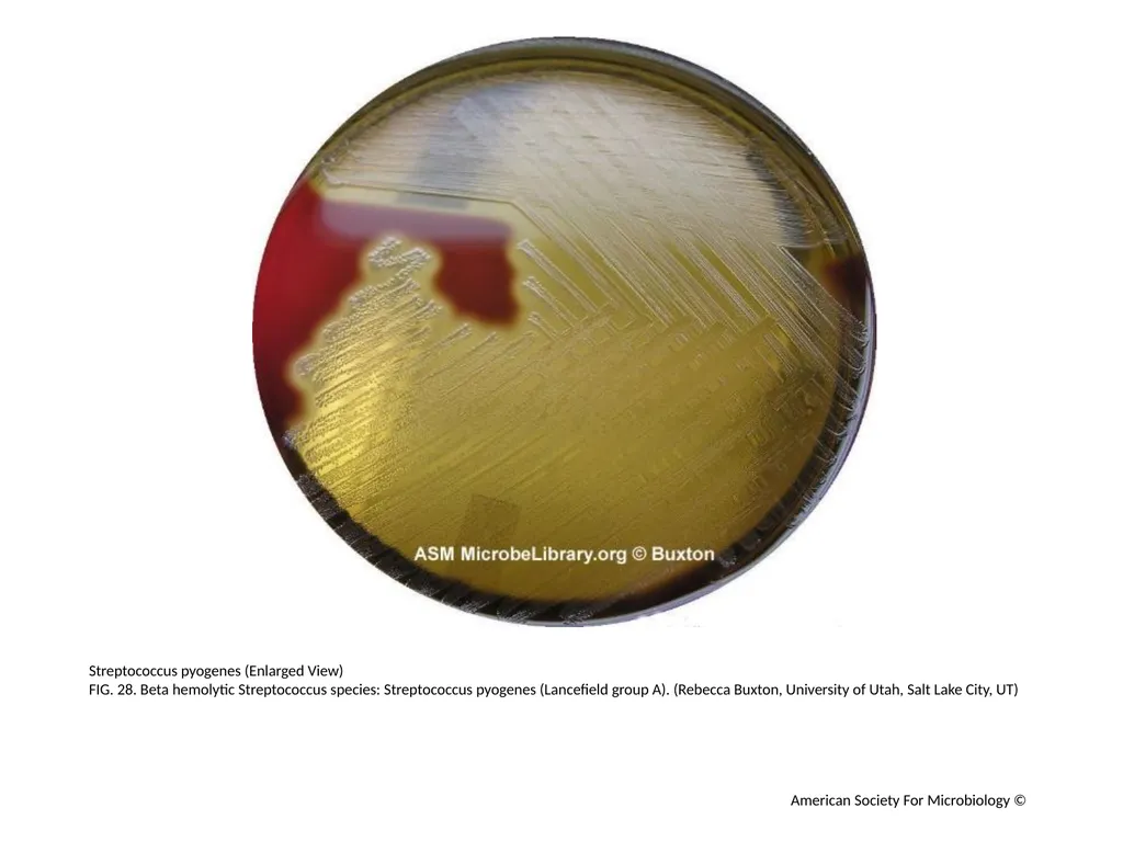
Streptococcus pyogenes (Enlarged View)
FIG. 28. Beta hemolytic Streptococcus species: Streptococcus pyogenes (Lancefield group A). (Rebecca Buxton, University of Utah, Salt Lake City, UT) American Society For Microbiology ©<br>
FIG. 28. Beta hemolytic Streptococcus species: Streptococcus pyogenes (Lancefield group A). (Rebecca Buxton, University of Utah, Salt Lake City, UT) American Society For Microbiology ©<br>
30
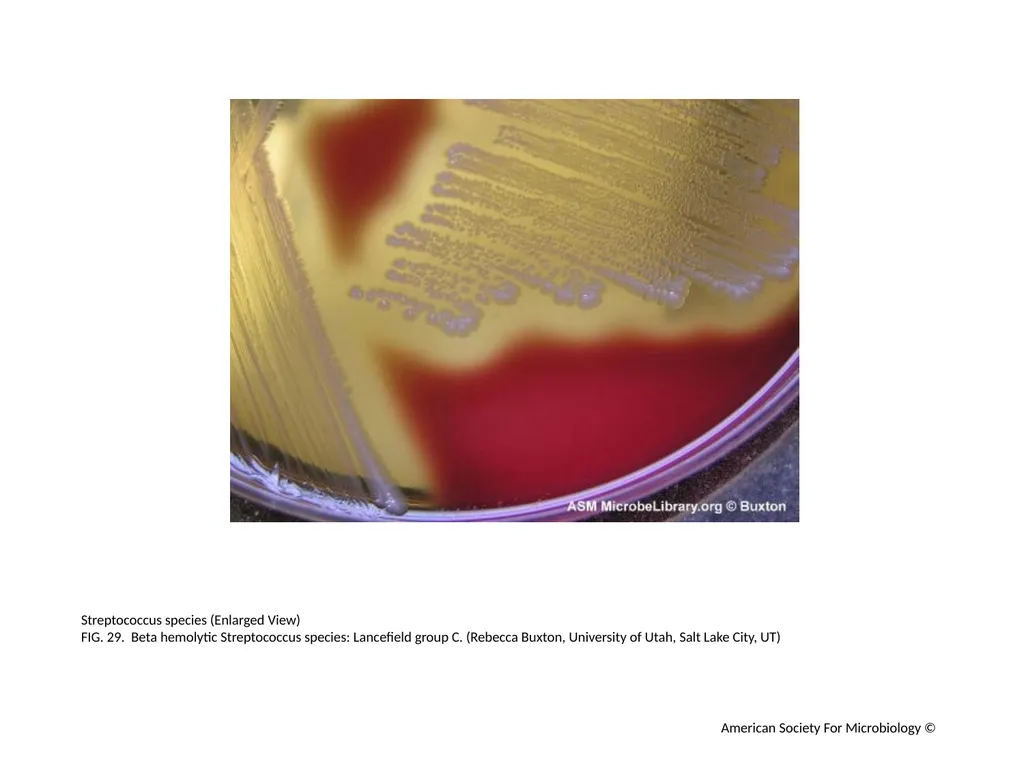
Streptococcus species (Enlarged View)
FIG. 29. Beta hemolytic Streptococcus species: Lancefield group C. (Rebecca Buxton, University of Utah, Salt Lake City, UT) American Society For Microbiology ©<br>
FIG. 29. Beta hemolytic Streptococcus species: Lancefield group C. (Rebecca Buxton, University of Utah, Salt Lake City, UT) American Society For Microbiology ©<br>
31
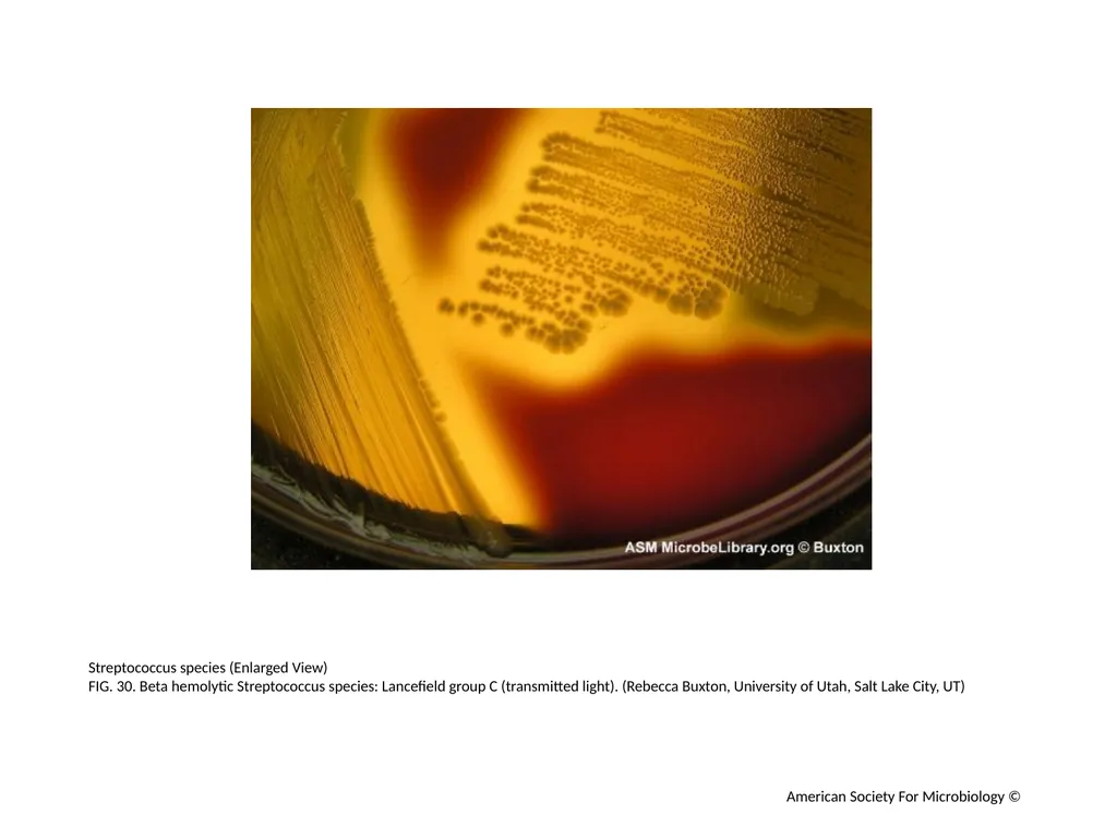
Streptococcus species (Enlarged View)
FIG. 30. Beta hemolytic Streptococcus species: Lancefield group C (transmitted light). (Rebecca Buxton, University of Utah, Salt Lake City, UT) American Society For Microbiology ©<br>
FIG. 30. Beta hemolytic Streptococcus species: Lancefield group C (transmitted light). (Rebecca Buxton, University of Utah, Salt Lake City, UT) American Society For Microbiology ©<br>
32
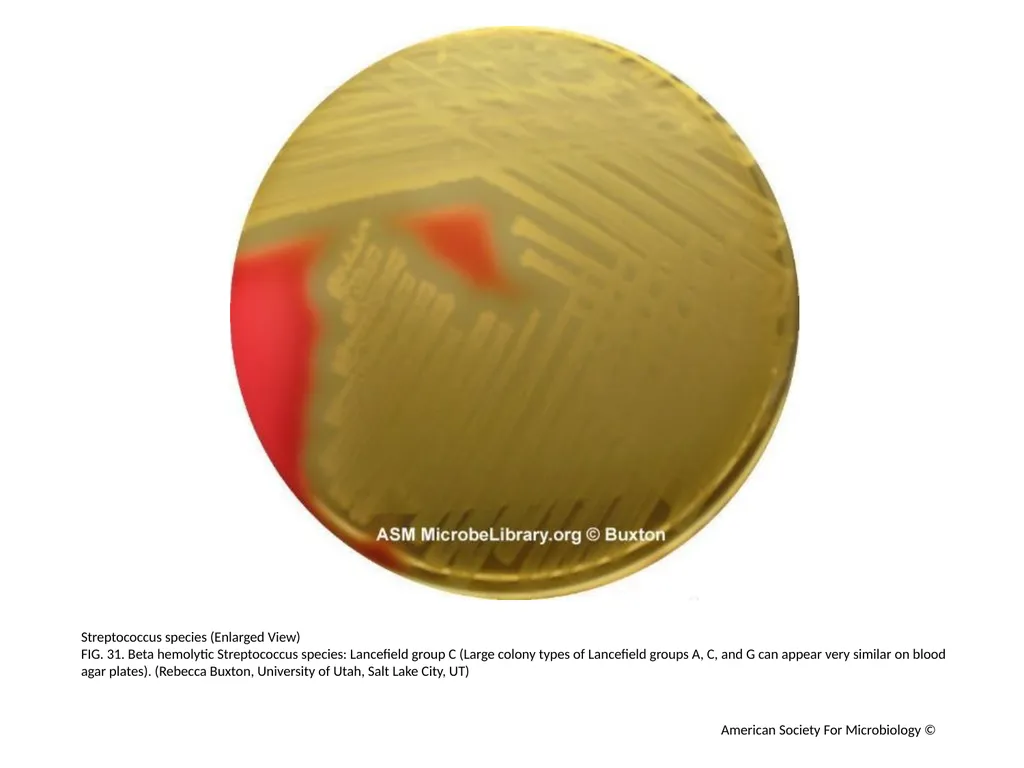
Streptococcus species (Enlarged View)
FIG. 31. Beta hemolytic Streptococcus species: Lancefield group C (Large colony types of Lancefield groups A, C, and G can appear very similar on blood agar plates). (Rebecca Buxton, University of Utah, Salt Lake City, UT) American Society For Microbiology ©<br>
FIG. 31. Beta hemolytic Streptococcus species: Lancefield group C (Large colony types of Lancefield groups A, C, and G can appear very similar on blood agar plates). (Rebecca Buxton, University of Utah, Salt Lake City, UT) American Society For Microbiology ©<br>
33
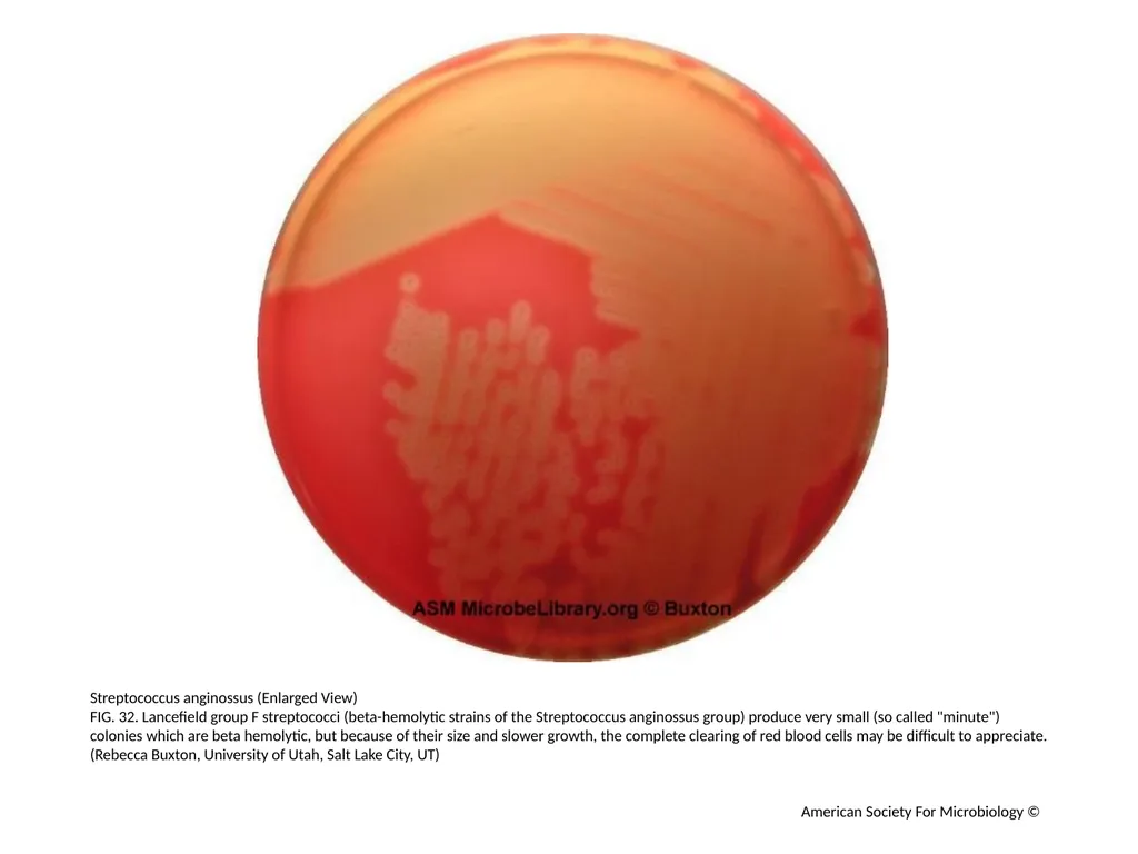
Streptococcus anginossus (Enlarged View)
FIG. 32. Lancefield group F streptococci (beta-hemolytic strains of the Streptococcus anginossus group) produce very small (so called "minute") colonies which are beta hemolytic, but because of their size and slower growth, the complete clearing of red blood cells may be difficult to appreciate. (Rebecca Buxton, University of Utah, Salt Lake City, UT) American Society For Microbiology ©<br>
FIG. 32. Lancefield group F streptococci (beta-hemolytic strains of the Streptococcus anginossus group) produce very small (so called "minute") colonies which are beta hemolytic, but because of their size and slower growth, the complete clearing of red blood cells may be difficult to appreciate. (Rebecca Buxton, University of Utah, Salt Lake City, UT) American Society For Microbiology ©<br>
34
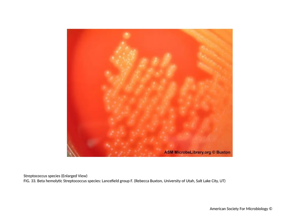
Streptococcus species (Enlarged View)
FIG. 33. Beta hemolytic Streptococcus species: Lancefield group F. (Rebecca Buxton, University of Utah, Salt Lake City, UT) American Society For Microbiology ©<br>
FIG. 33. Beta hemolytic Streptococcus species: Lancefield group F. (Rebecca Buxton, University of Utah, Salt Lake City, UT) American Society For Microbiology ©<br>
35
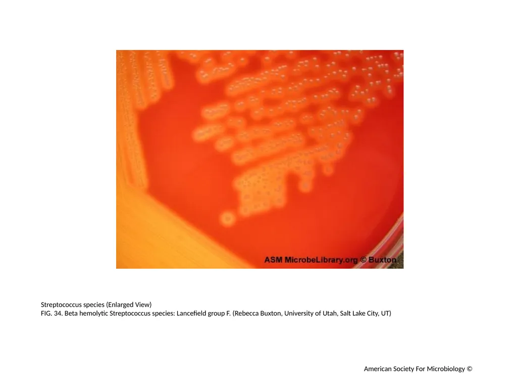
Streptococcus species (Enlarged View)
FIG. 34. Beta hemolytic Streptococcus species: Lancefield group F. (Rebecca Buxton, University of Utah, Salt Lake City, UT) American Society For Microbiology ©<br>
FIG. 34. Beta hemolytic Streptococcus species: Lancefield group F. (Rebecca Buxton, University of Utah, Salt Lake City, UT) American Society For Microbiology ©<br>
36
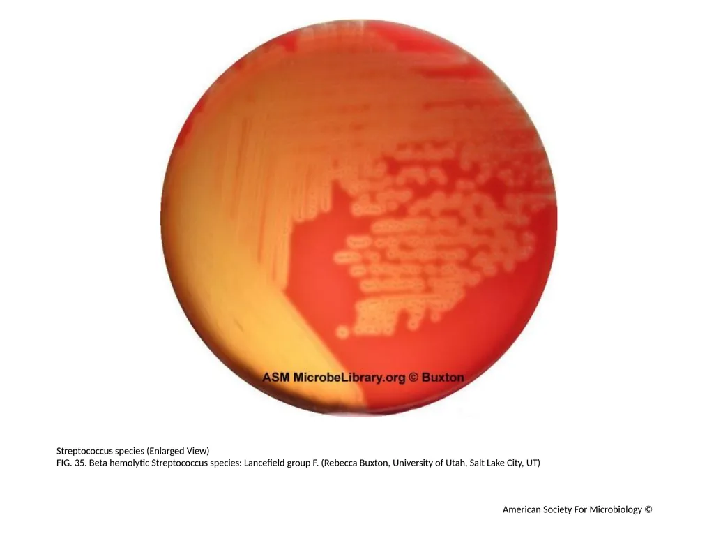
Streptococcus species (Enlarged View)
FIG. 35. Beta hemolytic Streptococcus species: Lancefield group F. (Rebecca Buxton, University of Utah, Salt Lake City, UT) American Society For Microbiology ©<br>
FIG. 35. Beta hemolytic Streptococcus species: Lancefield group F. (Rebecca Buxton, University of Utah, Salt Lake City, UT) American Society For Microbiology ©<br>
37
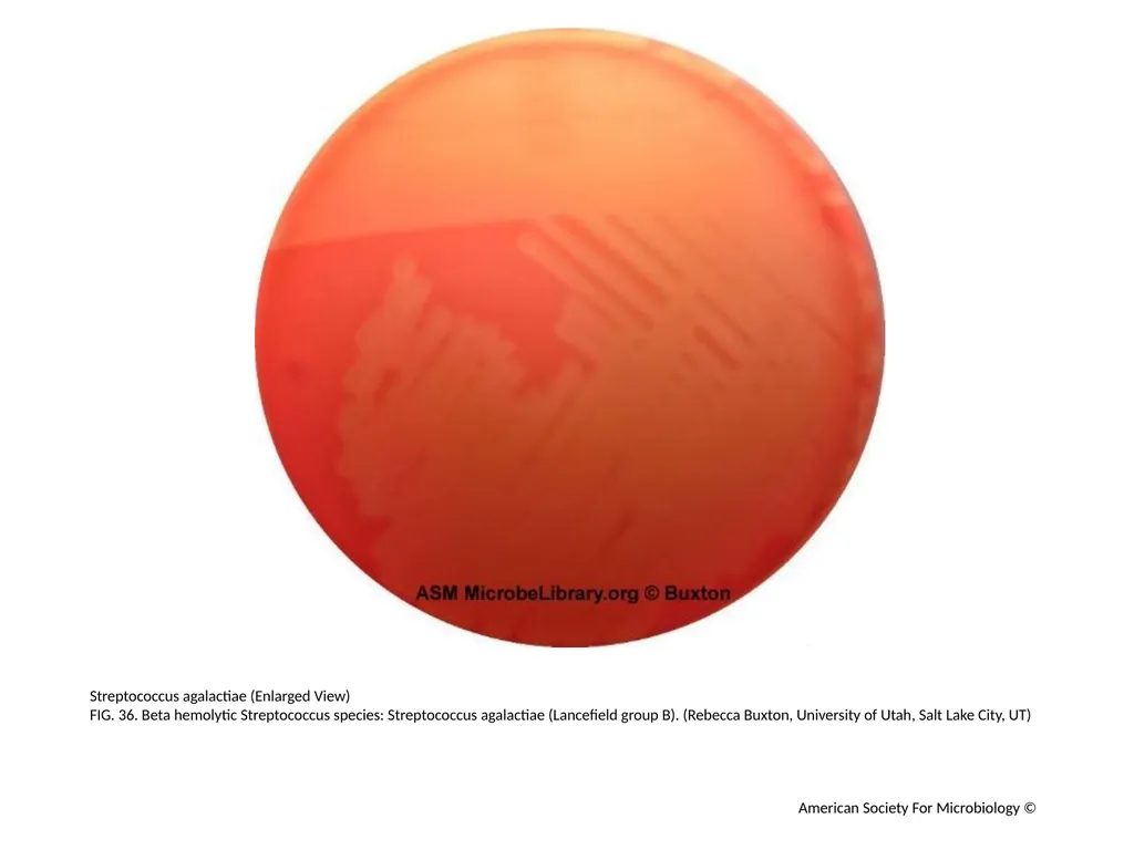
Streptococcus agalactiae (Enlarged View)
FIG. 36. Beta hemolytic Streptococcus species: Streptococcus agalactiae (Lancefield group B). (Rebecca Buxton, University of Utah, Salt Lake City, UT) American Society For Microbiology ©<br>
FIG. 36. Beta hemolytic Streptococcus species: Streptococcus agalactiae (Lancefield group B). (Rebecca Buxton, University of Utah, Salt Lake City, UT) American Society For Microbiology ©<br>
38
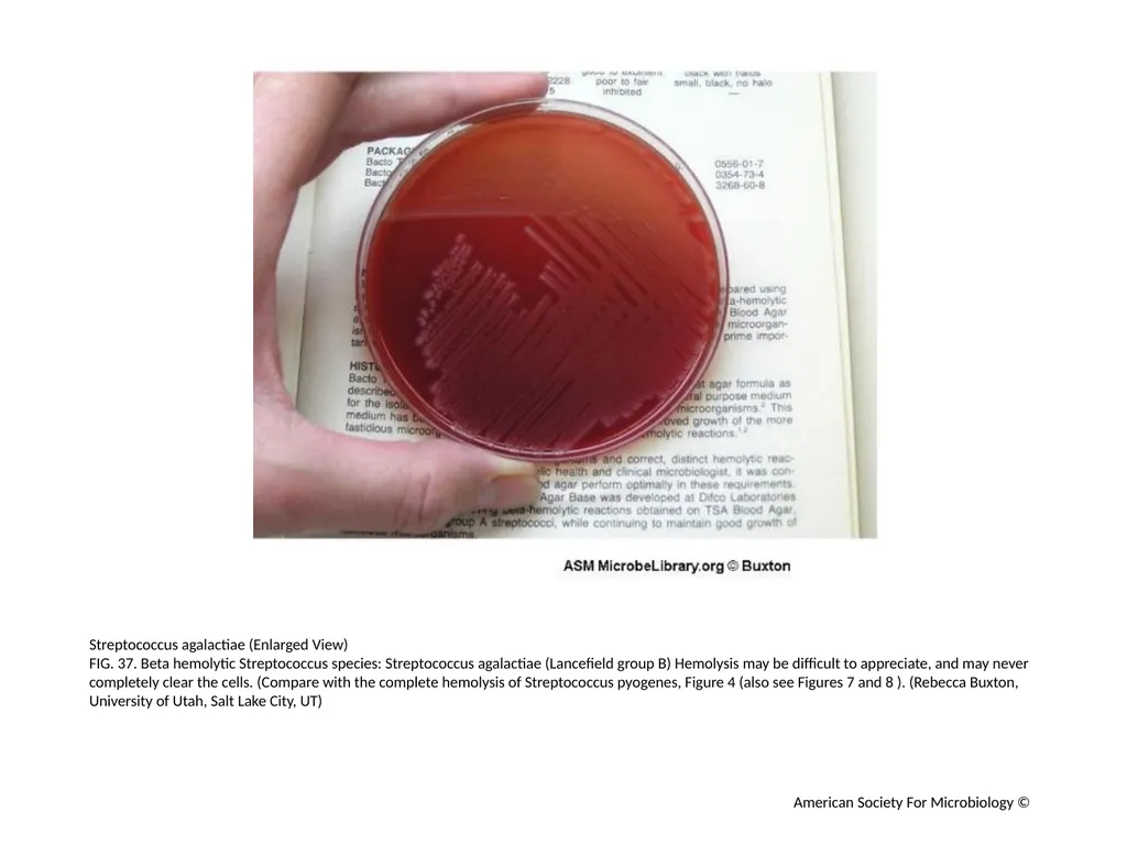
Streptococcus agalactiae (Enlarged View)
FIG. 37. Beta hemolytic Streptococcus species: Streptococcus agalactiae (Lancefield group B) Hemolysis may be difficult to appreciate, and may never completely clear the cells. (Compare with the complete hemolysis of Streptococcus pyogenes, Figure 4 (also see Figures 7 and 8 ). (Rebecca Buxton, University of Utah, Salt Lake City, UT) American Society For Microbiology ©<br>
FIG. 37. Beta hemolytic Streptococcus species: Streptococcus agalactiae (Lancefield group B) Hemolysis may be difficult to appreciate, and may never completely clear the cells. (Compare with the complete hemolysis of Streptococcus pyogenes, Figure 4 (also see Figures 7 and 8 ). (Rebecca Buxton, University of Utah, Salt Lake City, UT) American Society For Microbiology ©<br>
39
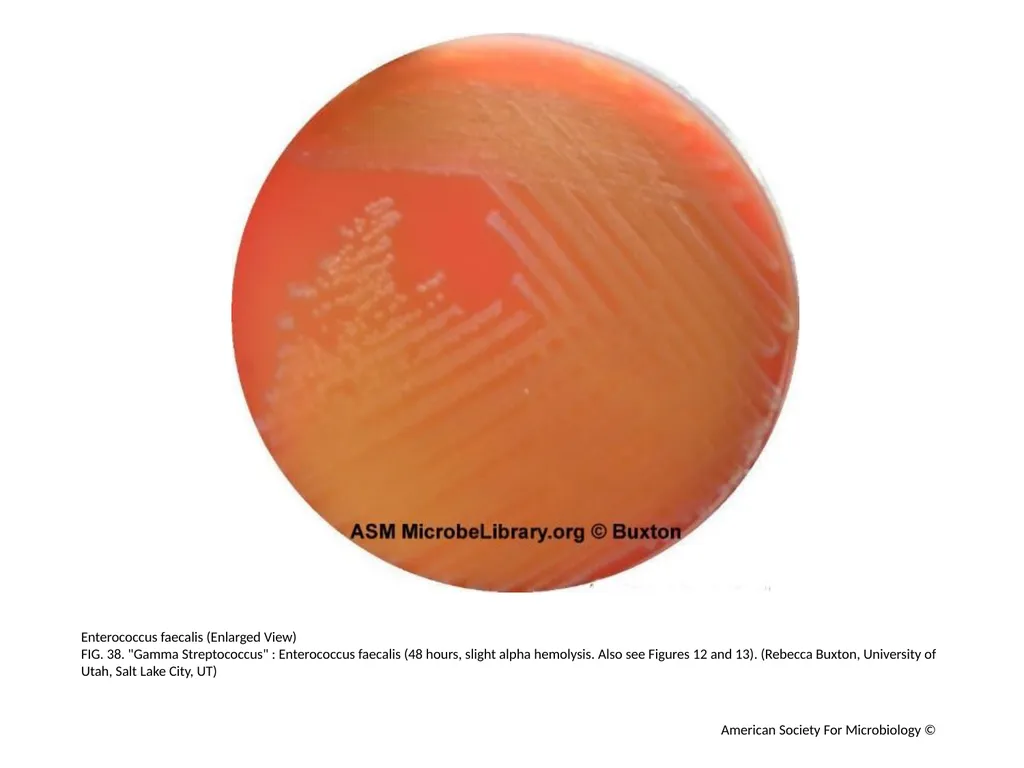
Enterococcus faecalis (Enlarged View)
FIG. 38. "Gamma Streptococcus" : Enterococcus faecalis (48 hours, slight alpha hemolysis. Also see Figures 12 and 13). (Rebecca Buxton, University of Utah, Salt Lake City, UT) American Society For Microbiology ©<br>
FIG. 38. "Gamma Streptococcus" : Enterococcus faecalis (48 hours, slight alpha hemolysis. Also see Figures 12 and 13). (Rebecca Buxton, University of Utah, Salt Lake City, UT) American Society For Microbiology ©<br>
40
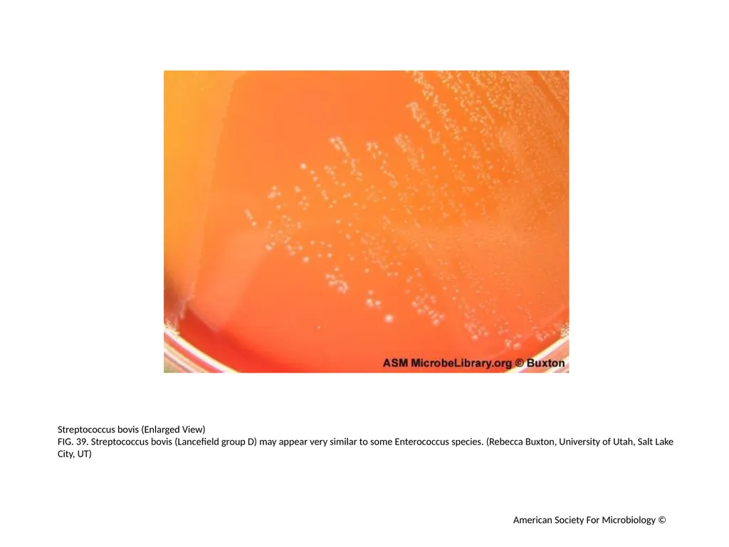
Streptococcus bovis (Enlarged View)
FIG. 39. Streptococcus bovis (Lancefield group D) may appear very similar to some Enterococcus species. (Rebecca Buxton, University of Utah, Salt Lake City, UT) American Society For Microbiology ©<br>
FIG. 39. Streptococcus bovis (Lancefield group D) may appear very similar to some Enterococcus species. (Rebecca Buxton, University of Utah, Salt Lake City, UT) American Society For Microbiology ©<br>
41
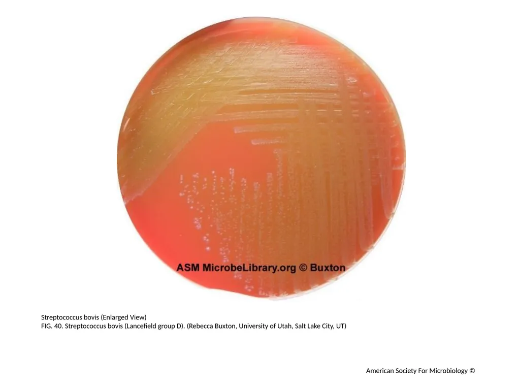
Streptococcus bovis (Enlarged View)
FIG. 40. Streptococcus bovis (Lancefield group D). (Rebecca Buxton, University of Utah, Salt Lake City, UT) American Society For Microbiology ©<br>
FIG. 40. Streptococcus bovis (Lancefield group D). (Rebecca Buxton, University of Utah, Salt Lake City, UT) American Society For Microbiology ©<br>
42
Streptococcus bovis (Enlarged View)
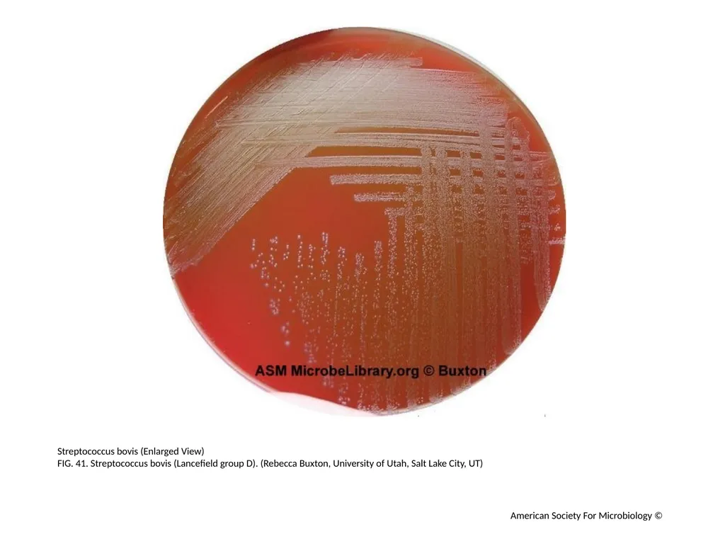
FIG. 41. Streptococcus bovis (Lancefield group D). (Rebecca Buxton, University of Utah, Salt Lake City, UT) American Society For Microbiology ©<br>
FIG. 41. Streptococcus bovis (Lancefield group D). (Rebecca Buxton, University of Utah, Salt Lake City, UT) American Society For Microbiology ©<br>
43
Streptococcus bovis (Enlarged View)
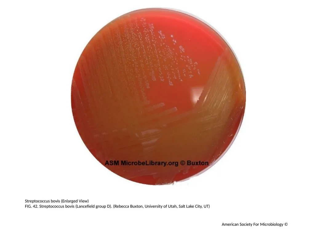
FIG. 42. Streptococcus bovis (Lancefield group D). (Rebecca Buxton, University of Utah, Salt Lake City, UT) American Society For Microbiology ©<br>
FIG. 42. Streptococcus bovis (Lancefield group D). (Rebecca Buxton, University of Utah, Salt Lake City, UT) American Society For Microbiology ©<br>
44
American Society For Microbiology ©<br>