04
History
Dyspnea
Chest pain
Palpitations
Syncope
Hemoptysis
Edema<br>
05
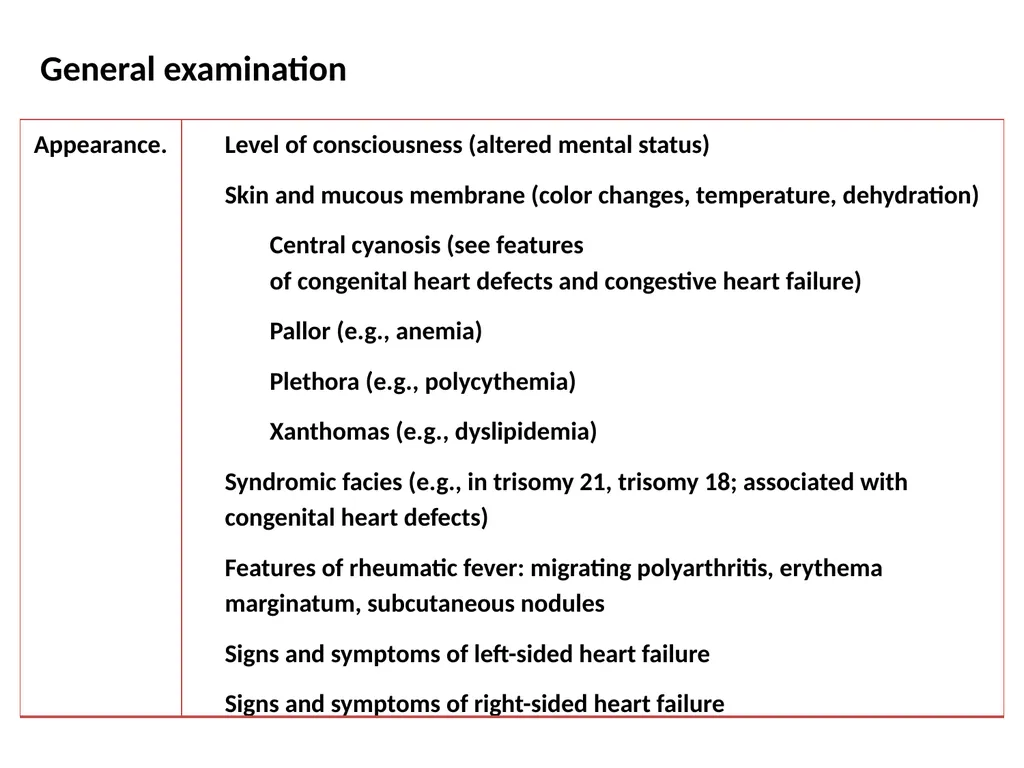
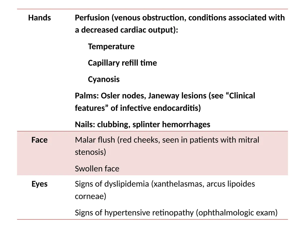
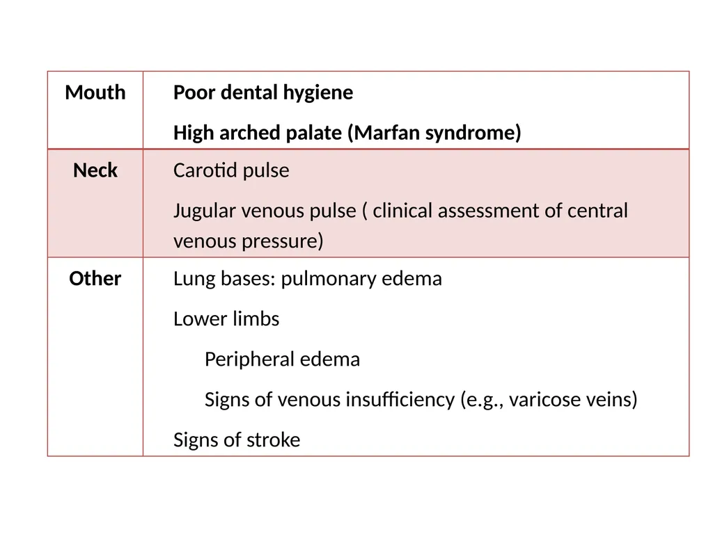
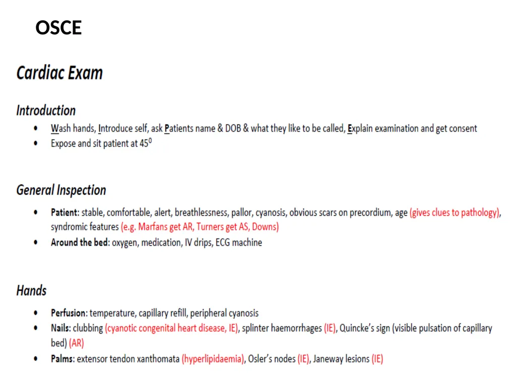
General examination<br>
06
Central venous pressure
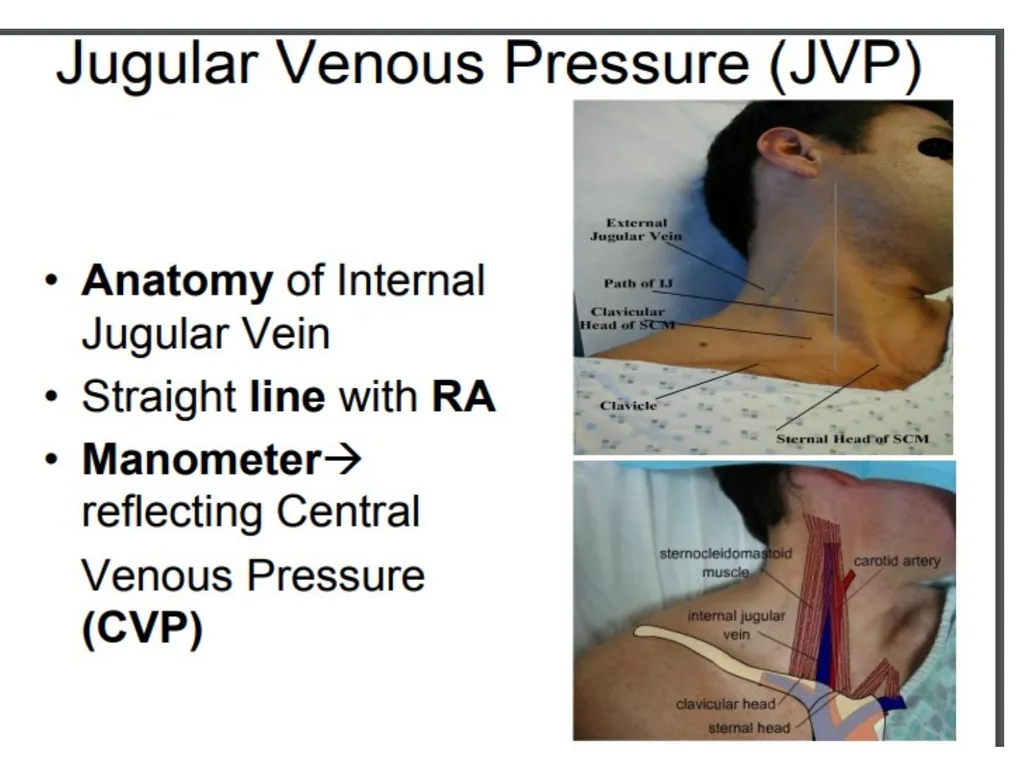
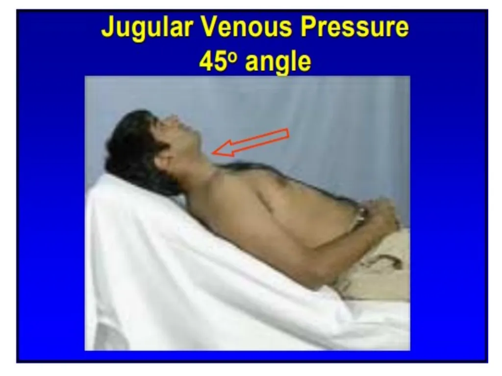
The jugular venous pulse (JVP) can be used to estimate the central venous pressure (CVP) and provides information about fluid status and cardiac function.<br>
07
JUGULAR VENOUS PULSE WHAT: VISIBLE PRESSURE CHANGES IN RIGHT ATRIUM
WHERE: UNDER STERNOCLEIDOMASTOID MUSCLE
WHY: DIAGNOSE HEART FAILURE, FLUID OVERLOAD, AV BLOCK<br>
08
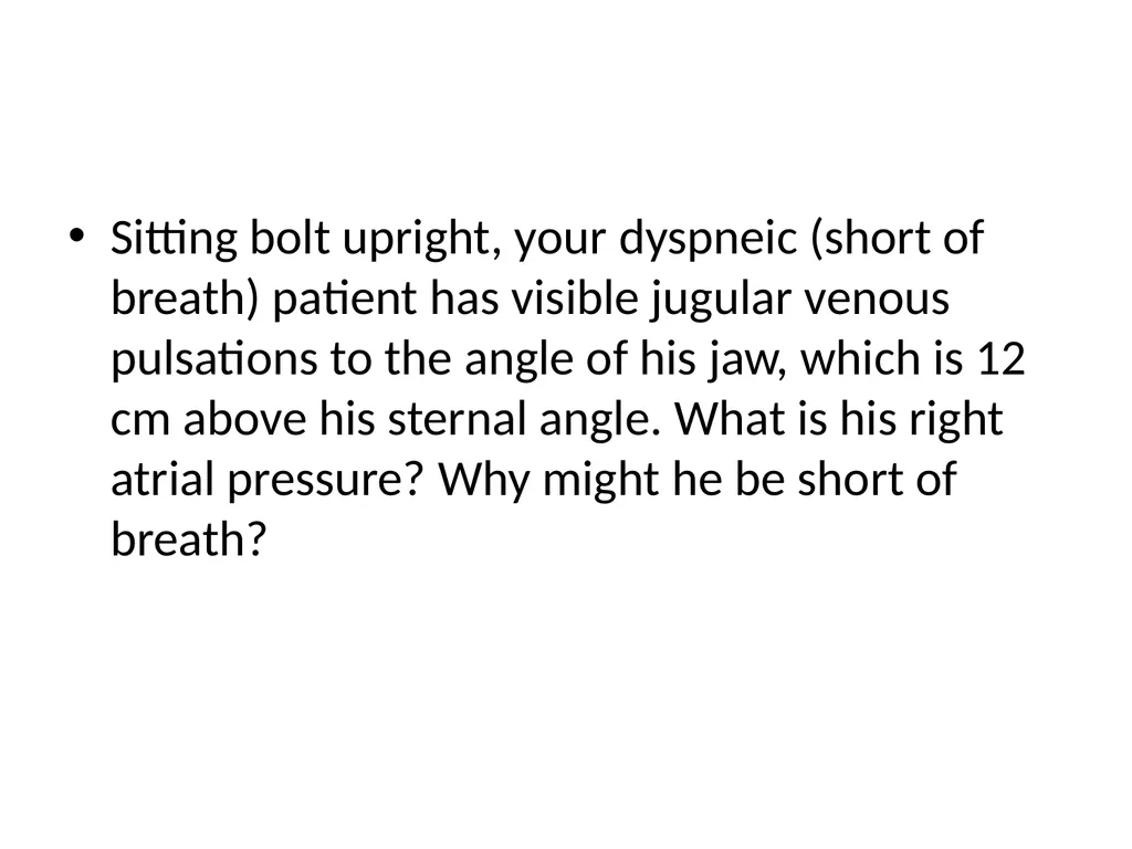
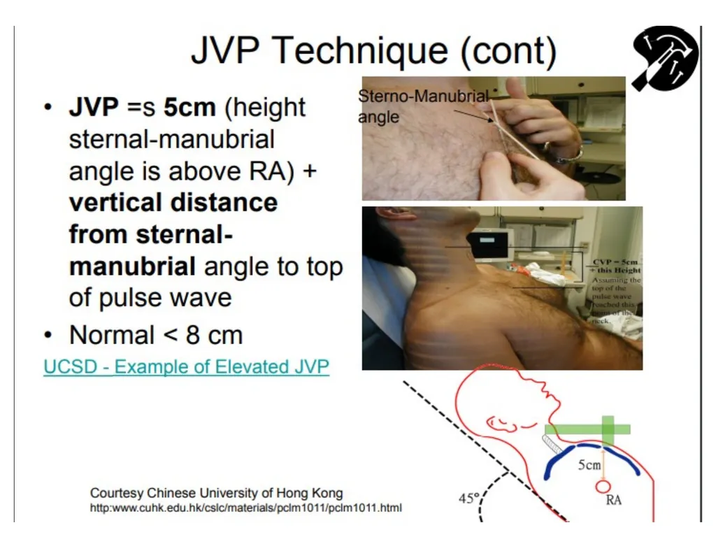
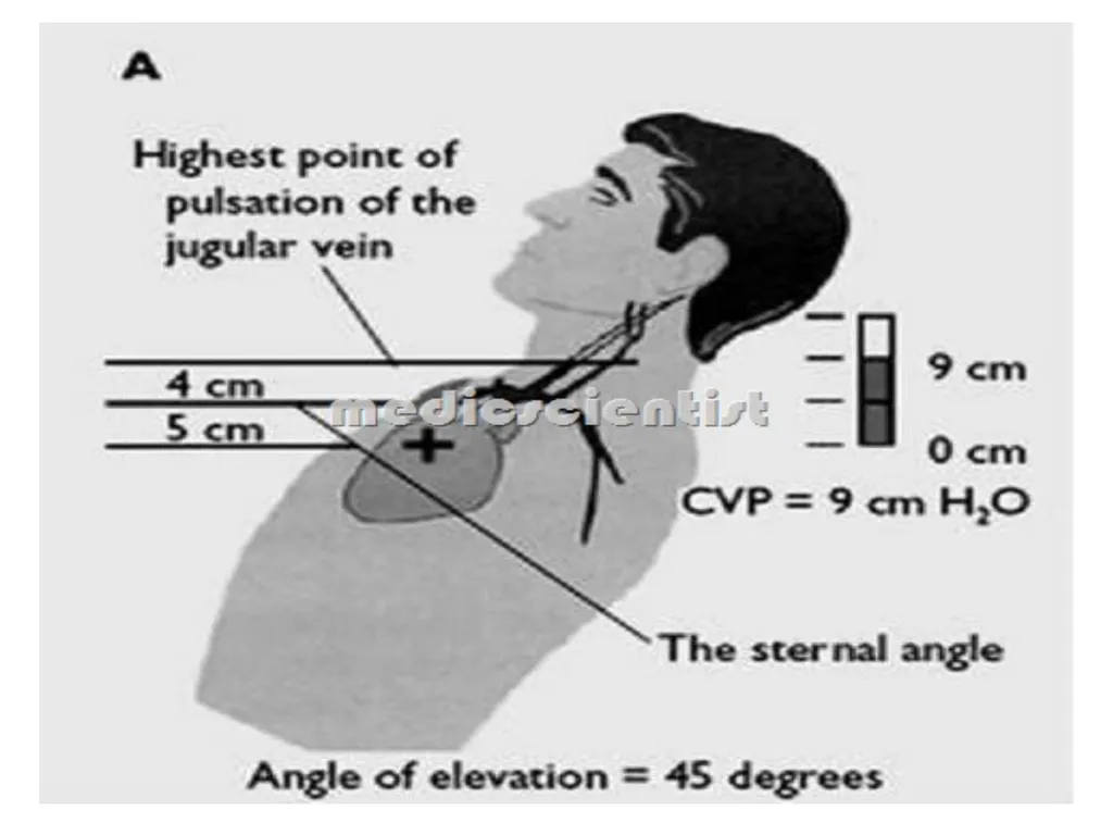
JUGULAR VENOUS PULSE STERNAL ANGLE IS 5 CM ABOVE RIGHT ATRIUM
RIGHT ATRIAL PRESSURE = HEIGHT OF JVP ABOVE STERNAL ANGLE + 5
NORMAL RA PRESSURE: 5-10 CM H2O<br>
09
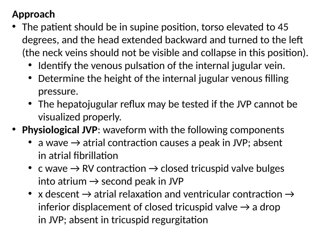
Approach
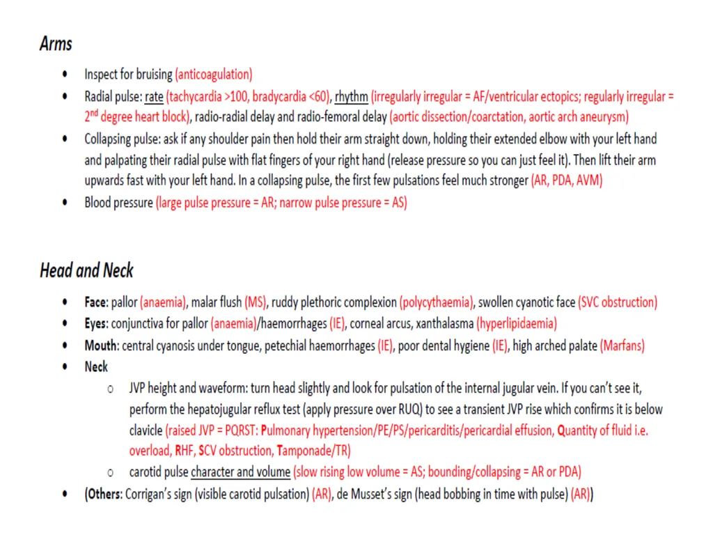
The patient should be in supine position, torso elevated to 45 degrees, and the head extended backward and turned to the left (the neck veins should not be visible and collapse in this position).
Identify the venous pulsation of the internal jugular vein.
Determine the height of the internal jugular venous filling pressure.
The hepatojugular reflux may be tested if the JVP cannot be visualized properly.
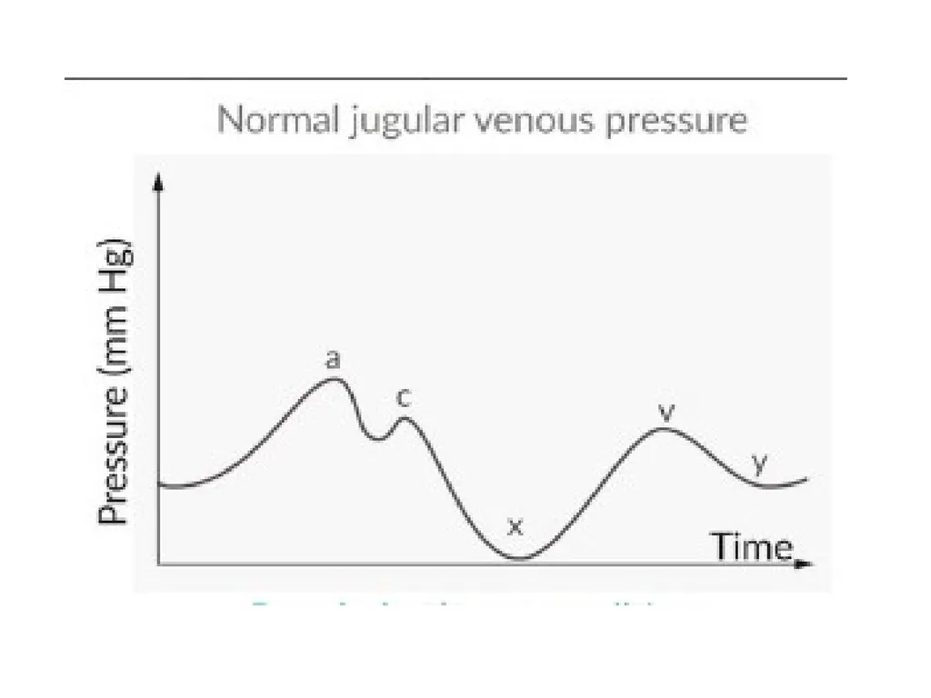
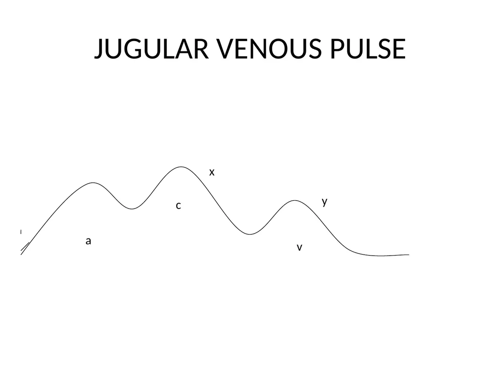
Physiological JVP: waveform with the following components
a wave → atrial contraction causes a peak in JVP; absent in atrial fibrillation
c wave → RV contraction → closed tricuspid valve bulges into atrium → second peak in JVP
x descent → atrial relaxation and ventricular contraction → inferior displacement of closed tricuspid valve → a drop in JVP; absent in tricuspid regurgitation<br>
10
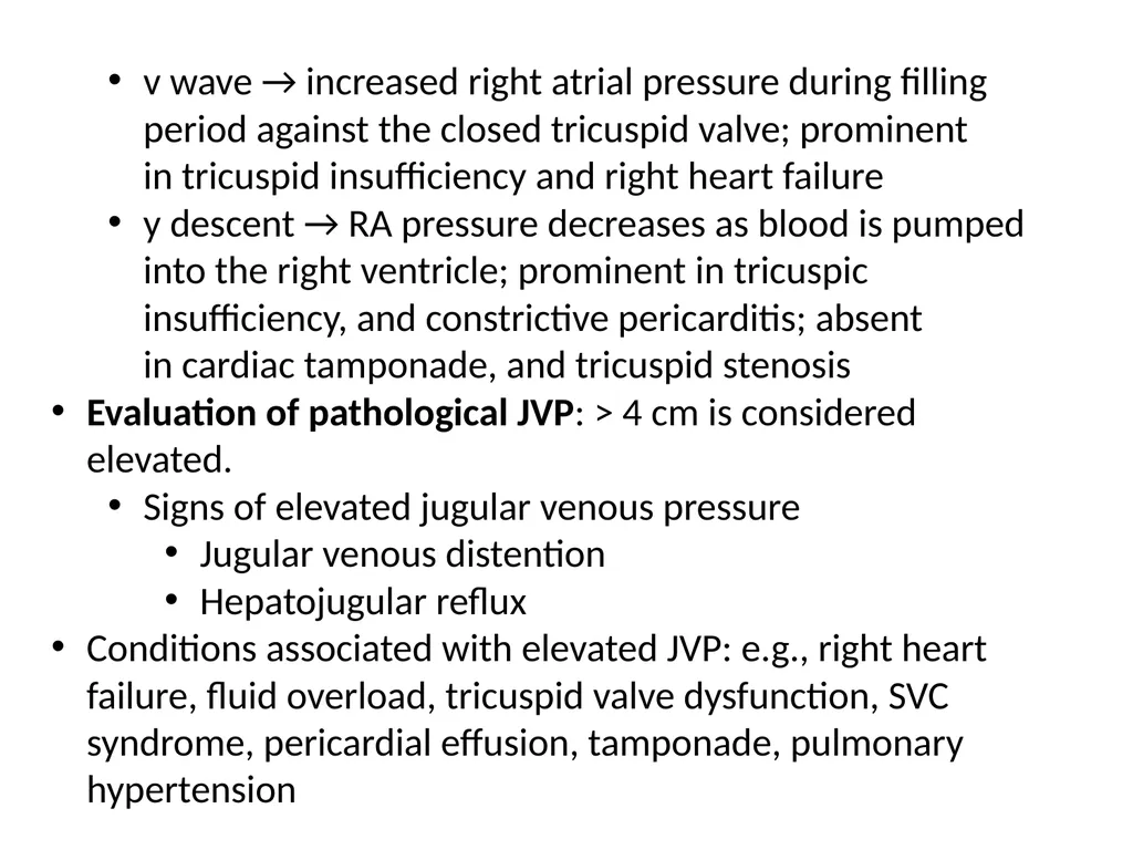
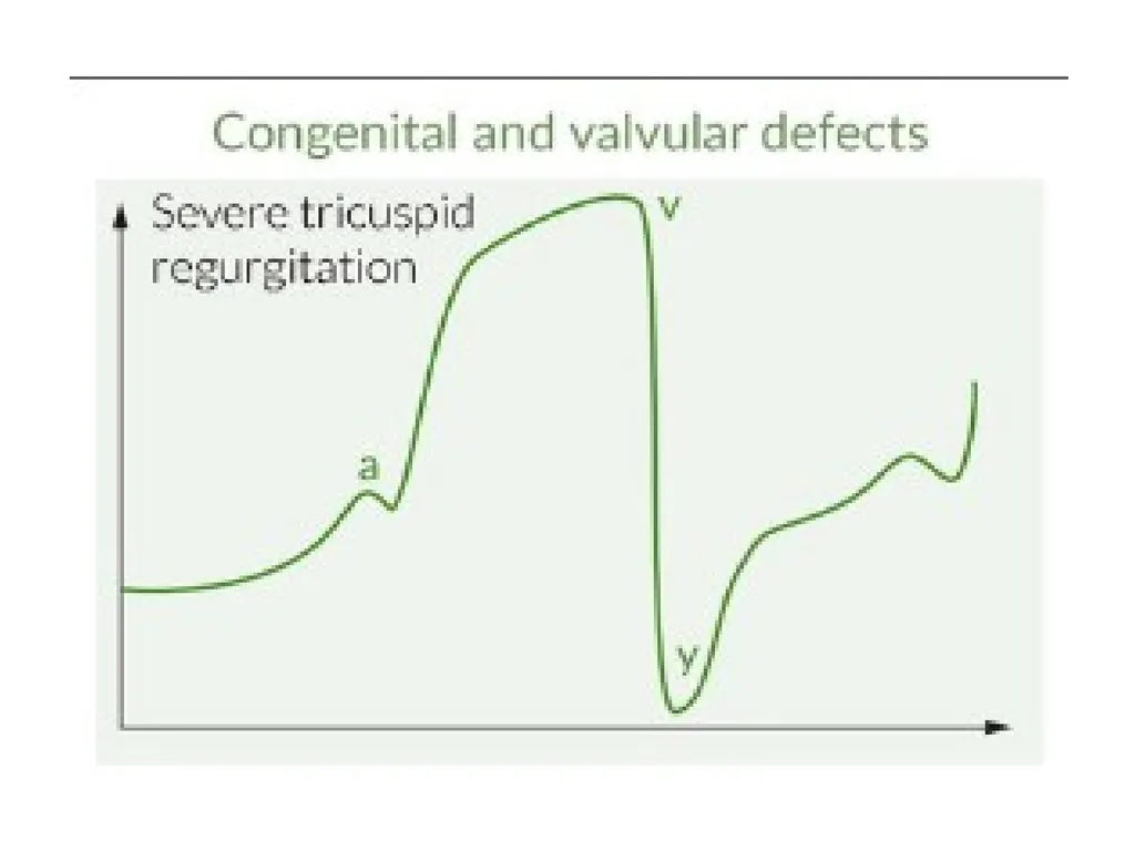
v wave → increased right atrial pressure during filling period against the closed tricuspid valve; prominent in tricuspid insufficiency and right heart failure
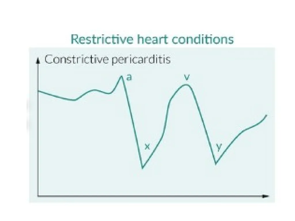
y descent → RA pressure decreases as blood is pumped into the right ventricle; prominent in tricuspic insufficiency, and constrictive pericarditis; absent in cardiac tamponade, and tricuspid stenosis
Evaluation of pathological JVP: > 4 cm is considered elevated.
Signs of elevated jugular venous pressure
Jugular venous distention
Hepatojugular reflux
Conditions associated with elevated JVP: e.g., right heart failure, fluid overload, tricuspid valve dysfunction, SVC syndrome, pericardial effusion, tamponade, pulmonary hypertension<br>
11
JUGULAR VENOUS PULSE a v c y x<br>
12
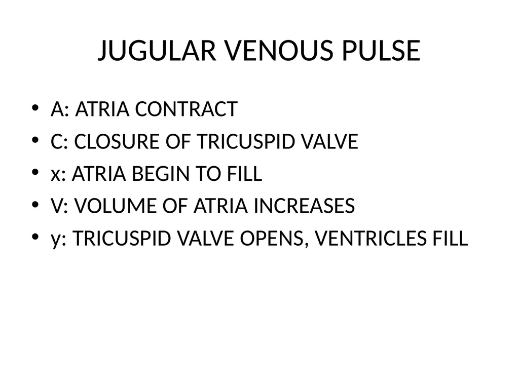
JUGULAR VENOUS PULSE A: ATRIA CONTRACT
C: CLOSURE OF TRICUSPID VALVE
x: ATRIA BEGIN TO FILL
V: VOLUME OF ATRIA INCREASES
y: TRICUSPID VALVE OPENS, VENTRICLES FILL<br>
13
WHICH POINT ON THE JUGULAR VENOUS PULSE OCCURS NEAR THE BEGINNING OF DIASTOLE?<br>
14
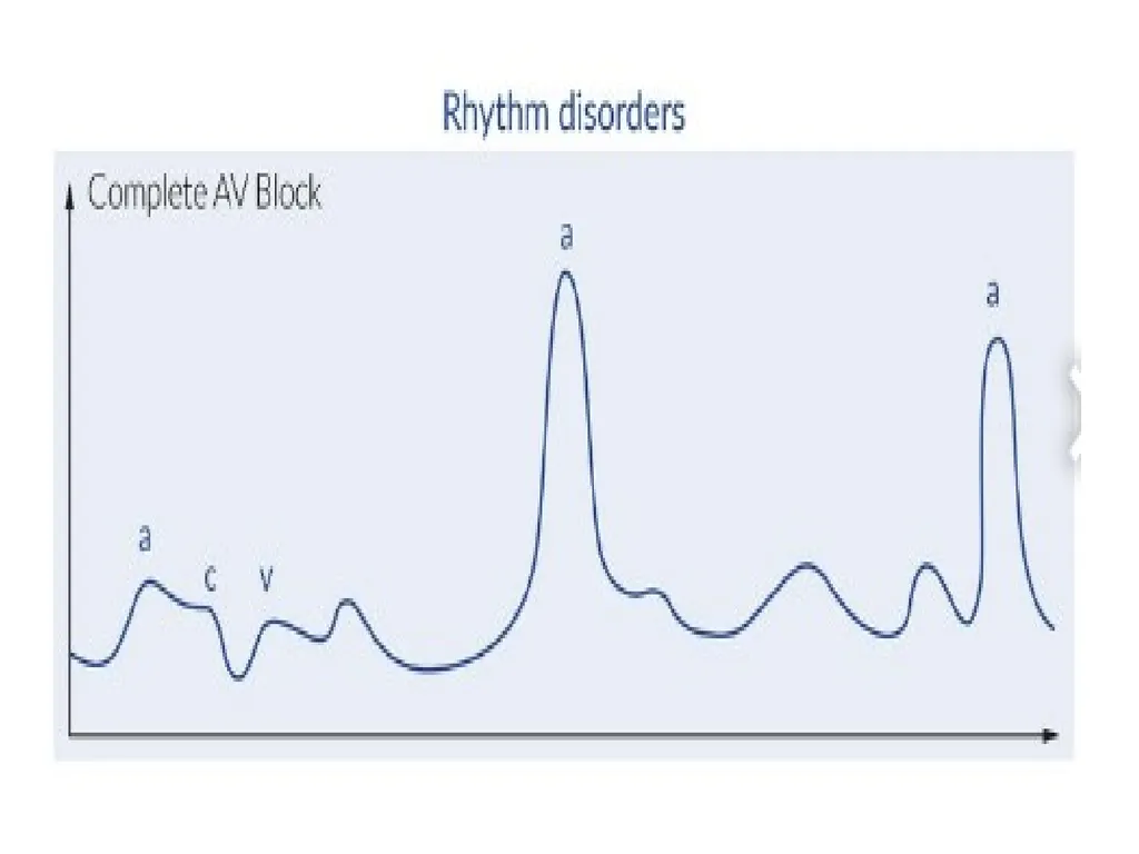
ATRIOVENTRICULAR DISSOCIATION/ AV BLOCK ATRIA AND VENTRICLES CONTRACT INDEPENDENTLY
ATRIA THUS CONTRACT AGAINST CLOSED AV VALVES
CANNON A WAVES<br>
15
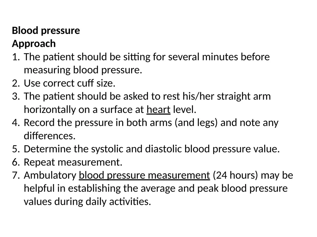
Blood pressure
Approach
The patient should be sitting for several minutes before measuring blood pressure.
Use correct cuff size.
The patient should be asked to rest his/her straight arm horizontally on a surface at heart level.
Record the pressure in both arms (and legs) and note any differences.
Determine the systolic and diastolic blood pressure value.
Repeat measurement.
Ambulatory blood pressure measurement (24 hours) may be helpful in establishing the average and peak blood pressure values during daily activities.<br>
16
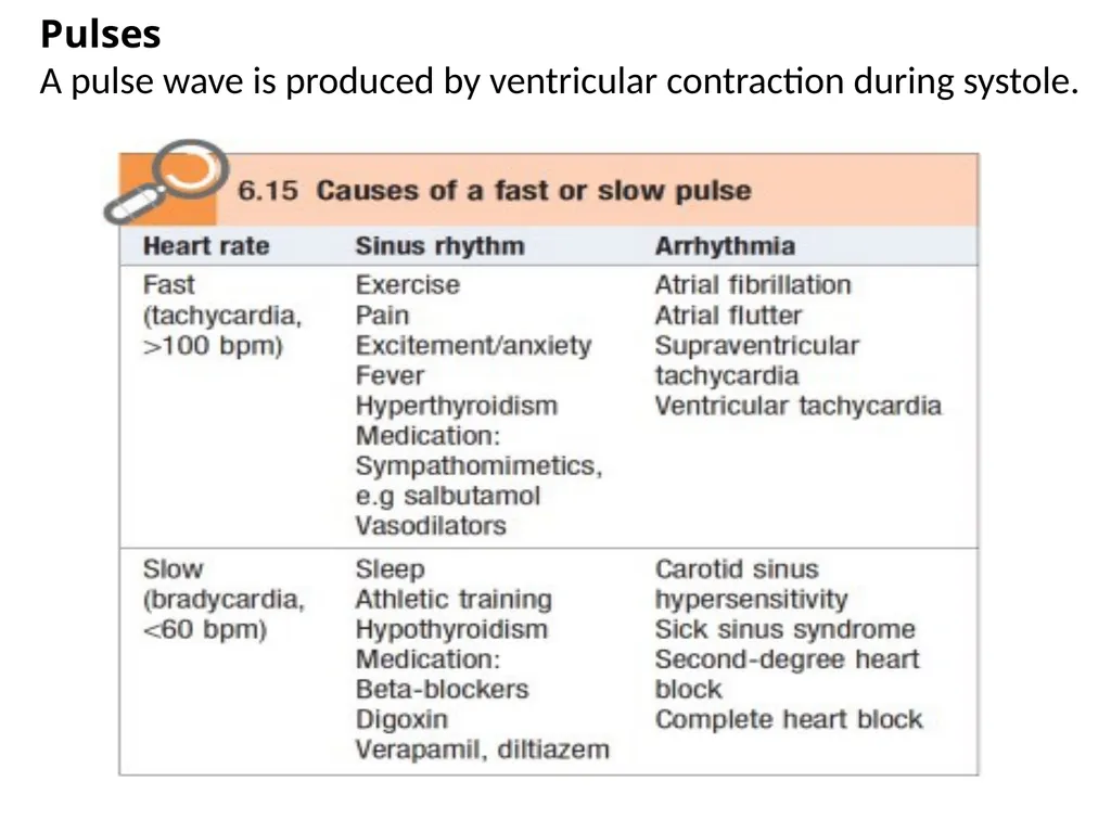
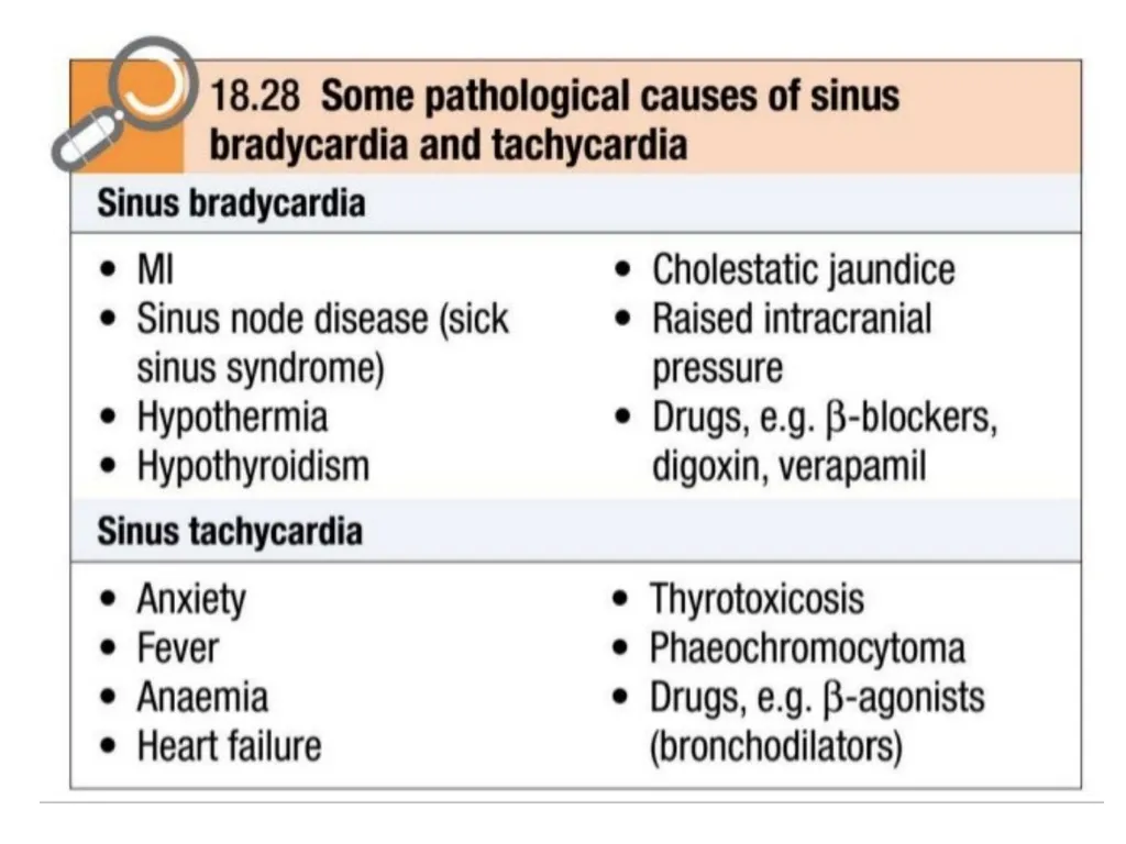
Pulses
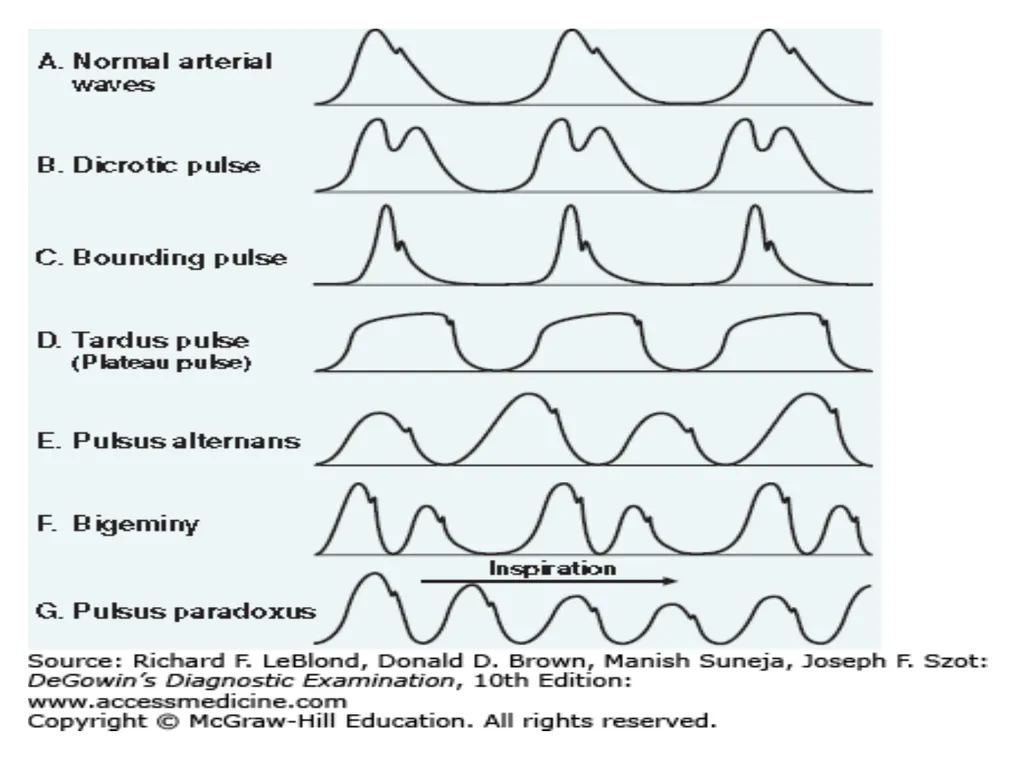
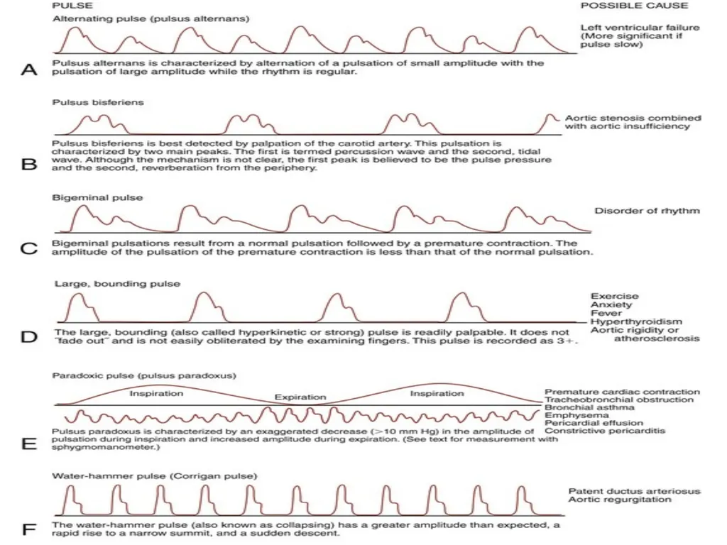
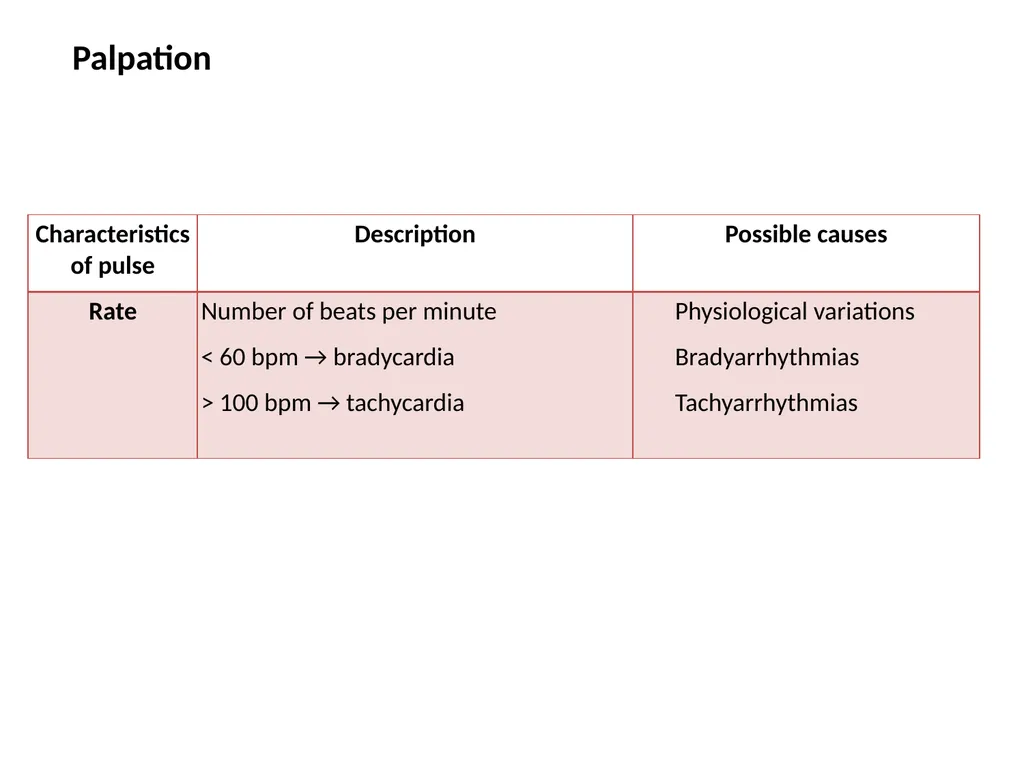
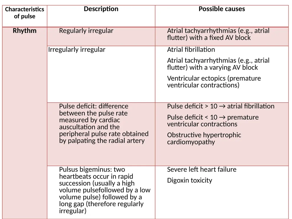
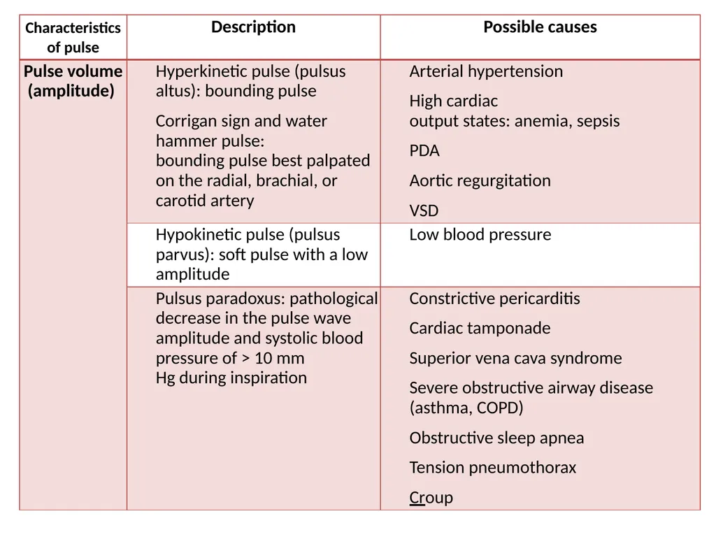
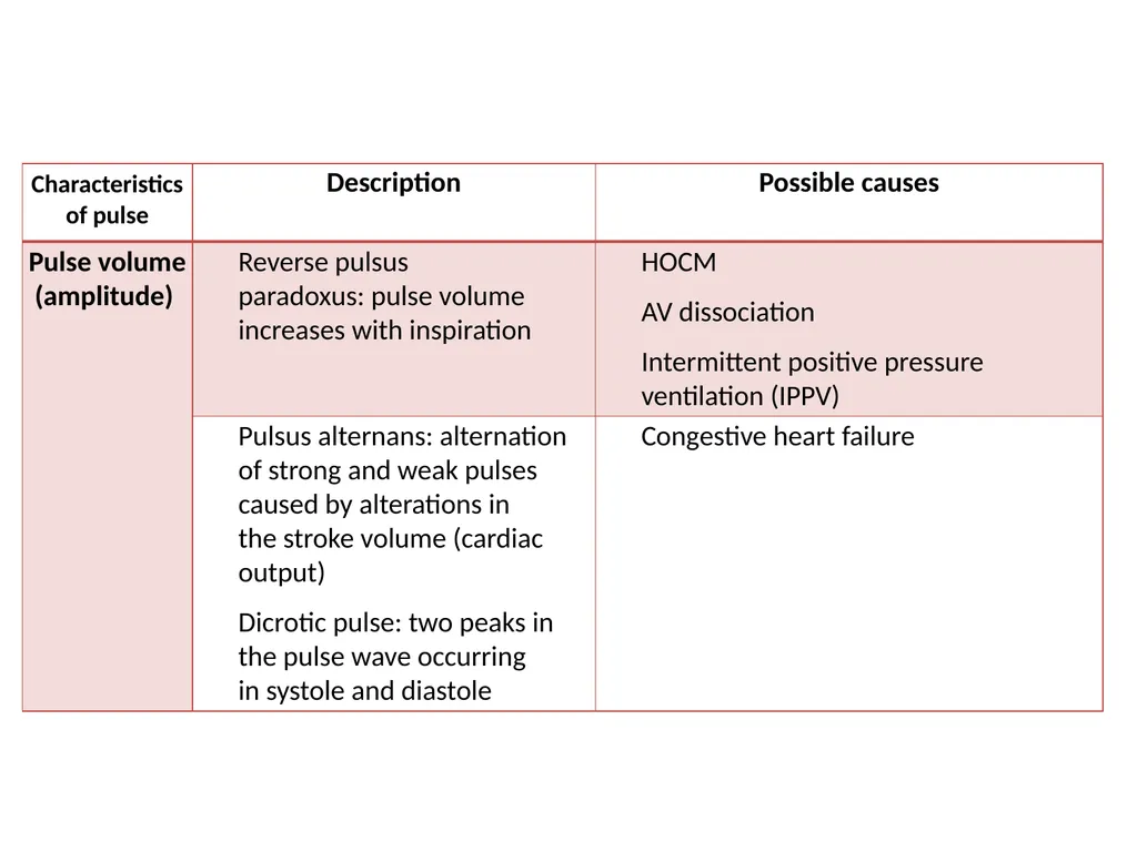
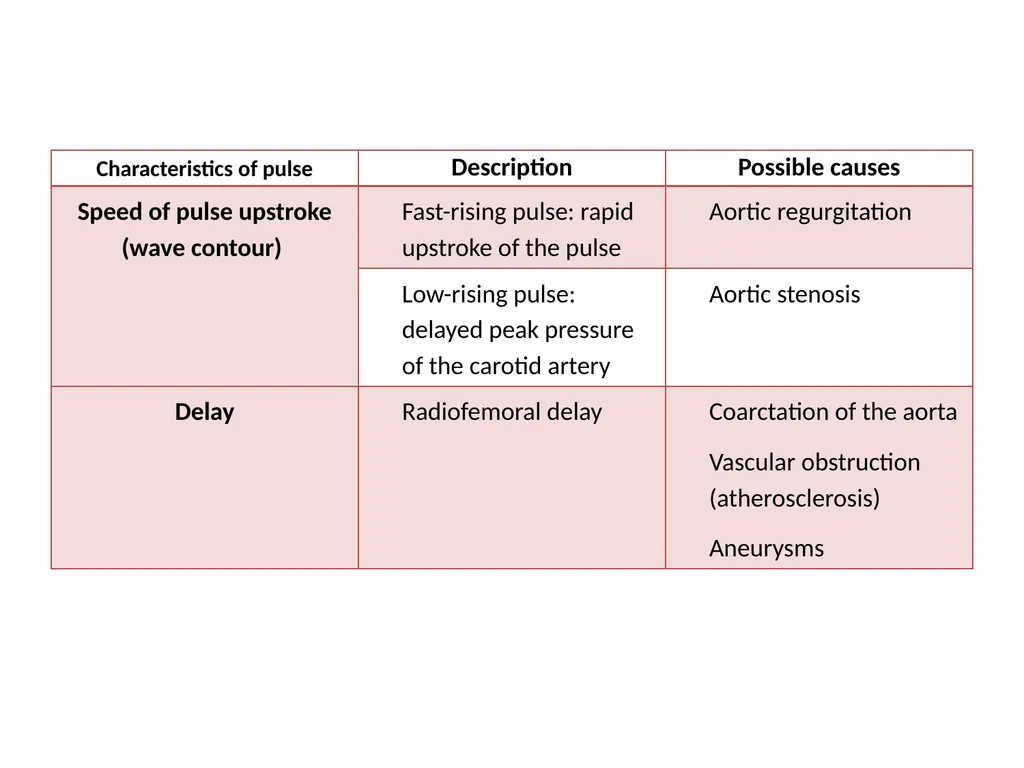
A pulse wave is produced by ventricular contraction during systole.<br>
17
Approach
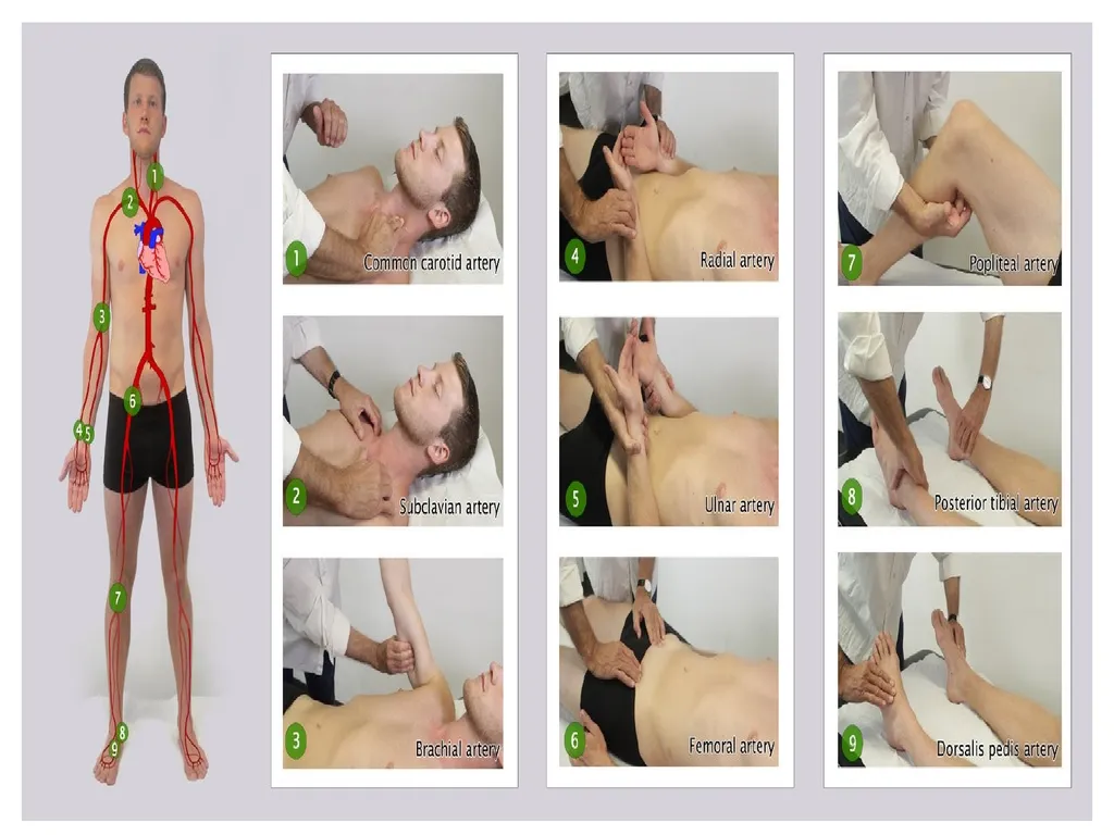
Three finger method: palpation with 2nd–4th fingertips
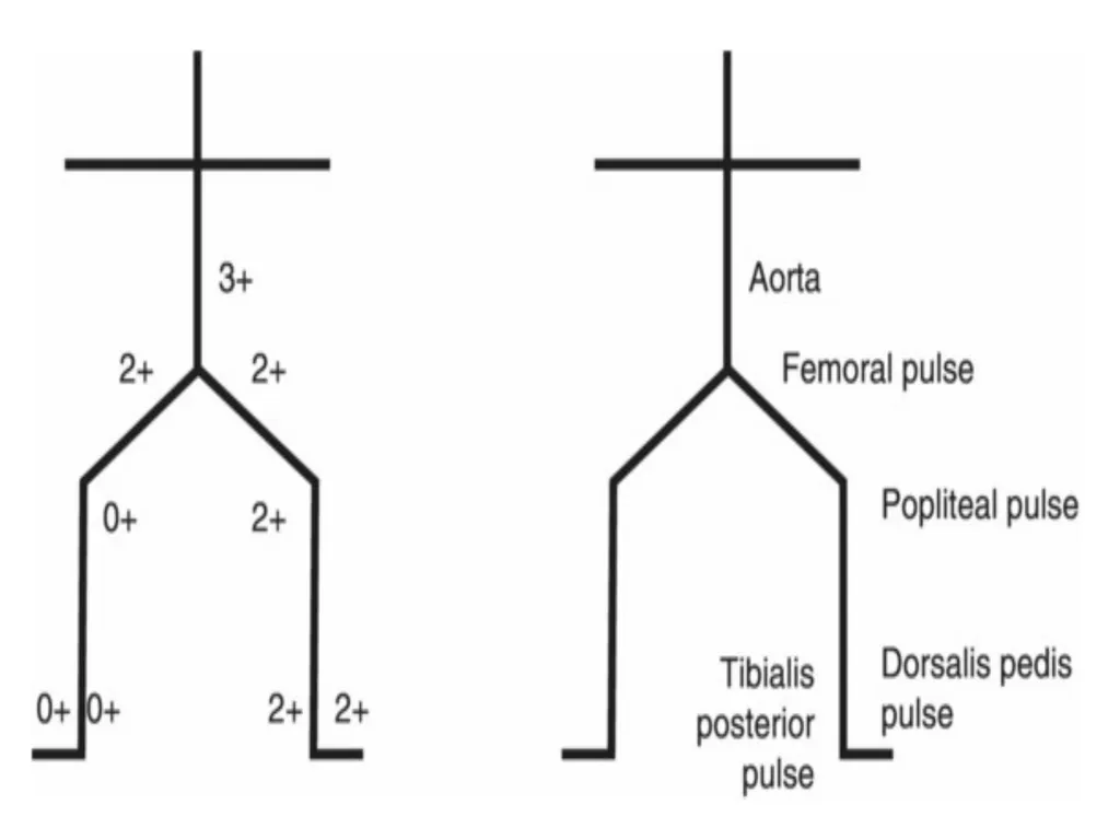
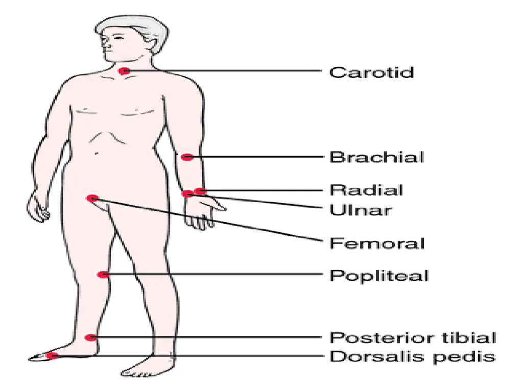
Palpation of the common carotid artery, radial artery, abdominal aorta, femoral artery, popliteal artery, tibialis posterior artery, and dorsalis pedis artery.
The pulse of the carotid artery should NEVER be palpated bilaterally and simultaneously!
Risk of compression of vessels → cerebral hypoperfusion → syncope
Risk of hyperstimulation of the carotid sinus reflex → bradycardia/low blood pressure → cerebral hypoperfusion → syncope
The thumb of the examiner should never be used to take the pulse as it has its own strong pulse, which might be mistaken for the patient's pulse!<br>
18
POINT OF MAXIMUM IMPULSE (PMI) CONTRACTION OF LEFT VENTRICLE
FIFTH INTERCOSTAL SPACE, MIDCLAVICULAR LINE
BRIEF; IF SUSTAINED, SUGGESTS HEART FAILURE<br>
19
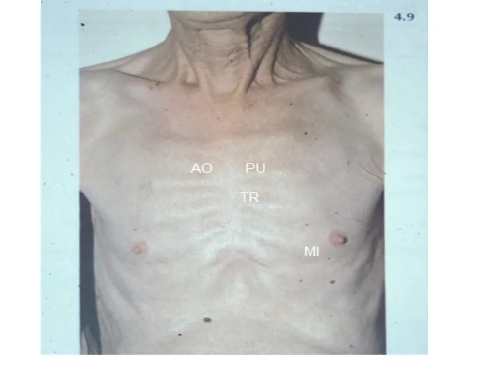
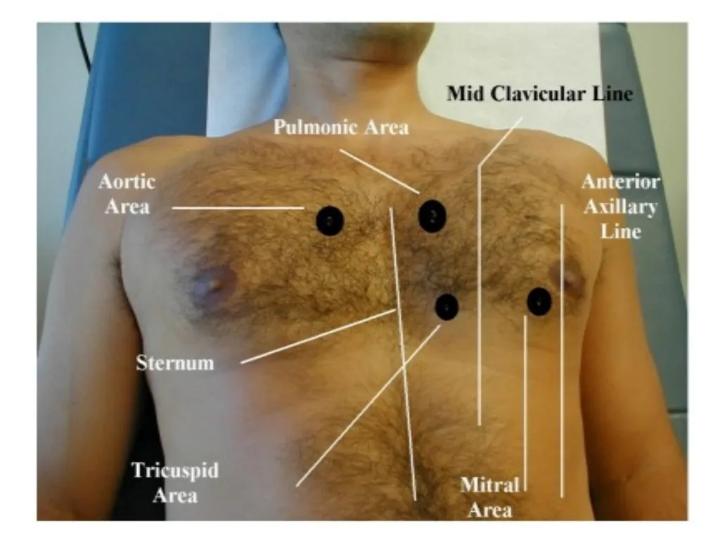
FOUR VALVE AREAS AORTIC: RIGHT STERNAL BORDER
PULMONIC: LEFT UPPER STERNAL
TRICUSPID: LEFT FOURTH INTERCOSTAL SPACE
MITRAL: APEX (5TH INTERCOSTAL SPACE, MIDCLAVICULAR LINE)<br>
20
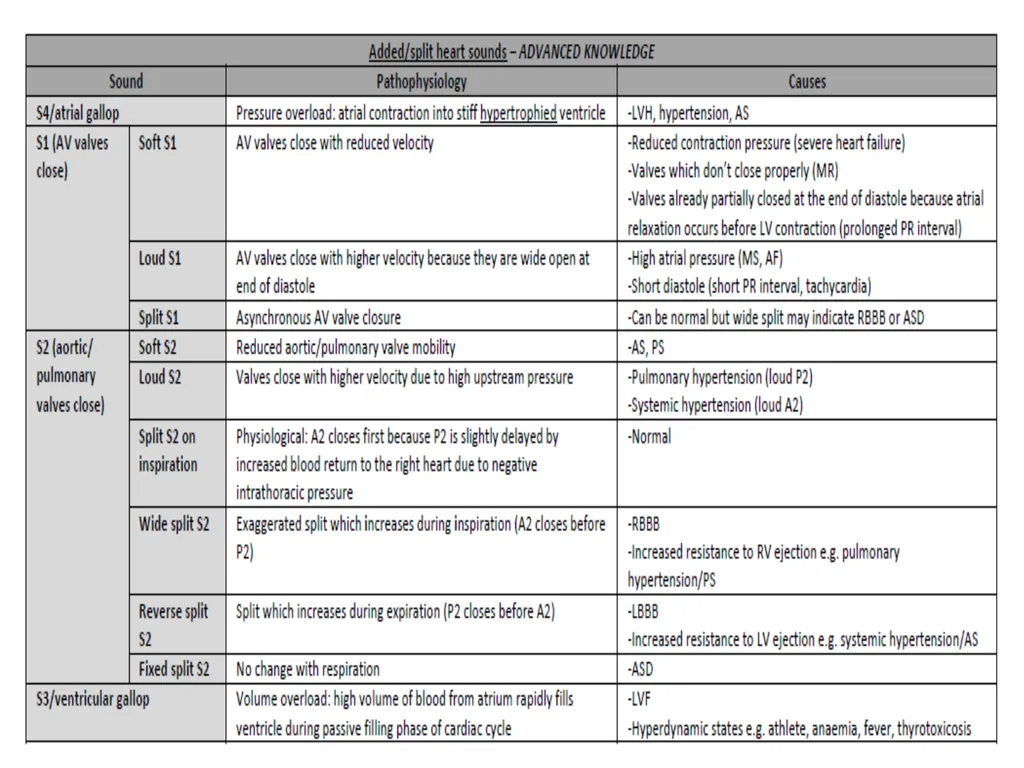
WHAT MAKES NOISES? VALVES CLOSING: S1, S2
BLOOD STRIKING LEFT VENTRICULAR WALL: S3, S4
TURBULENCE: MURMURS<br>
21
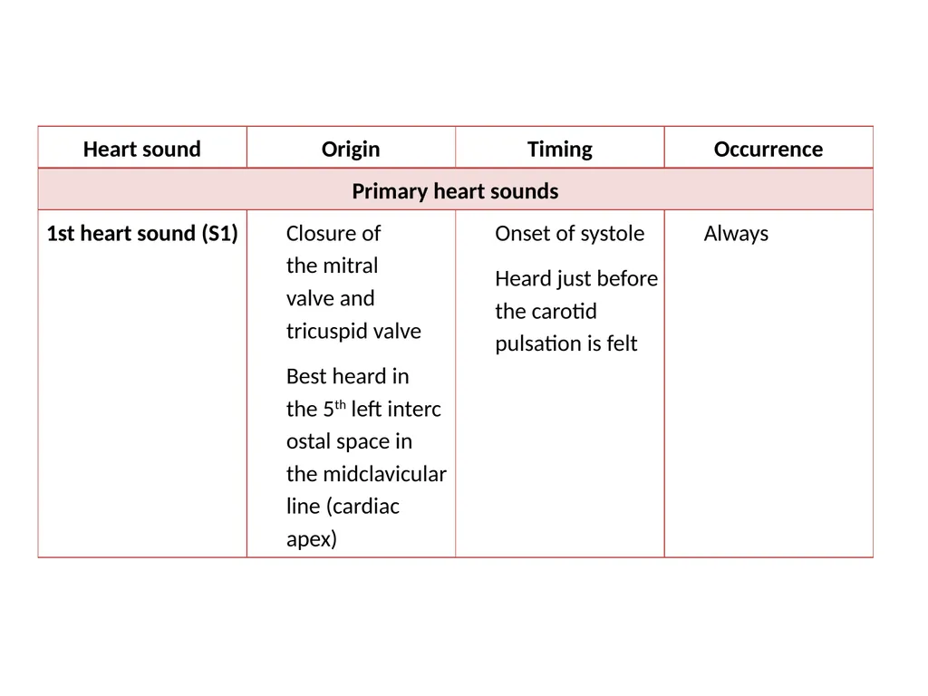
S1 AV VALVES CLOSING (MITRAL AND TRICUSPID)
START OF SYSTOLE
LOUDEST AT APEX<br>
22
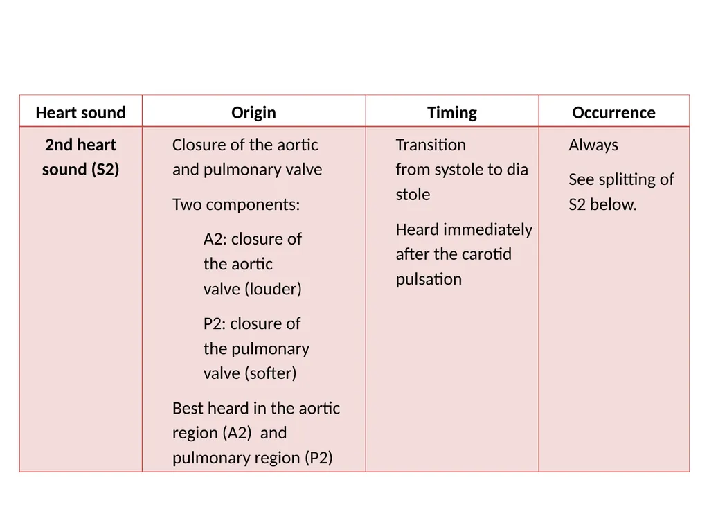
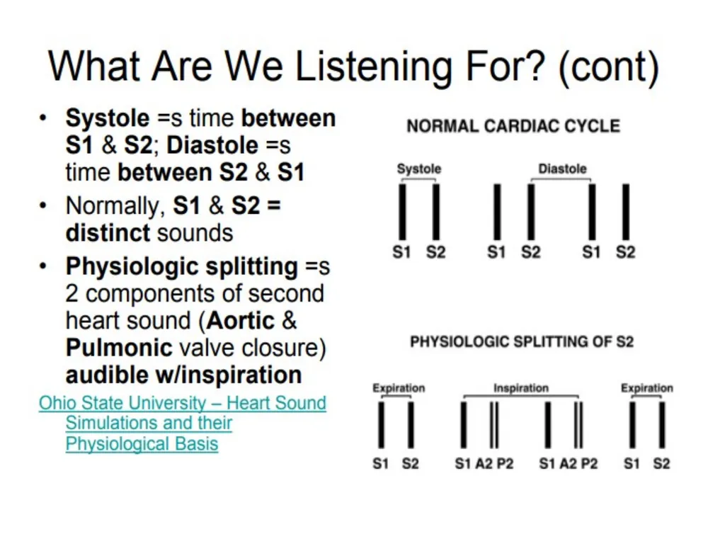
S2 SEMILUNAR VALVES CLOSING: AORTIC AND PULMONIC
A2 BEFORE P2
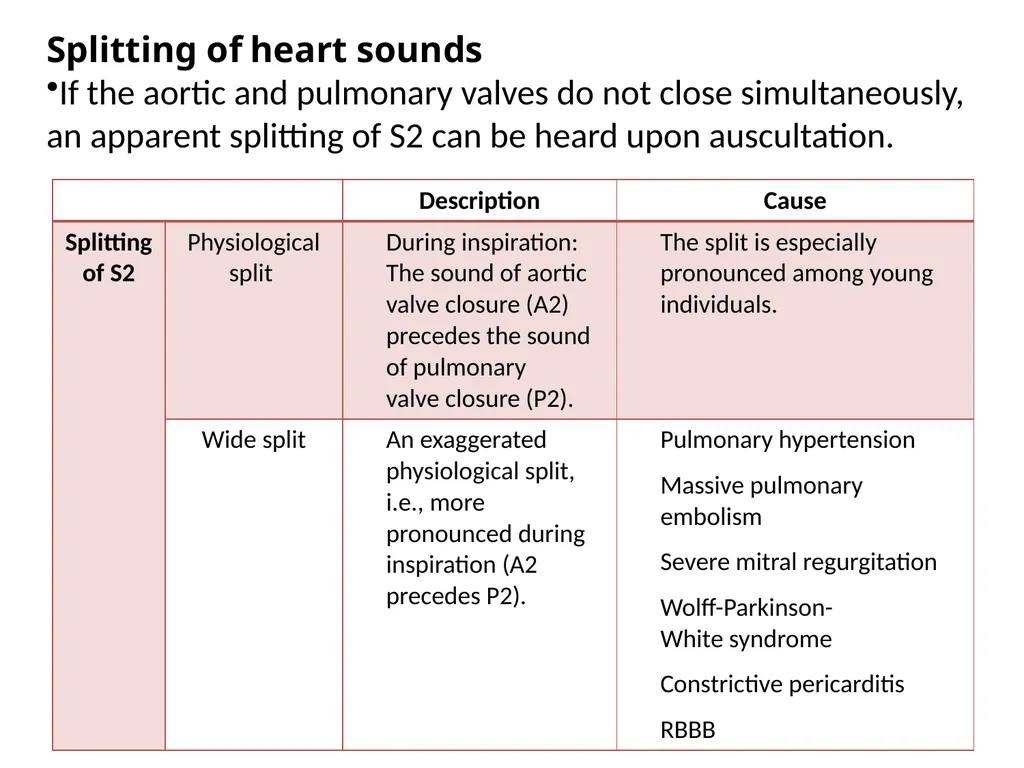
SPLITS WITH INSPIRATION AT PULMONIC AREA (LUSB)
LOUDEST AT BASE (TOP OF HEART)<br>
23
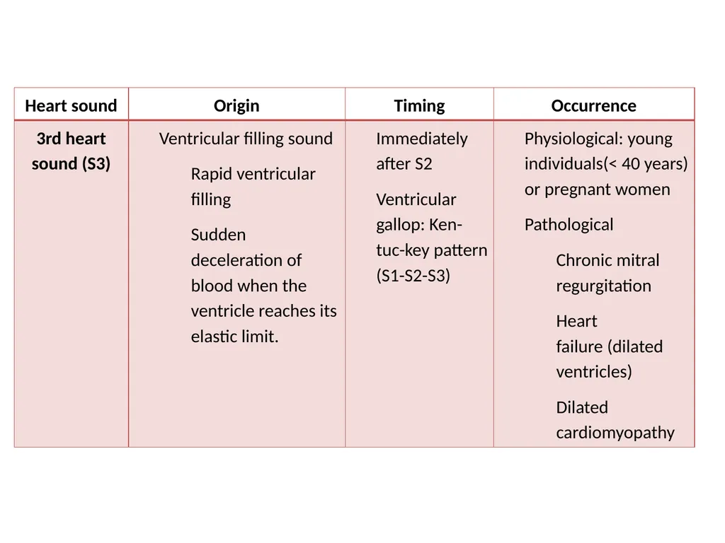
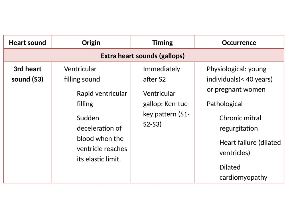
S3 EARLY DIASTOLE (SOON AFTER S2)
BLOOD RUSHES IN JUST AFTER MITRAL VALVE OPENS, STRIKING LV WALL (PALPABLE)
AT APEX ONLY
CONGESTIVE HEART FAILURE (OR HEALTHY YOUNG PERSON)<br>
24
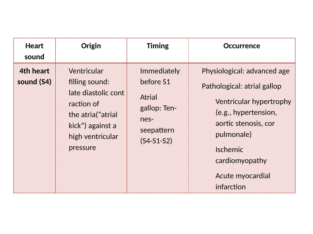
S4 ATRIAL CONTRACTION
JUST BEFORE S1 (MITRAL VALVE CLOSURE) – LATE IN DIASTOLE
BLOOD STRIKES STIFF LEFT VENTRICLE (PALPABLE, AT APEX)
SIGN OF HIGH BLOOD PRESSURE OR HEART ATTACK (MI)<br>
25
S4 QUESTION SHORTLY AFTER S3?
HEALTHY ATHLETES?
REDUCED VENTRICULAR ELASTICITY
INTERMITTENT IN ATRIAL FIB?<br>
26
HEART MURMURS TURBULENCE
INCREASED FLOW ACROSS VALVE
TIGHT VALVE (STENOSIS)
LEAKY VALVE (REGURGITATION)
HOLE (SEPTAL DEFECT)<br>
27
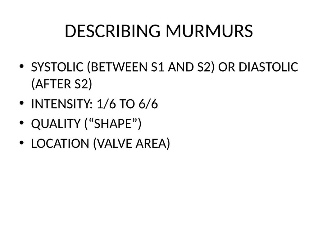
DESCRIBING MURMURS SYSTOLIC (BETWEEN S1 AND S2) OR DIASTOLIC (AFTER S2)
INTENSITY: 1/6 TO 6/6
QUALITY (“SHAPE”)
LOCATION (VALVE AREA)<br>
28
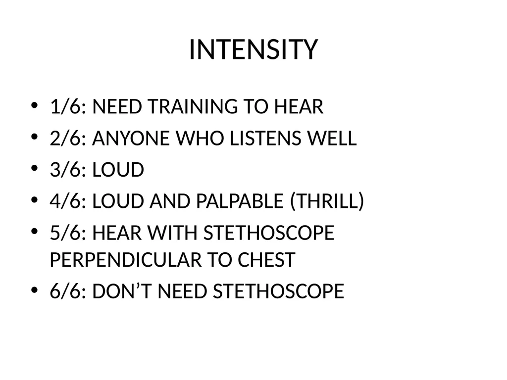
INTENSITY 1/6: NEED TRAINING TO HEAR
2/6: ANYONE WHO LISTENS WELL
3/6: LOUD
4/6: LOUD AND PALPABLE (THRILL)
5/6: HEAR WITH STETHOSCOPE PERPENDICULAR TO CHEST
6/6: DON’T NEED STETHOSCOPE<br>
29
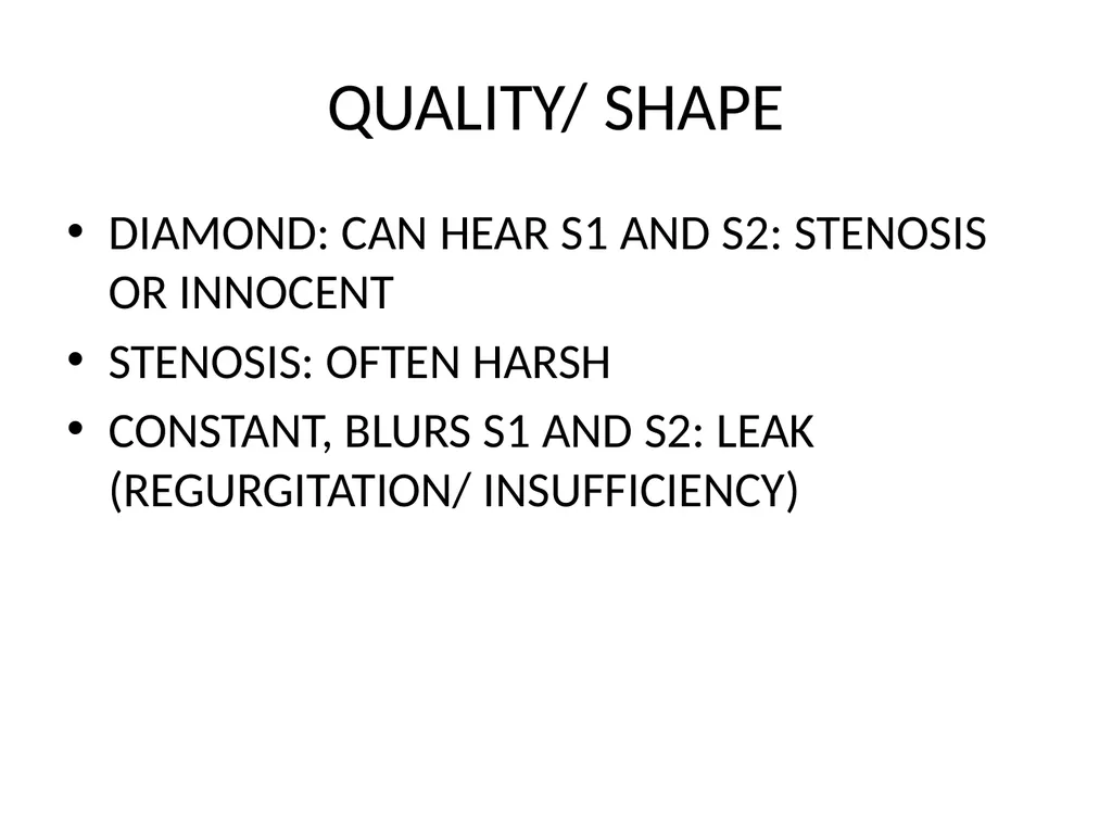
QUALITY/ SHAPE DIAMOND: CAN HEAR S1 AND S2: STENOSIS OR INNOCENT
STENOSIS: OFTEN HARSH
CONSTANT, BLURS S1 AND S2: LEAK (REGURGITATION/ INSUFFICIENCY)<br>
30
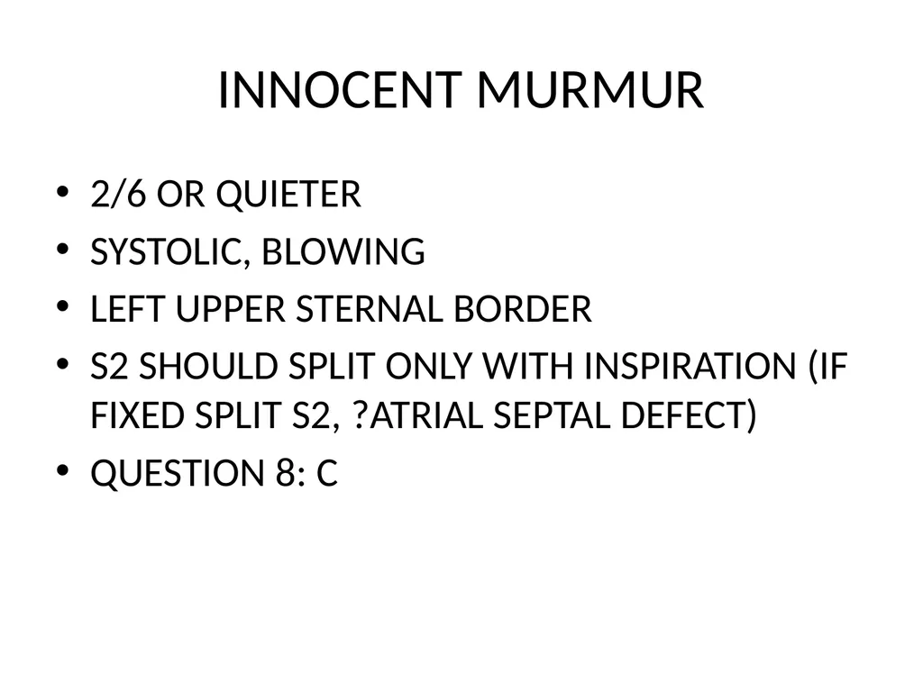
INNOCENT MURMUR 2/6 OR QUIETER
SYSTOLIC, BLOWING
LEFT UPPER STERNAL BORDER
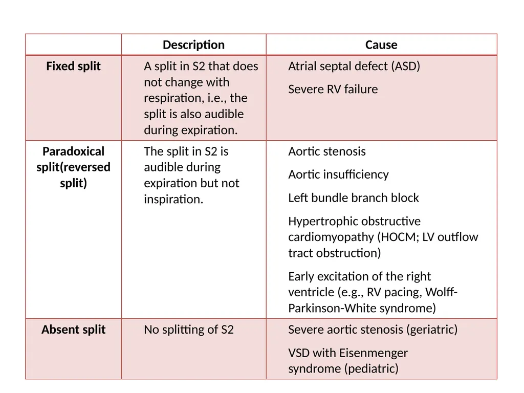
S2 SHOULD SPLIT ONLY WITH INSPIRATION (IF FIXED SPLIT S2, ?ATRIAL SEPTAL DEFECT)
QUESTION 8: C<br>
31
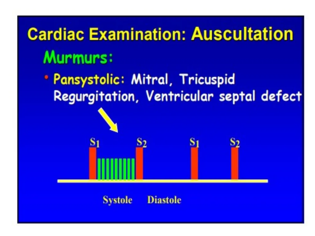
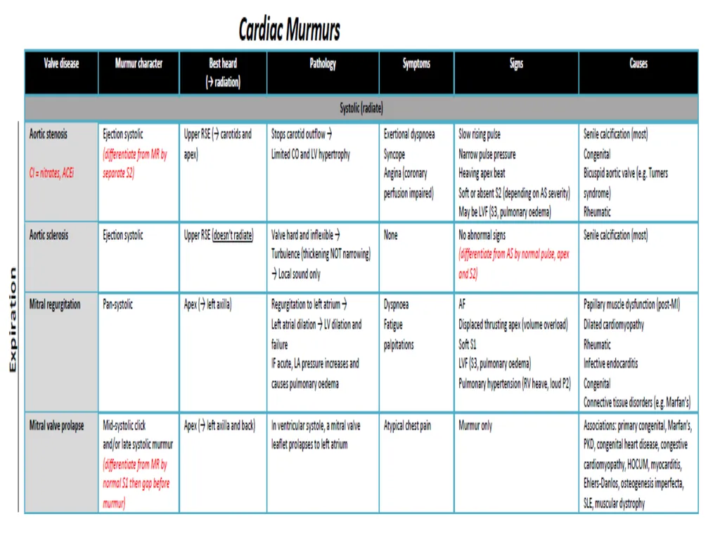
MITRAL INSUFFICIENCY HOLOSYSTOLIC (BLURS S1 AND S2)
BLOWING
AT APEX; RADIATES TO AXILLA<br>
32
AORTIC STENOSIS HARSH, RIGHT STERNAL BORDER
SOFTER S2 (WHY?)
DIAMOND-SHAPED, PEAKS LATER
DELAY IN CAROTID PULSE
RADIATES TO CAROTID ARTERY
FAILURE TO RADIATE MAKES AORTIC STENOSIS LESS LIKELY (QUESTION 9)<br>
33
SUMMARY S1(AV),SYSTOLE, S2(TR/AO),DIASTOLE
S3 (SLOSHING IN), S4 (A STIFF WALL)
LOCATION,TIMING,QUALITY,INTENSITY
INNOCENT MURMUR (LUSB)
MITRAL REGURGITATION (APEX)
AORTIC STENOSIS (HARSH, RSB)<br>
34
Auscultation
For detection of vascular murmurs in the carotid artery, abdominal aorta, renal artery, and femoral artery
Causes of vascular murmurs: alteration of local vessels and vascular walls
Fistulae
Aneurysms
Vascular stenosis
Auscultation of high-frequency, pulse-synchronous sounds proximal to the stenosis
Hypercirculation and increased cardiac output
E.g., pregnancy, fever, anemia, hyperthyroidism
May occur physiologically in adolescents<br>
35
Chest inspection
Approach
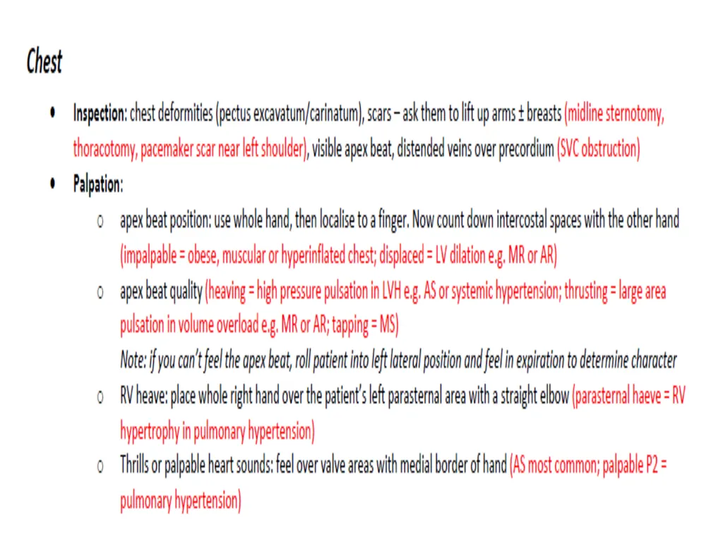
The patient is initially requested to remove their upper body attire to identify:
Scars (e.g., heart surgery, pacemakers)
Chest deformities (pectus carinatum or pectus excavatum)
Visible apex beat
Distended veins on chest (e.g., in SVC obstruction)
De Musset sign: head bobbing in time with pulse (seen in aortic regurgitation)<br>
36
Chest palpation
Apex impulse
The apex impulse (apex beat) is the outermost and lowermost cardiac impulse on the chest wall that is definitely palpable.
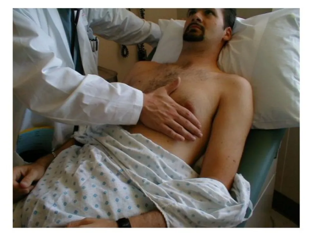
The examiner places their flat hand on the cardiac apex to locate the apex beat; it is further localized and assessed by palpating with 2–3 fingers.
If the apex beat is not initially palpable, the patient should be positioned on his/her left lateral side and the cardiac apex should be palpated during expiration.<br>
37
No conclusion can be drawn from the location of the apex beat in the presence of spinal or chest wall deformities or a tracheal deviation (mediastinal shift)!<br>
38
Other impulses
Parasternal heave: a heaving motion felt over the left parasternal area (palpate with right hand and straightened elbow)
Suggests RV hypertrophy (e.g., pulmonary hypertension)
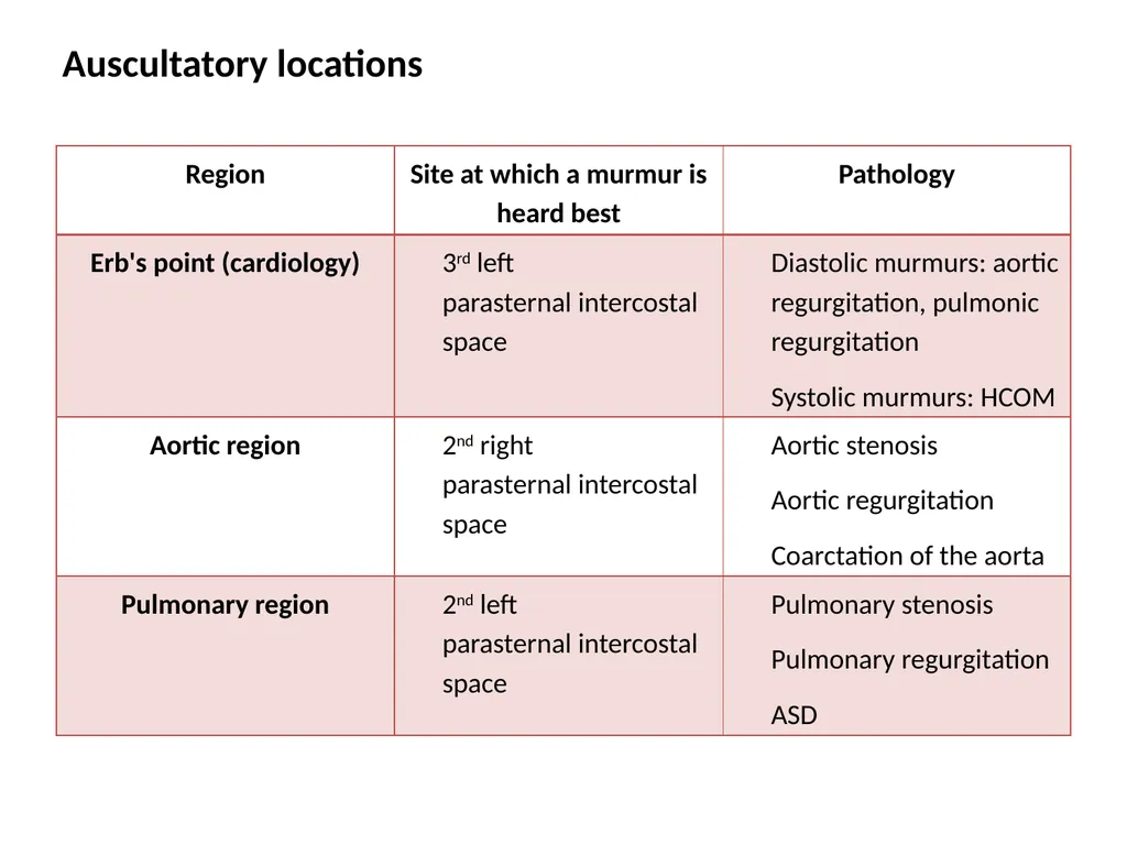
Thrills: a palpable murmur, usually over the region where the heart murmur is heard best (see auscultatory locations below)
Palpable heart sounds: palpate over valve areas (e.g., palpable P2: see auscultatory locations below)<br>
39
Chest percussion
Although cardiac percussion can provide some information about the size and shape of the heart, it is very unreliable and dependent on the examiner and is thus of limited clinical use.<br>
40
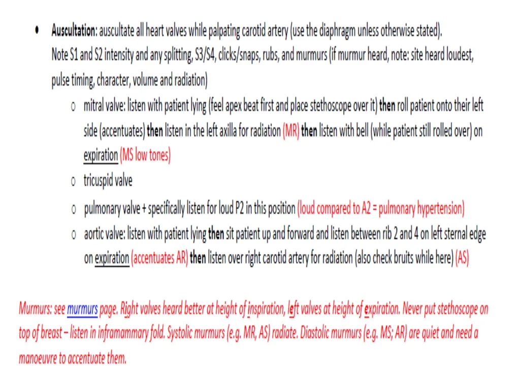
Chest auscultation
Approach
Performed in the supine position with slight elevation of the torso
Politely ask the patient to refrain from speaking while the heart sounds are being assessed.
The pulse should be simultaneously palpated during auscultation (mainly the radial artery).
If heart sounds are weakly audible, request that the patient holds their breath for a moment after expiration (respiratory rest position).
Assess the following:
Location, timing, changes in intensity, and splitting of heart sounds
Abnormal heart sounds
Murmurs<br>
41
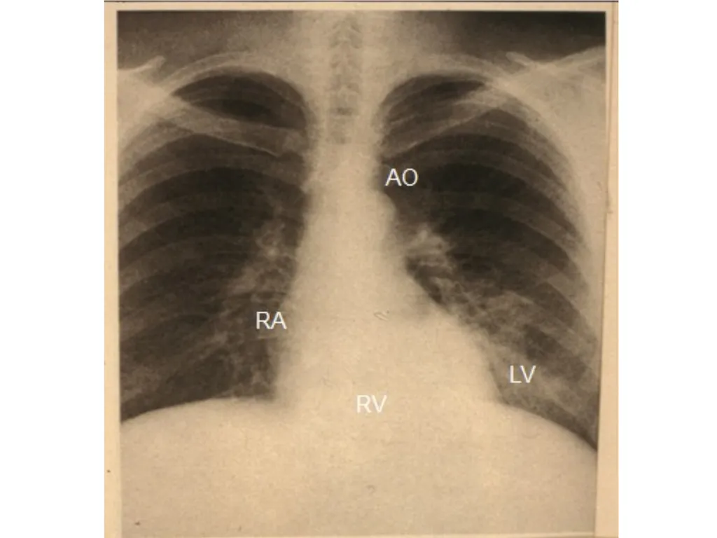
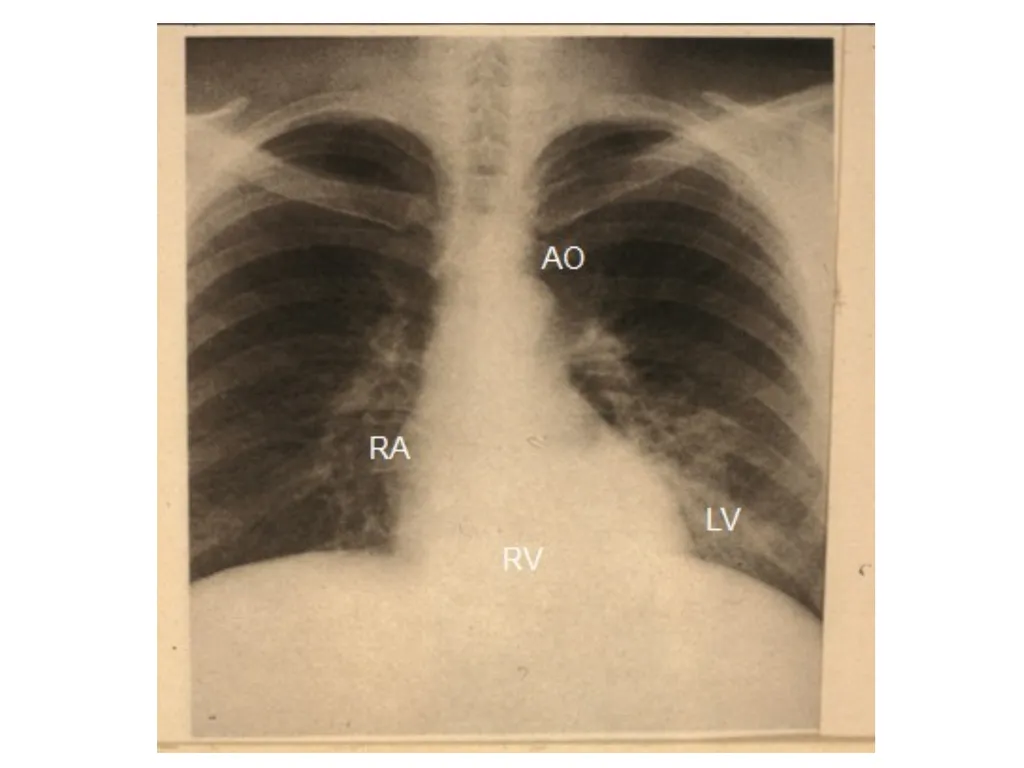
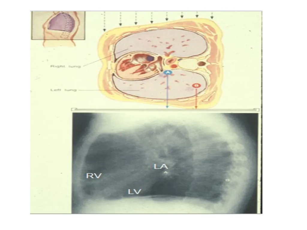
LOCATION OF CHAMBERS RIGHT VENTRICLE: ANTERIOR
LEFT VENTRICLE: LEFT HEART BORDER/ APEX/ POSTERIOR
RIGHT ATRIUM: RIGHT HEART BORDER
LEFT ATRIUM: POSTERIOR<br>
42
Location and timing
The first (S1) and second (S2) heart sounds are physiological sounds that are heard in all healthy individuals.
The third (S3) and fourth (S4) heart sounds may be physiological (particularly in young adults, pregnant women, and the elderly) or indicate an underlying pathology.<br>
43
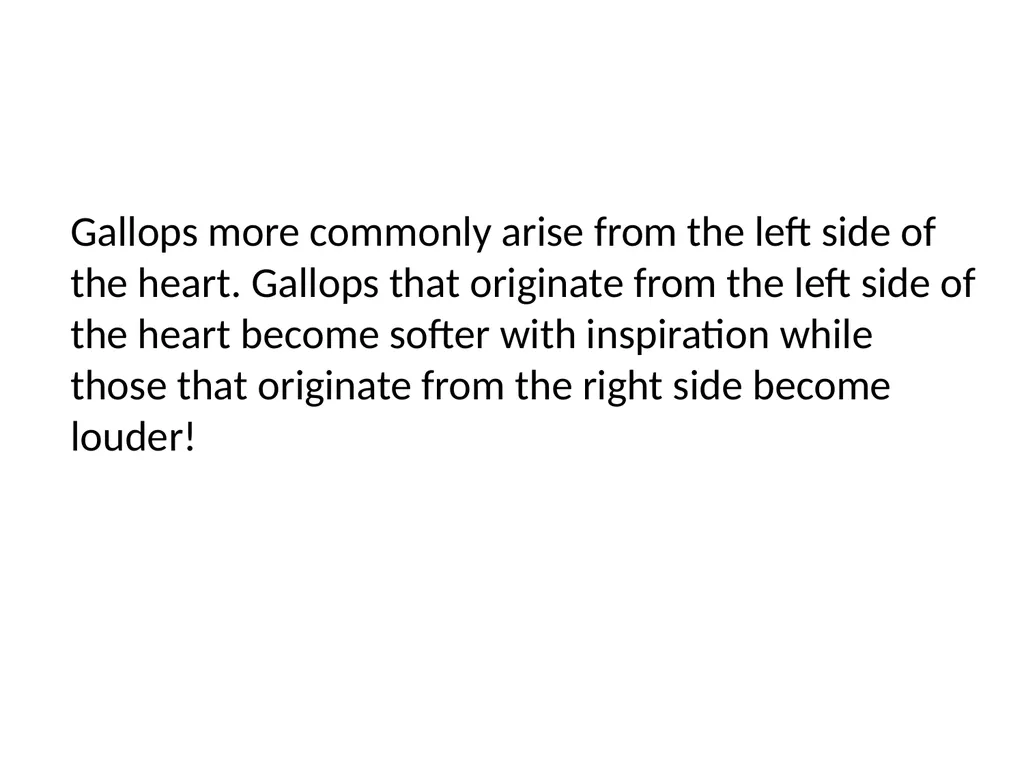
Gallops more commonly arise from the left side of the heart. Gallops that originate from the left side of the heart become softer with inspiration while those that originate from the right side become louder!<br>
44
Splitting of heart sounds
If the aortic and pulmonary valves do not close simultaneously, an apparent splitting of S2 can be heard upon auscultation.<br>
45
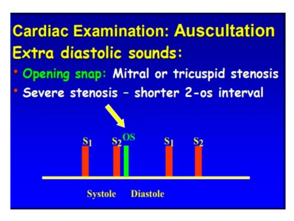
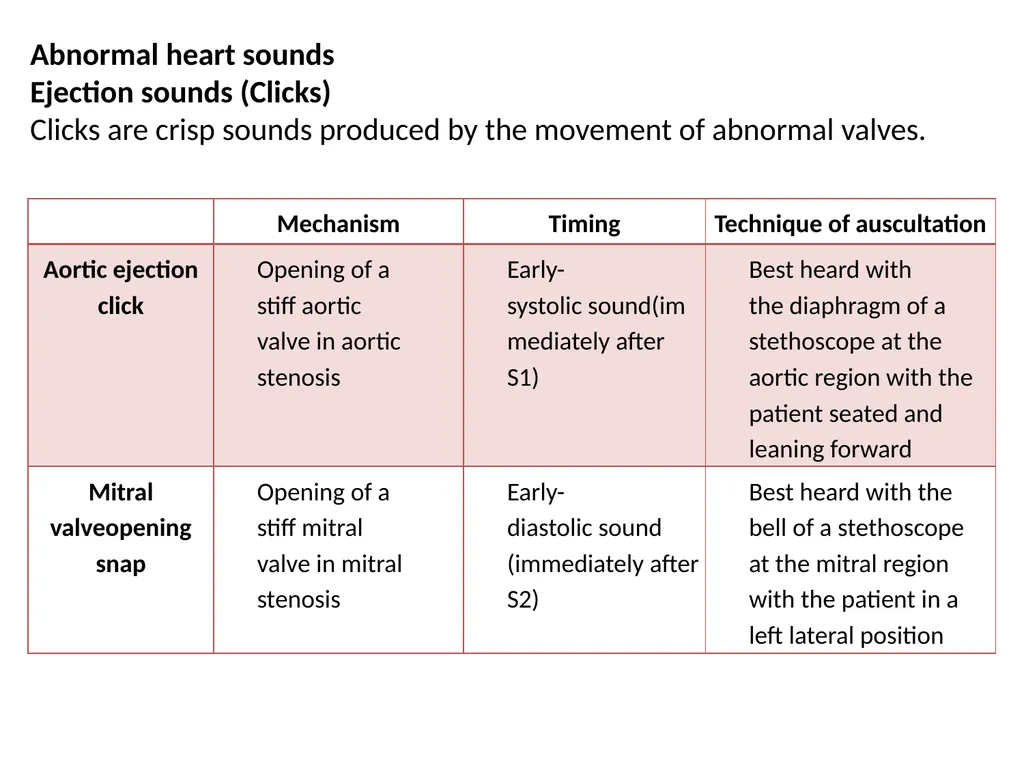
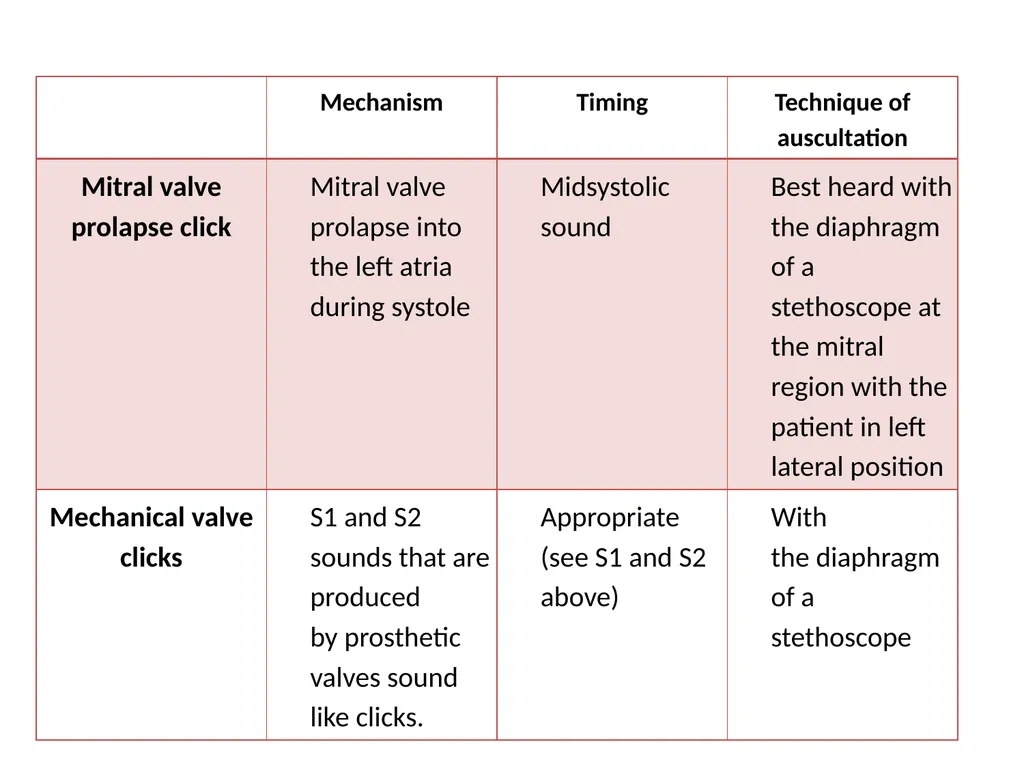
Abnormal heart sounds
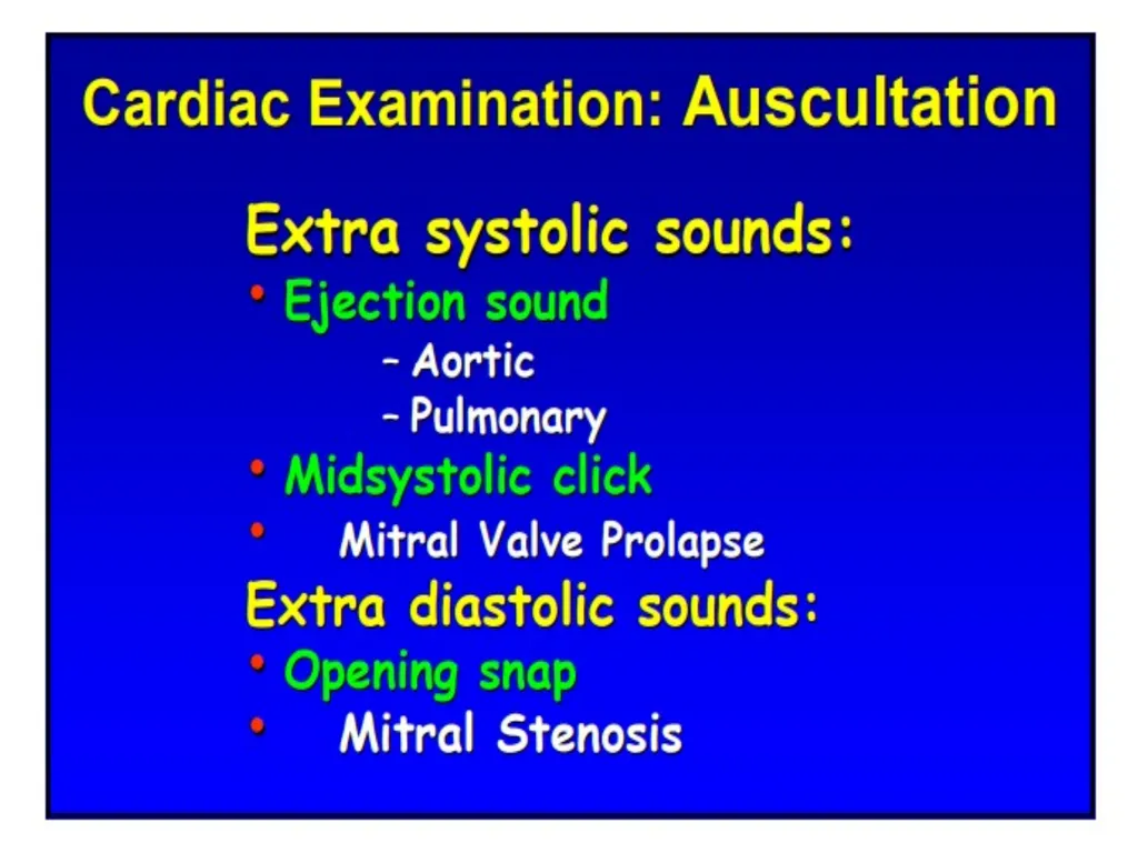
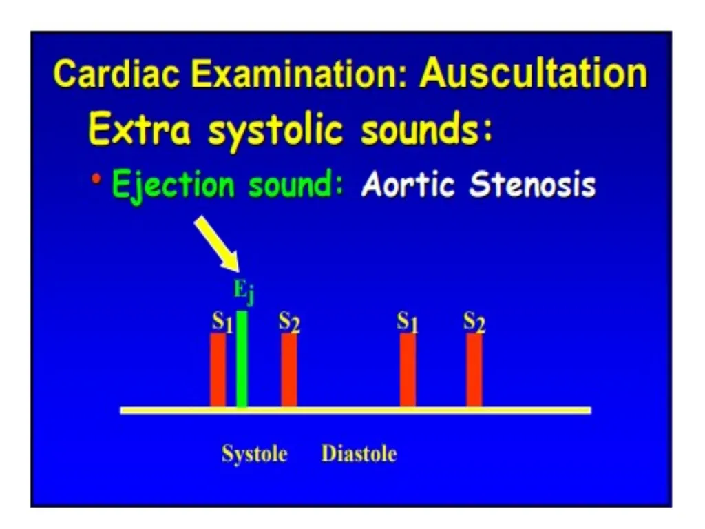
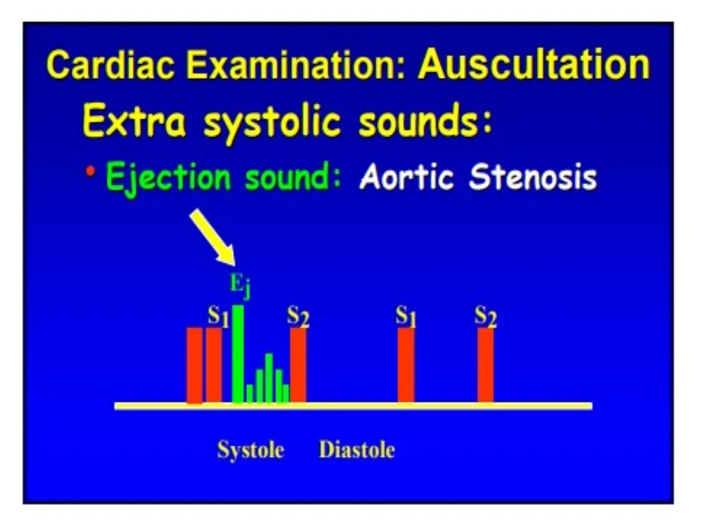
Ejection sounds (Clicks)
Clicks are crisp sounds produced by the movement of abnormal valves.<br>
46
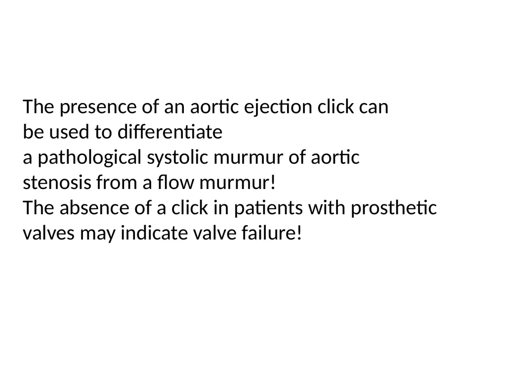
The presence of an aortic ejection click can
be used to differentiate
a pathological systolic murmur of aortic stenosis from a flow murmur!
The absence of a click in patients with prosthetic valves may indicate valve failure!<br>
47
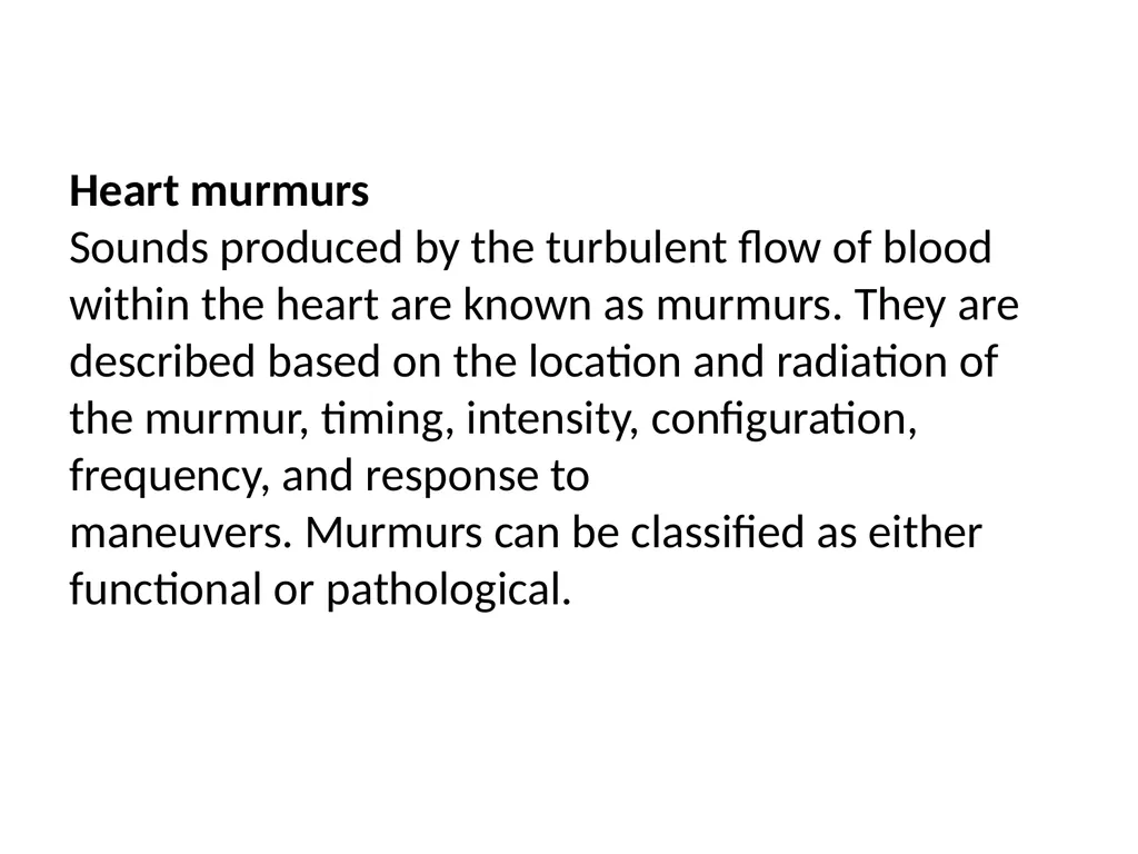
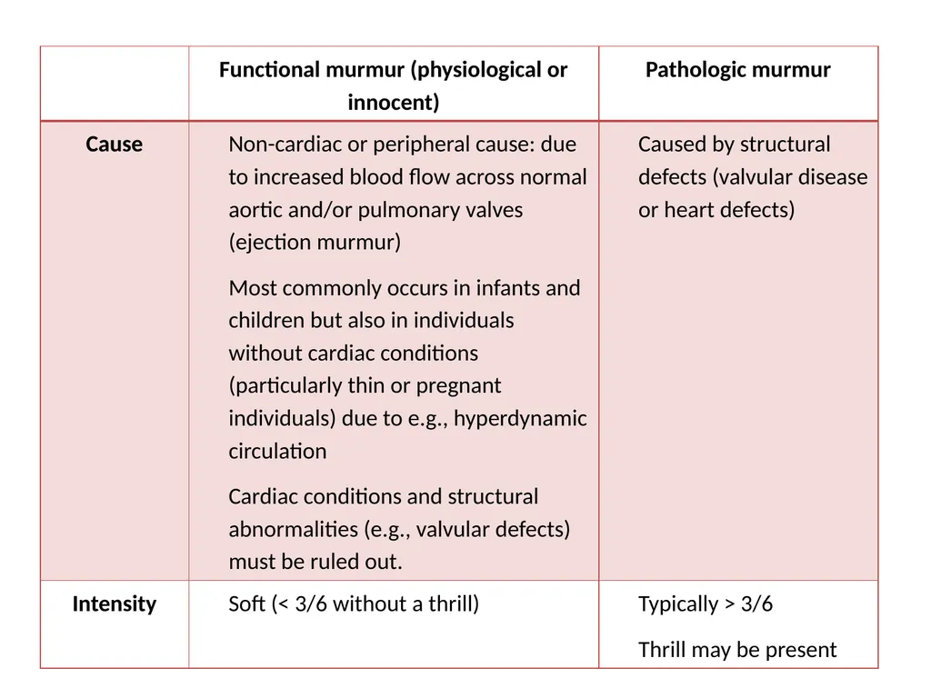
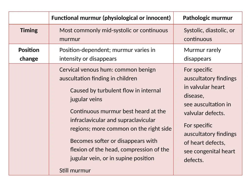
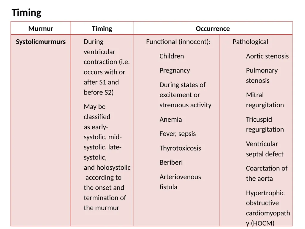
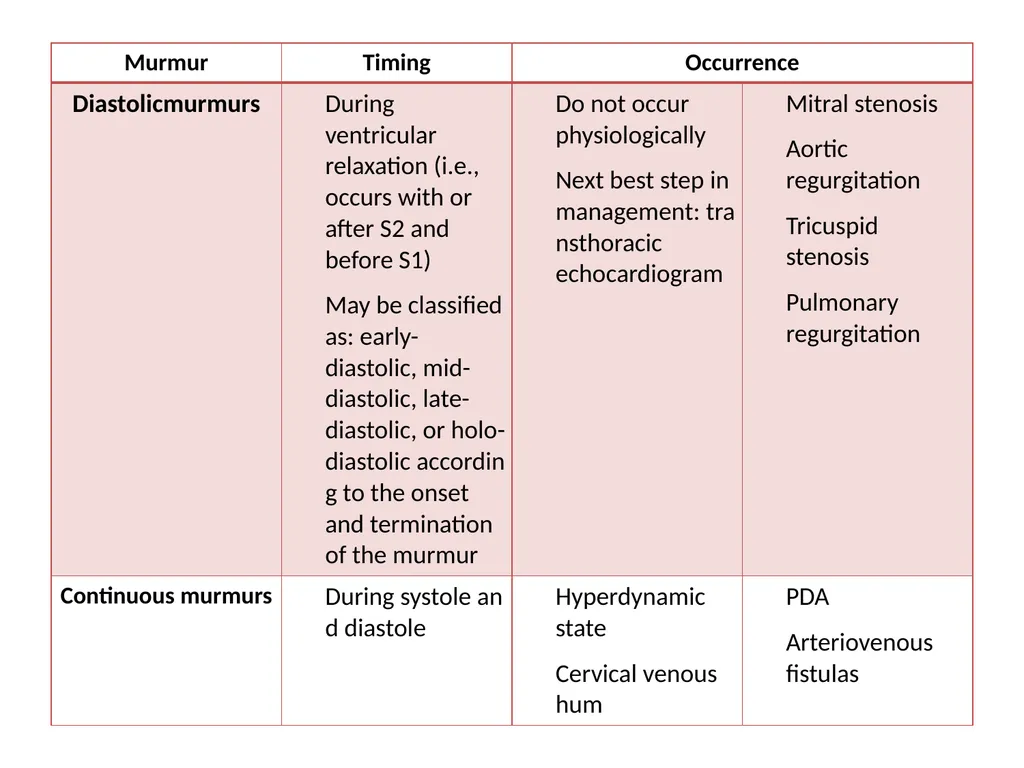
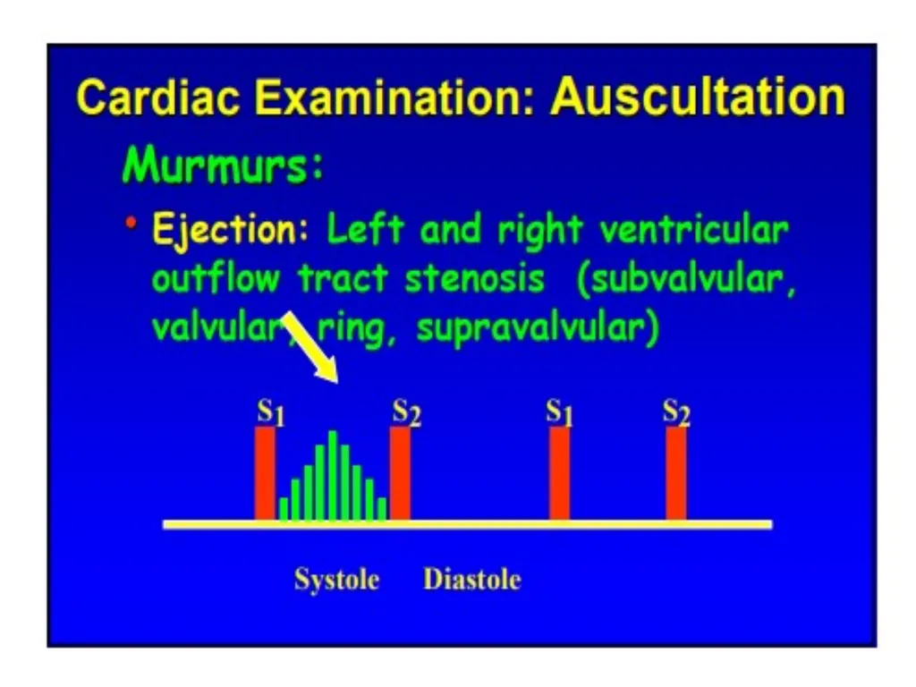
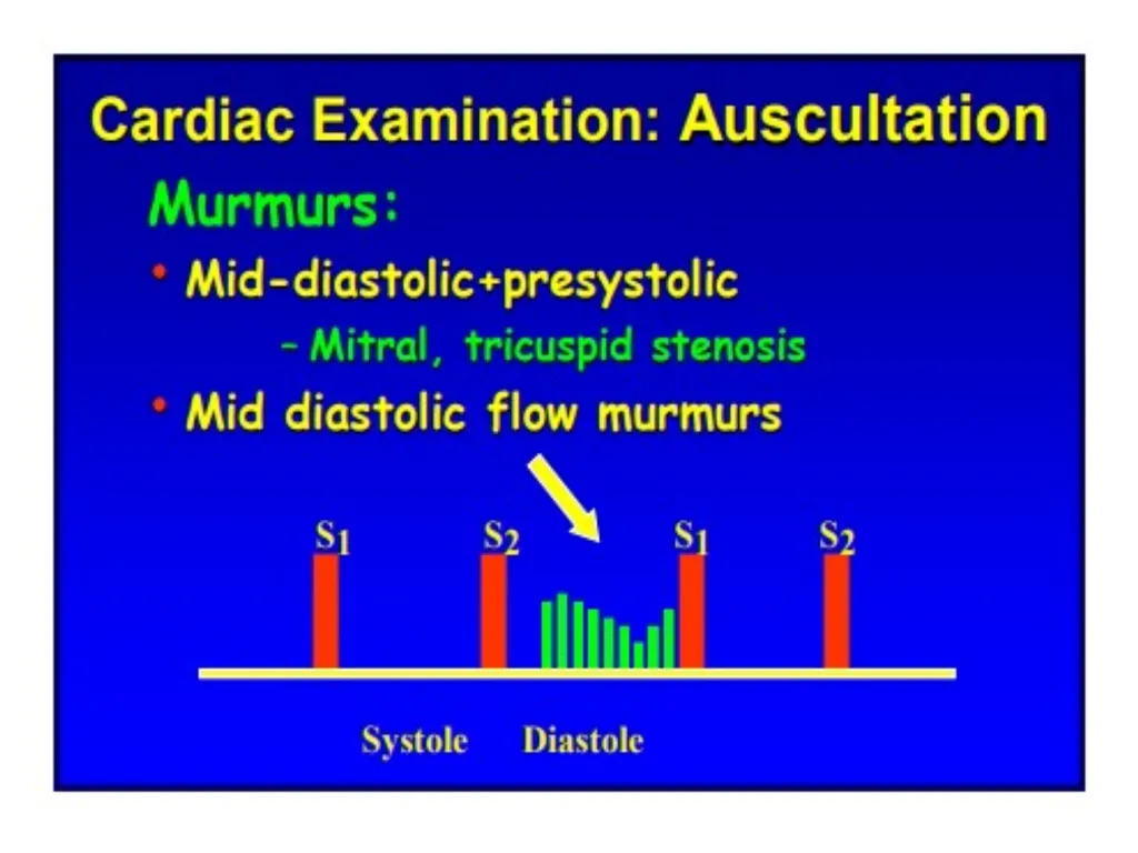
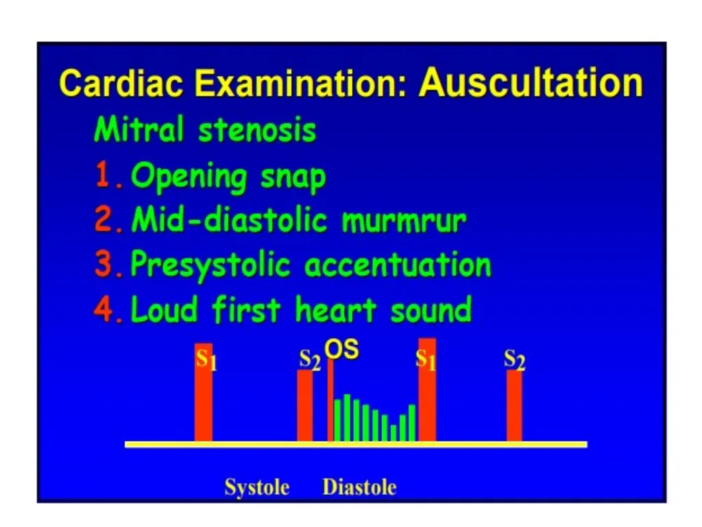
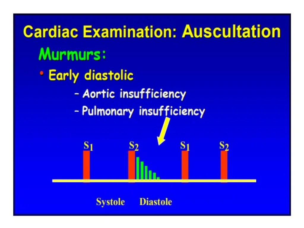
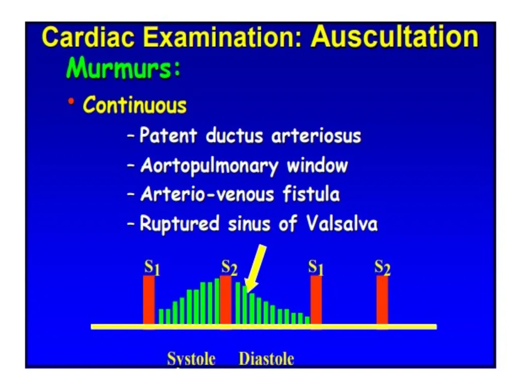
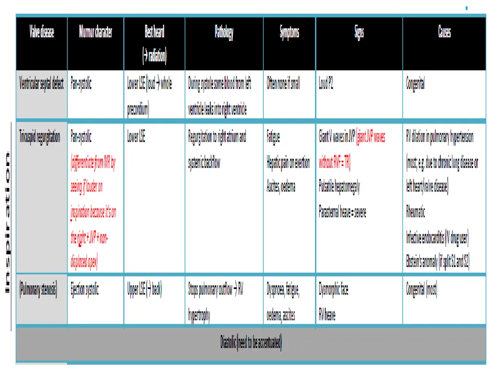
Heart murmurs
Sounds produced by the turbulent flow of blood within the heart are known as murmurs. They are described based on the location and radiation of the murmur, timing, intensity, configuration, frequency, and response to maneuvers. Murmurs can be classified as either functional or pathological.<br>
48
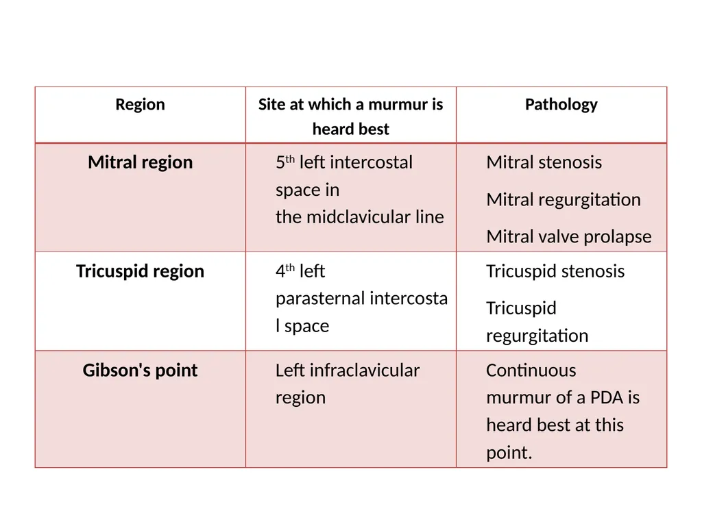
Auscultatory locations<br>
50
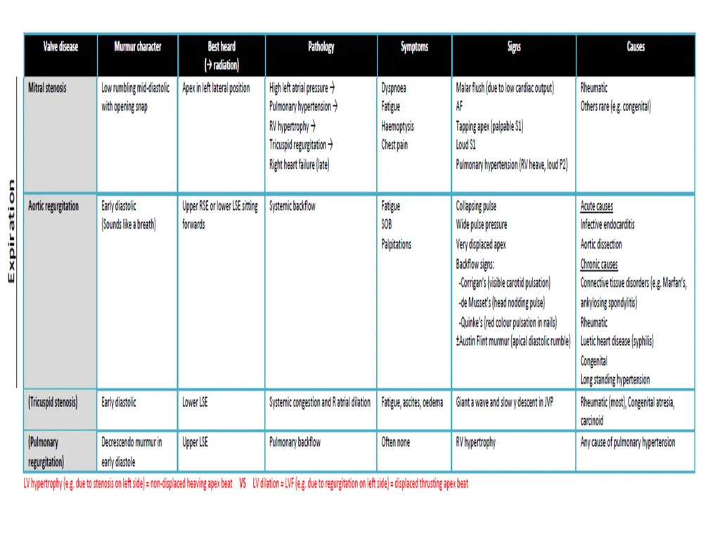
A mid-systolic murmur in an asymptomatic individual is most likely physiological! Unlike systolic murmurs, diastolic murmurs are almost always pathological!
Diastolic murmurs may require that certain maneuvers be performed to make them more apparent.<br>
51
Radiation
A murmur may be auscultated at a site that does not lie directly over the heart.
Aortic stenosis: systolic murmur radiates to carotid arteries
Mitral regurgitation: systolic murmur radiates to the left axilla (in the left lateral recumbent position)
Pulmonary stenosis: systolic murmur radiates to the interscapular region<br>