
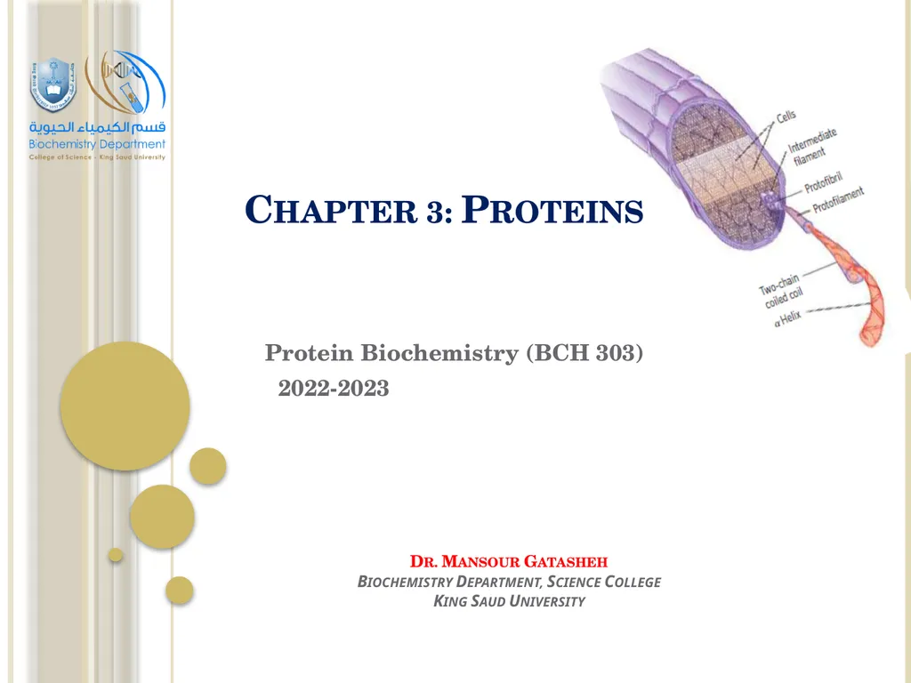
Chapter 3: Proteins Protein Biochemistry (BCH 303)
CS
Published · 107 slides · 0 views

1 / 1


Description
Chapter 3: Proteins Protein Biochemistry (BCH 303) 2022-2023 Dr. Mansour Gatasheh Biochemistry Department, Science College King Saud University Proteins are made up of large numbers of amino acids linked into chains by peptide bonds joining
Related Topics
Share
Embed code
Download this presentation From Below
"Chapter 3: Proteins Protein Biochemistry (BCH 303)" is the property of its rightful owner. Permission is granted to download and print the materials on this website for personal, non-commercial use only, and to display it on your personal computer provided you do not modify the materials and that you retain all copyright notices contained in the materials. By downloading content from our website, you accept the terms of this agreement.
Presentation Transcript
01
Chapter 3: Proteins Protein Biochemistry (BCH 303)
2022-2023 Dr. Mansour Gatasheh
Biochemistry Department, Science College
King Saud University<br>
2022-2023 Dr. Mansour Gatasheh
Biochemistry Department, Science College
King Saud University<br>
02
Proteins are made up of large numbers of amino acids linked into chains by peptide bonds joining the amino group of one amino acid to the carboxyl of the next.
The number of amino acids present varies from about a hundred to several thousands in different proteins.
Some proteins are composed of only one polypeptide chain while others are composed of two or more polypeptide chains (multisubunit proteins) held together by non-covalent bonds. Proteins<br>
The number of amino acids present varies from about a hundred to several thousands in different proteins.
Some proteins are composed of only one polypeptide chain while others are composed of two or more polypeptide chains (multisubunit proteins) held together by non-covalent bonds. Proteins<br>
03
The individual polypeptide chains in a multisubunit protein may be identical or different.
If at least two are identical, the protein is said to be oligomeric and the identical units are referred to as protomers.
- Each protein has a unique amino acid sequence and a well- defined three-dimensional structure known as the conformation.<br>
If at least two are identical, the protein is said to be oligomeric and the identical units are referred to as protomers.
- Each protein has a unique amino acid sequence and a well- defined three-dimensional structure known as the conformation.<br>
04
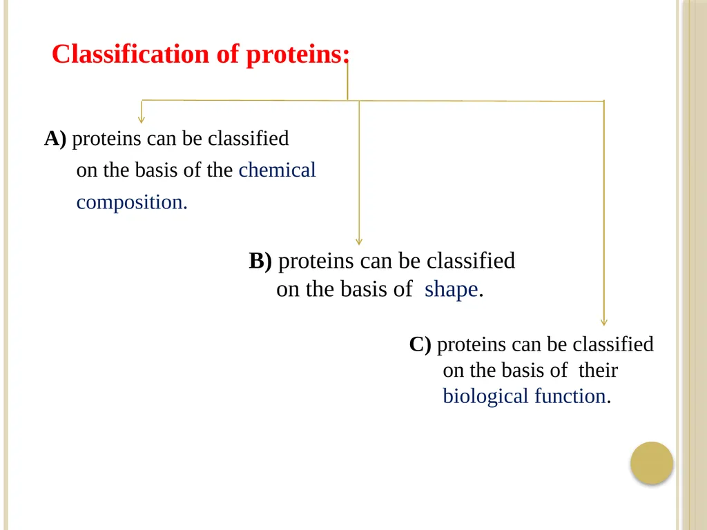
A) proteins can be classified
on the basis of the chemical
composition. C) proteins can be classified on the basis of their biological function. B) proteins can be classified on the basis of shape. Classification of proteins:<br>
on the basis of the chemical
composition. C) proteins can be classified on the basis of their biological function. B) proteins can be classified on the basis of shape. Classification of proteins:<br>
05
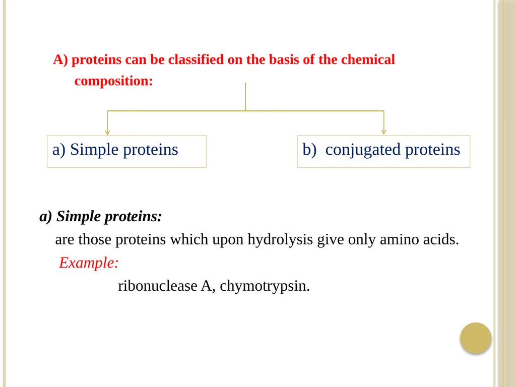
A) proteins can be classified on the basis of the chemical
composition: a) Simple proteins b) conjugated proteins a) Simple proteins:
are those proteins which upon hydrolysis give only amino acids.
Example:
ribonuclease A, chymotrypsin.<br>
composition: a) Simple proteins b) conjugated proteins a) Simple proteins:
are those proteins which upon hydrolysis give only amino acids.
Example:
ribonuclease A, chymotrypsin.<br>
06
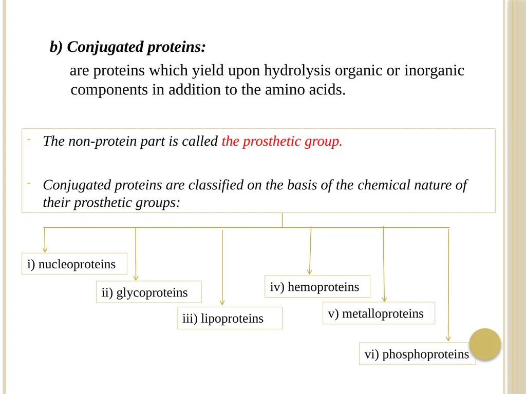
The non-protein part is called the prosthetic group.
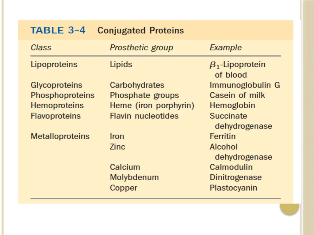
Conjugated proteins are classified on the basis of the chemical nature of their prosthetic groups: b) Conjugated proteins:
are proteins which yield upon hydrolysis organic or inorganic components in addition to the amino acids. i) nucleoproteins ii) glycoproteins iii) lipoproteins iv) hemoproteins v) metalloproteins vi) phosphoproteins<br>
07
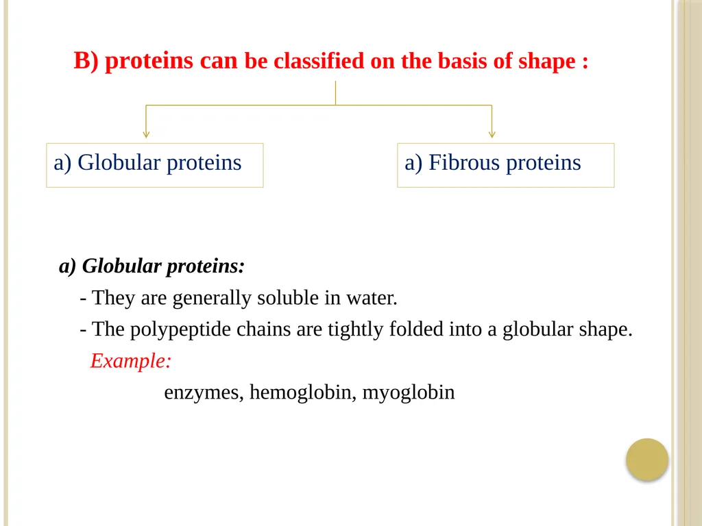
a) Globular proteins:
- They are generally soluble in water.
- The polypeptide chains are tightly folded into a globular shape.
Example:
enzymes, hemoglobin, myoglobin B) proteins can be classified on the basis of shape : a) Globular proteins a) Fibrous proteins<br>
- They are generally soluble in water.
- The polypeptide chains are tightly folded into a globular shape.
Example:
enzymes, hemoglobin, myoglobin B) proteins can be classified on the basis of shape : a) Globular proteins a) Fibrous proteins<br>
08
b) Fibrous proteins:
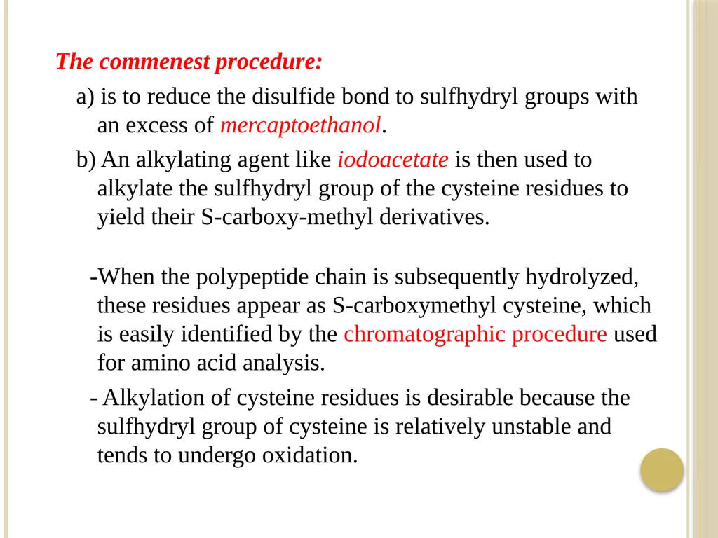
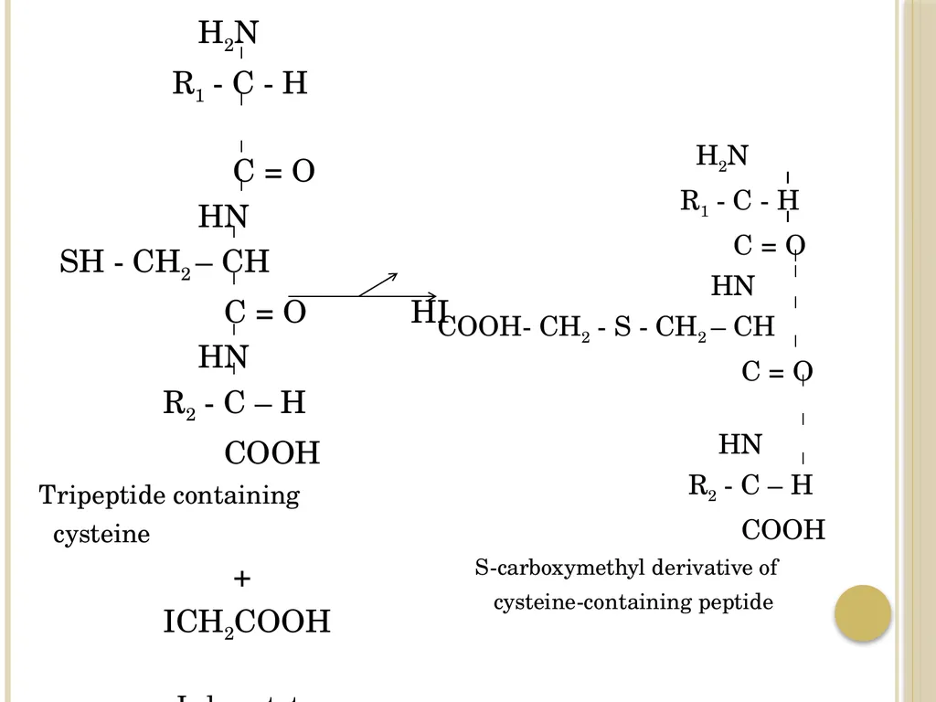
- They are insoluble in water
- Their polypeptide chains are arranged in long strands
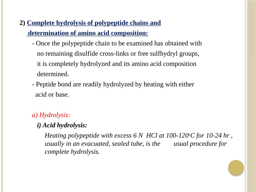
(elongated in the form of fibers).
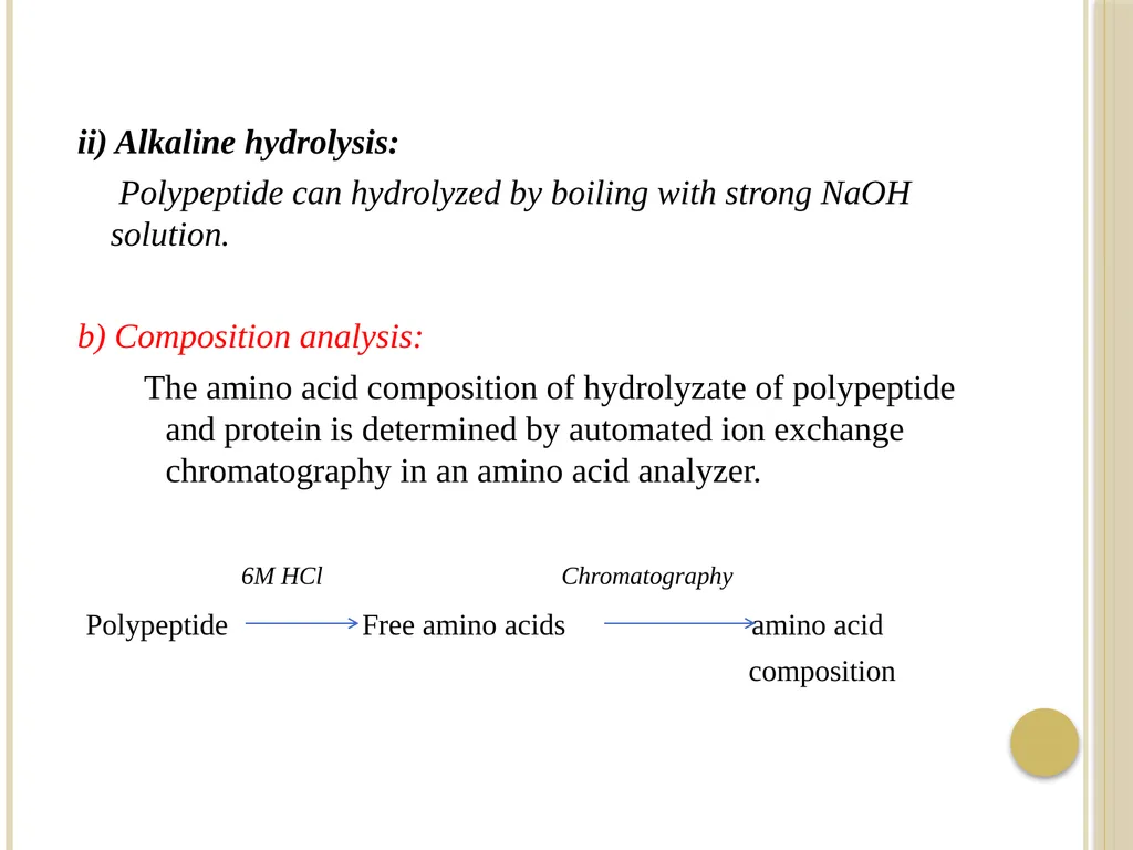
Example:
Collagen, keratin<br>
- They are insoluble in water
- Their polypeptide chains are arranged in long strands
(elongated in the form of fibers).
Example:
Collagen, keratin<br>
09
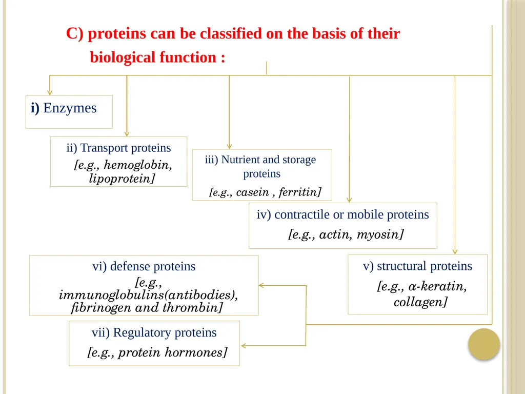
C) proteins can be classified on the basis of their
biological function : i) Enzymes ii) Transport proteins
[e.g., hemoglobin, lipoprotein] iii) Nutrient and storage proteins
[e.g., casein , ferritin] iv) contractile or mobile proteins
[e.g., actin, myosin] v) structural proteins
[e.g., α-keratin, collagen] vi) defense proteins
[e.g., immunoglobulins(antibodies), fibrinogen and thrombin] vii) Regulatory proteins
[e.g., protein hormones]<br>
10
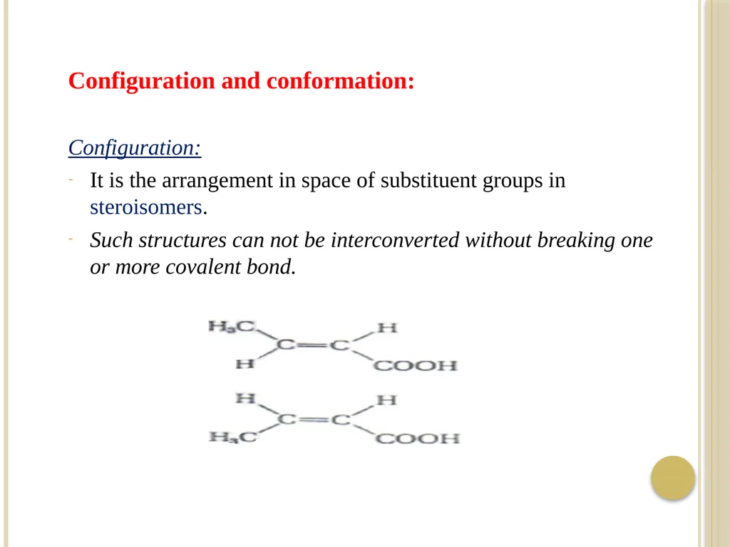
Configuration and conformation:
Configuration:
It is the arrangement in space of substituent groups in steroisomers.
Such structures can not be interconverted without breaking one or more covalent bond.<br>
Configuration:
It is the arrangement in space of substituent groups in steroisomers.
Such structures can not be interconverted without breaking one or more covalent bond.<br>
11
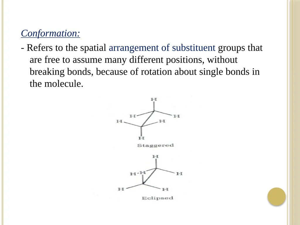
Conformation:
- Refers to the spatial arrangement of substituent groups that are free to assume many different positions, without breaking bonds, because of rotation about single bonds in the molecule.<br>
- Refers to the spatial arrangement of substituent groups that are free to assume many different positions, without breaking bonds, because of rotation about single bonds in the molecule.<br>
12
- Of the many conformation, one or (more commonly) a few
generally predominant under biological conditions.
[the need for multiple stable conformations reflects the changes that must take place in most proteins as they bind to other molecules or catalyze reaction].
- Proteins in any of their functional, folded conformations are
called native proteins.<br>
13
Structure of proteins:
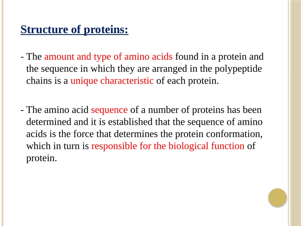
- The amount and type of amino acids found in a protein and the sequence in which they are arranged in the polypeptide chains is a unique characteristic of each protein.
- The amino acid sequence of a number of proteins has been determined and it is established that the sequence of amino acids is the force that determines the protein conformation, which in turn is responsible for the biological function of protein.<br>
14
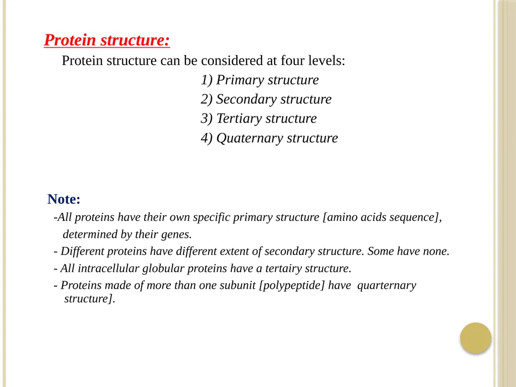
Protein structure:
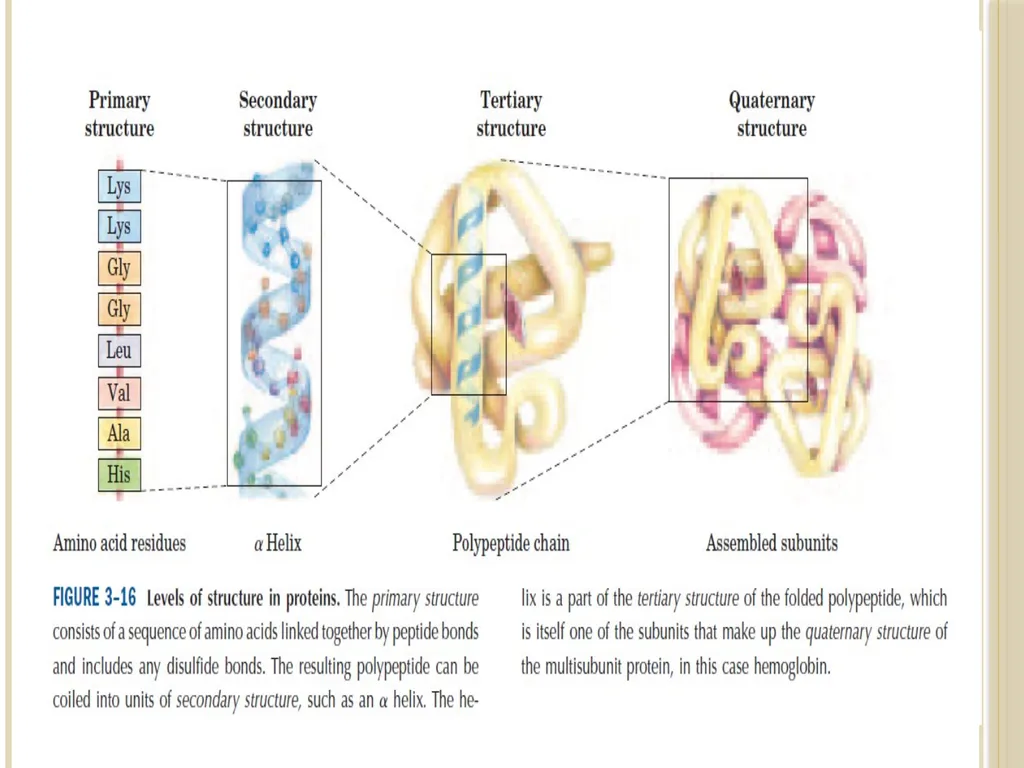
Protein structure can be considered at four levels:
1) Primary structure
2) Secondary structure
3) Tertiary structure
4) Quaternary structure
Note:
-All proteins have their own specific primary structure [amino acids sequence],
determined by their genes.
- Different proteins have different extent of secondary structure. Some have none.
- All intracellular globular proteins have a tertairy structure.
- Proteins made of more than one subunit [polypeptide] have quarternary structure].<br>
15
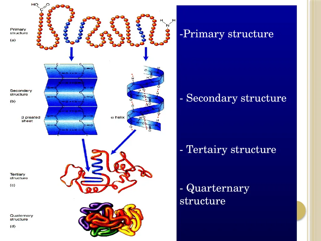
-Primary structure
- Secondary structure
- Tertairy structure
- Quarternary structure<br>
- Secondary structure
- Tertairy structure
- Quarternary structure<br>
16
1) Primary structure:
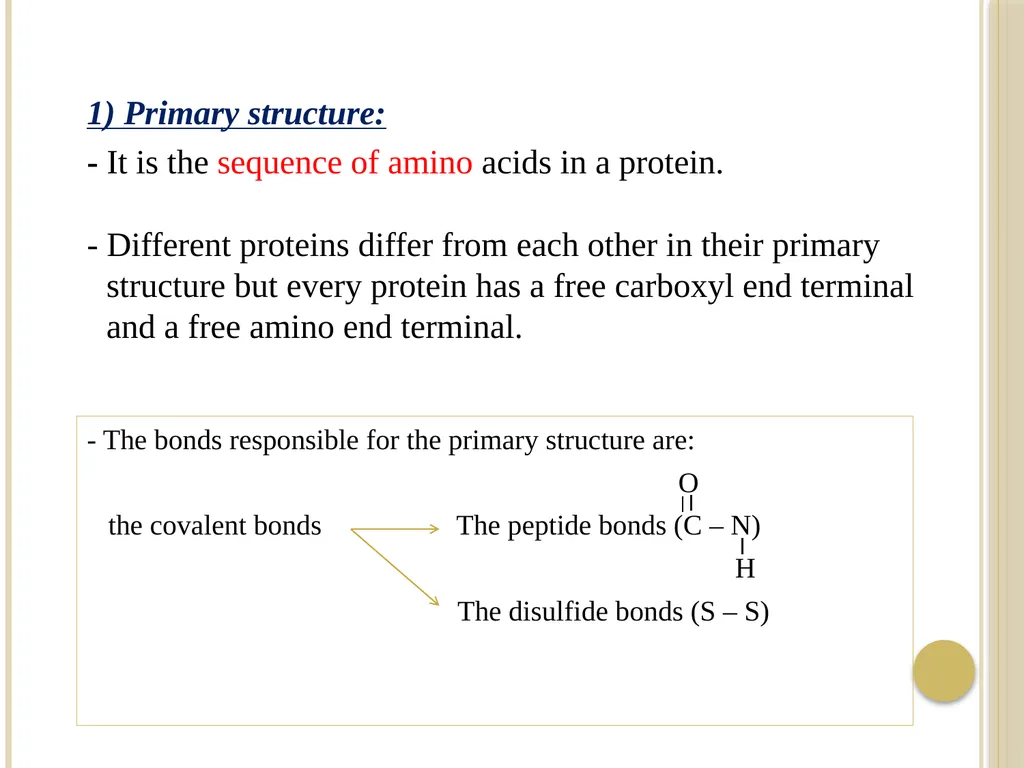
- It is the sequence of amino acids in a protein.
- Different proteins differ from each other in their primary structure but every protein has a free carboxyl end terminal and a free amino end terminal. - The bonds responsible for the primary structure are:
O
the covalent bonds The peptide bonds (C – N)
H
The disulfide bonds (S – S)<br>
17
- The disulfide bonds (if present) between two cysteine
residues of the same polypeptide chain or between different
polypeptide chains.
The primary structure is therefore very stable due to these bonds.
- Peptide bonds are not broken by normal handling nor that
conditions that denature proteins such as heating.
- Prolonged exposure to strong acid or base at elevated
temperature is required to hydrolyze these bonds (peptide
bond), non-enzymatically.
- Treatment with strong reducing agents disrupts the disulfide
bridges and effects the biological properties of the proteins.<br>
18
Characteristics of the peptide bond:
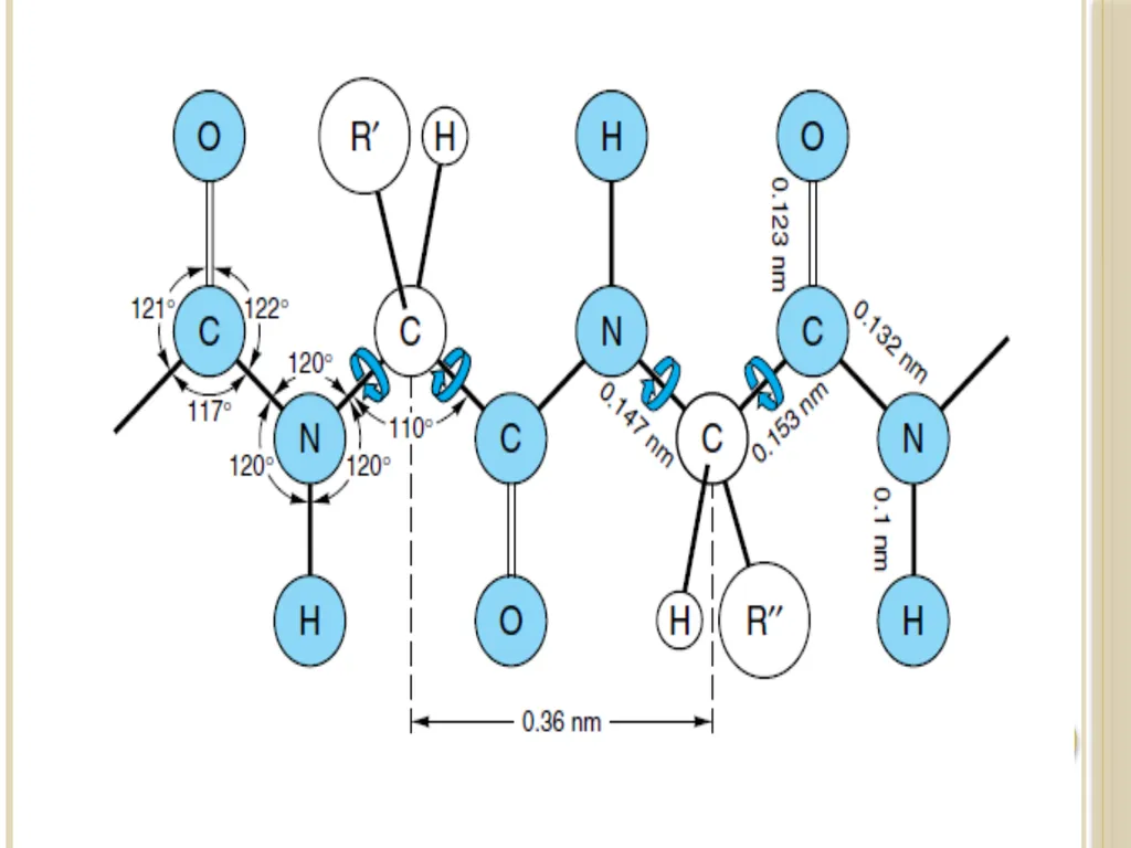
a) The peptide bond is rigid and planar:
- The peptide bond has a partial double bond character – that
is, it is shorter than a single bond [i.e peptide bond is
intermediate in bond length (0.132 nm ) between a single
bond (0.147 nm) and a double bond (0.123 nm)] and is,
therefore, rigid and planar.
This prevents free rotation around the bond between the carbonyl carbon and the nitrogen of the peptide bond.
- The rigid peptide bonds limit the number of conformations by polypeptide chains.
[ However, the bond between α-carbon and the α-amino or
α-carboxyl groups can be freely rotated].<br>
19
b) Trans configuration:
The peptide bond is generally a trans bond (instead of cis), because of steric interference of the R-groups when in the cis position.
c) Uncharged but polar:
( The - C = O and –NH groups of the peptide bond are polar and are involved in hydrogen bonds).<br>
The peptide bond is generally a trans bond (instead of cis), because of steric interference of the R-groups when in the cis position.
c) Uncharged but polar:
( The - C = O and –NH groups of the peptide bond are polar and are involved in hydrogen bonds).<br>
20
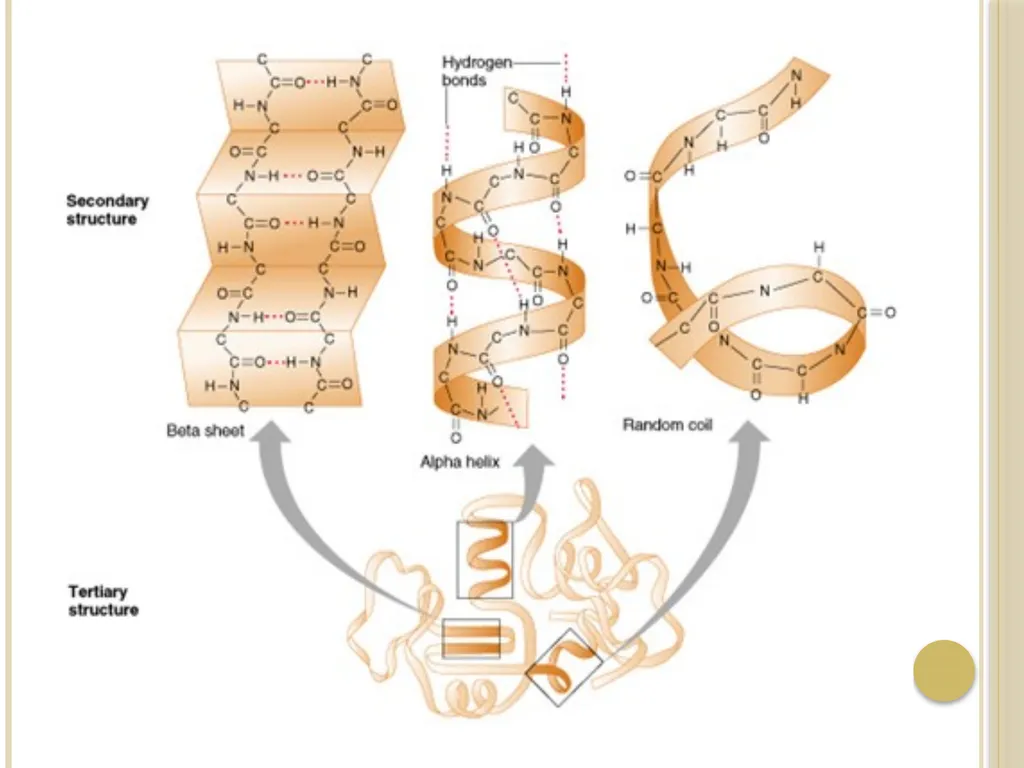
2) Secondary structure:
- It is the spatial arrangement of amino acid residues that are near one another in the linear sequence of the polypeptide chain.
- The secondary structure are of three types:
a) The α-helix
b) β-pleated sheets
c) Collagen helix<br>
- It is the spatial arrangement of amino acid residues that are near one another in the linear sequence of the polypeptide chain.
- The secondary structure are of three types:
a) The α-helix
b) β-pleated sheets
c) Collagen helix<br>
21
The bonds responsible for stability of secondary structure:
- The secondary structure is stabilized by a hydrogen bonds between peptide bond groups
(i.e. between NH group of one amino acid residue and the carbonyl oxygen (C=O) of other amino acid).<br>
- The secondary structure is stabilized by a hydrogen bonds between peptide bond groups
(i.e. between NH group of one amino acid residue and the carbonyl oxygen (C=O) of other amino acid).<br>
22
a) α-helix:
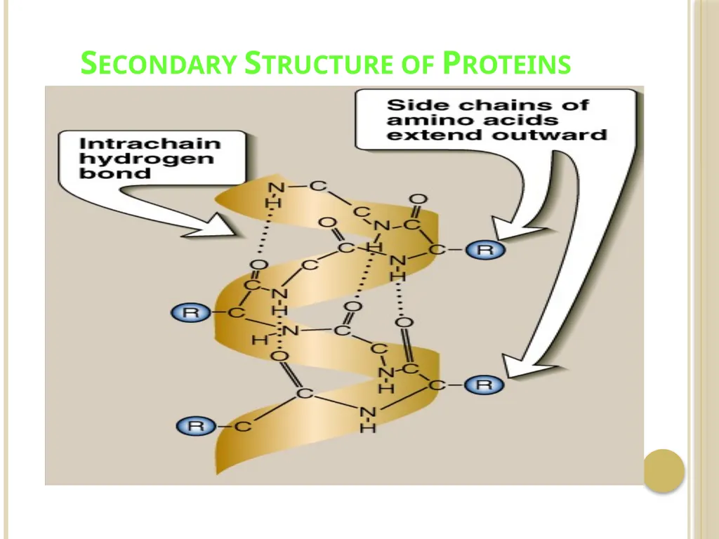
- It is a spiral structure.
- The polypeptide backbone is tightly wound around the long axis of the molecule and the R-groups of amino acid
residues protrude outward from the helical backbone.
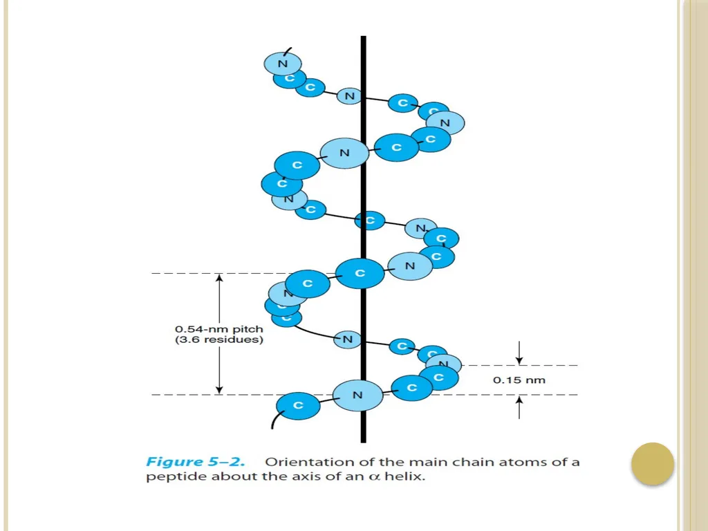
- The repeating unit is a single turn of the helix, which extends about 5.4 ͦA along the long axis.
-Each helical turn includes 3.6 amino acid residues
- In all proteins, the helical twist of the α-helix is right-handed.
The bonds responsible for stability of α-helix :
It is stabilized by a hydrogen bonds between peptide bondgroups.<br>
23
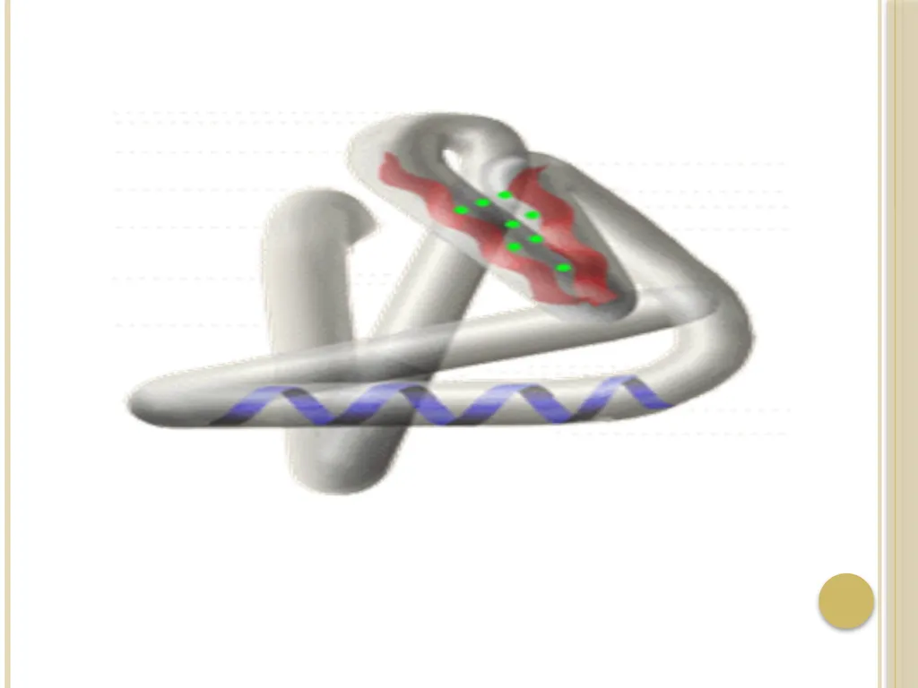
Secondary Structure of Proteins<br>
24
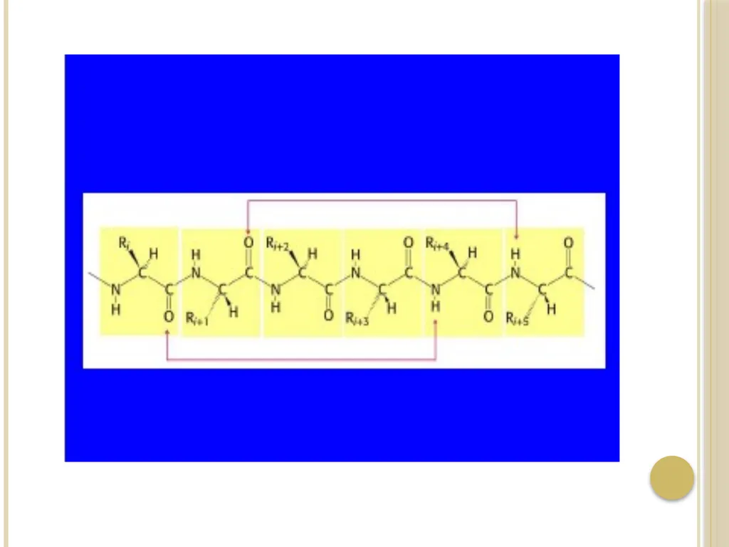
- An α-helix permits the formation of intrachain hydrogen
bonds between successive coils of the helix, parallel to the
long axis of the helix.
-The hydrogen bond formed between the hydrogen atom
attached to the electronegative nitrogen atom of each peptide
linkage and the electronegative carbonyl oxygen atom of the
fourth amino acid on the amino terminal side of it in the
helix.
- Every polypeptide bond of the chain participates in such
hydrogen bonding.
- Each successive turn of the α-helix is held to the adjacent
turns by three to four hydrogen bonds, conferring significant
stability in the overall structure.<br>
25
- Hydrogen bonds are individually weak but collectively serve to stabilize the helix.
Example:
● α-keratin:
- it is a fibrous protein whose structure is entirely
α-helix
- it is rich in cysteine residues.
- major component of tissues such as hair and
skin.
● hemoglobin:
- it is a globular protein.
- 80% of hemoglobin structure is α-helix.<br>
Example:
● α-keratin:
- it is a fibrous protein whose structure is entirely
α-helix
- it is rich in cysteine residues.
- major component of tissues such as hair and
skin.
● hemoglobin:
- it is a globular protein.
- 80% of hemoglobin structure is α-helix.<br>
26
Factors that affect the stability of α-helix:
a) Electrostatic repulsion (or attraction) between amino acid
residues with charged R-groups.
[ e.g., if there is a large numbers of charged amino acids (for example, glutamate, aspartate, histidine, lysine, or arginine) they disrupt the helix by forming ionic bonds, or by electrostatically repelling each other].
b) The bulkiness of adjacent R-groups.
[e.g., tryptophan, or amino acids, such as valine or isoleucine, that branch at the β-carbon (the first carbon in the R-group, next to the α-carbon) can interfere with formation of the α-helix if they are present in large numbers and close to each others].<br>
27
c) The interaction between amino acid side chains spaced three (or four) residues apart.
[e.g two aromatic amino acids are resulting in hydrophobic interaction].
d) The occurrence of proline residues:
- In proline the nitrogen atom is part of a rigid ring and the rotation around N-α C bond is not possible .
- In addition , the nitrogen atom of a proline residue in peptide linkage has no substitutent hydrogen to form hydrogen bond with other residues
So it inserts a kink in the chain that disrupts the smooth, helical structure.<br>
28
b) β-pleated sheet:
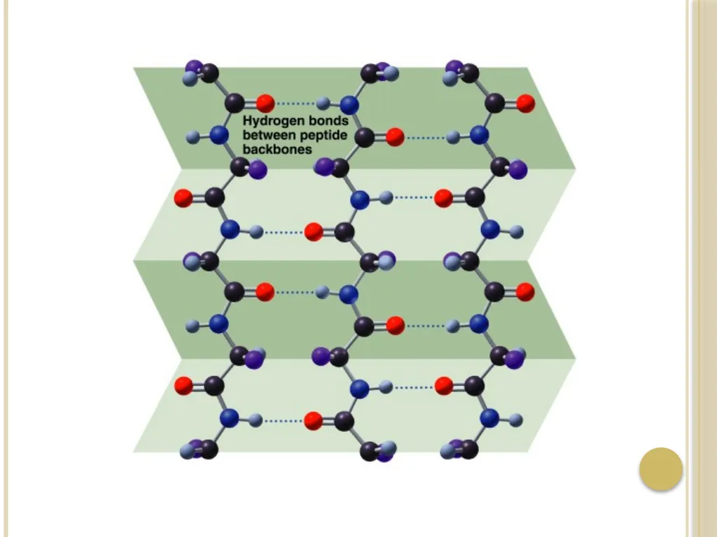
In the β-conformation, the backbone of the polypeptide chain is extended into a zigzag rather than a helical structure.
The zigzag polypeptide chains can be arranged side by side to form a structure resembling a series of pleats, such a structure is called a β-pleated sheet.
The R-groups of adjacent amino acids protrude from the zigzag structure in opposite directions, creating the alternating pattern.
Hydrogen bonds form between adjacent segments of polypeptide chain β- pleated sheet are composed of two or more β-strands.<br>
29
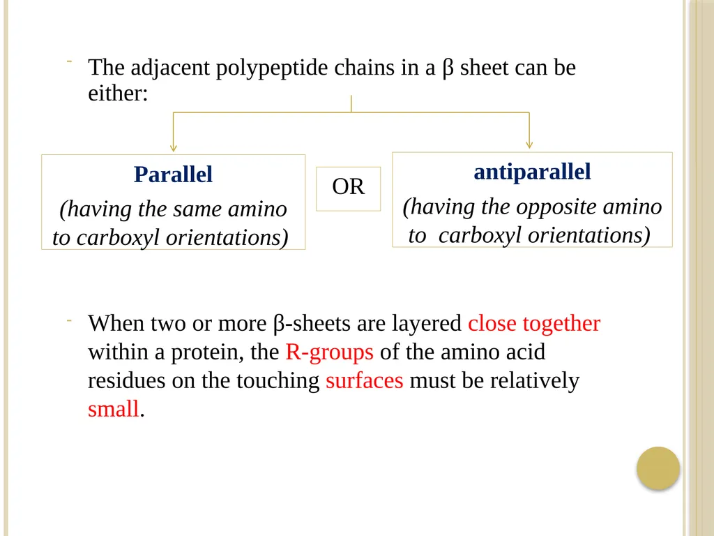
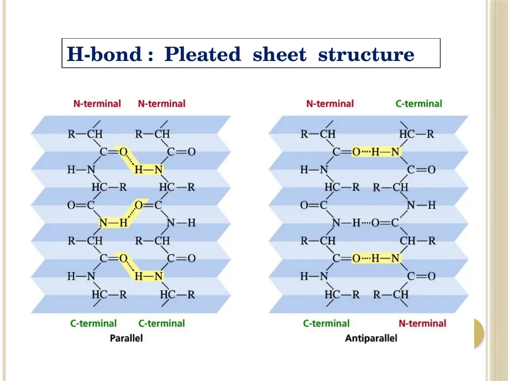
The adjacent polypeptide chains in a β sheet can be either: When two or more β-sheets are layered close together within a protein, the R-groups of the amino acid residues on the touching surfaces must be relatively small. Parallel
(having the same amino to carboxyl orientations) antiparallel
(having the opposite amino to carboxyl orientations) OR<br>
(having the same amino to carboxyl orientations) antiparallel
(having the opposite amino to carboxyl orientations) OR<br>
30
H-bond : Pleated sheet structure<br>
31
The bonds responsible for stability of β-pleated sheet :
- It is stabilized by a hydrogen bonds between peptide bond groups. Example:
β-keratins (such as silk fibroin):
- Consist Almost Entirely Stacks Of Antiparallel Β-sheets.
- Rich In Amino Acids Having Relatively Small R-groups, Particularly Glycine And Alanine.
- The β-keratin such as fibroin, have no S–S cross linkages.<br>
32
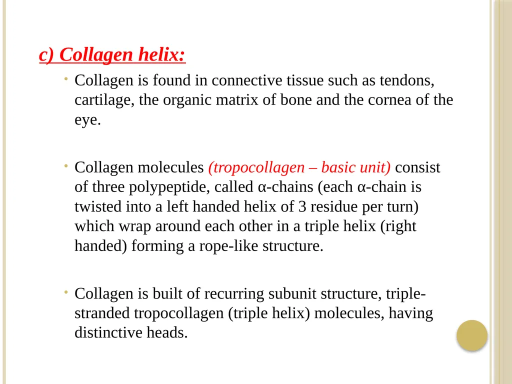
c) Collagen helix:
Collagen is found in connective tissue such as tendons, cartilage, the organic matrix of bone and the cornea of the eye.
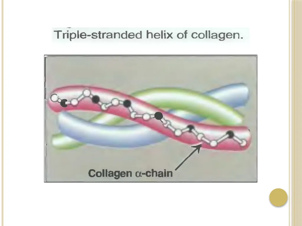
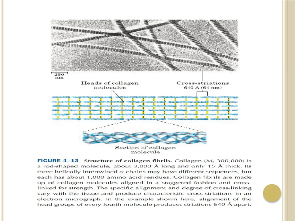
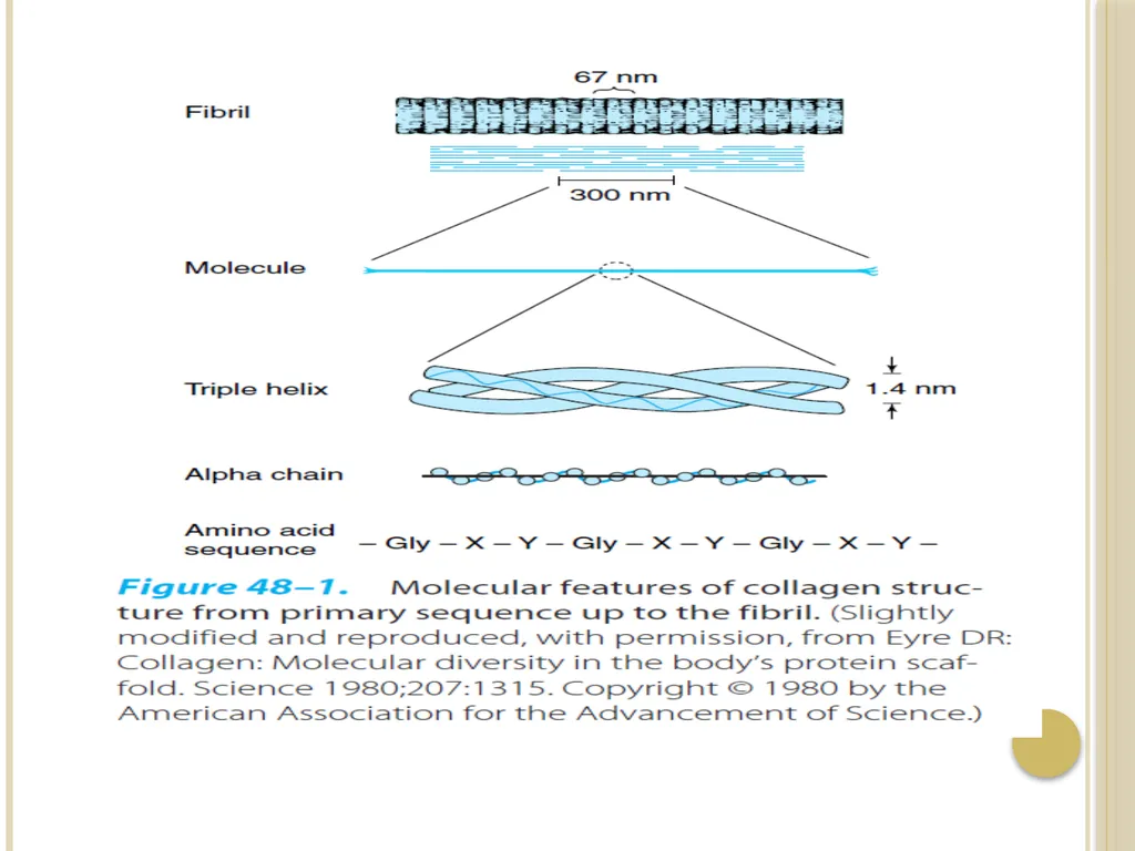
Collagen molecules (tropocollagen – basic unit) consist of three polypeptide, called α-chains (each α-chain is twisted into a left handed helix of 3 residue per turn) which wrap around each other in a triple helix (right handed) forming a rope-like structure.
Collagen is built of recurring subunit structure, triple-stranded tropocollagen (triple helix) molecules, having distinctive heads.<br>
33
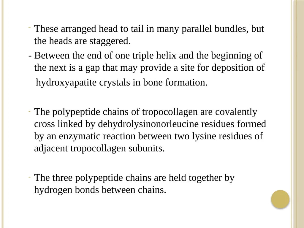
These arranged head to tail in many parallel bundles, but the heads are staggered.
- Between the end of one triple helix and the beginning of the next is a gap that may provide a site for deposition of
hydroxyapatite crystals in bone formation.
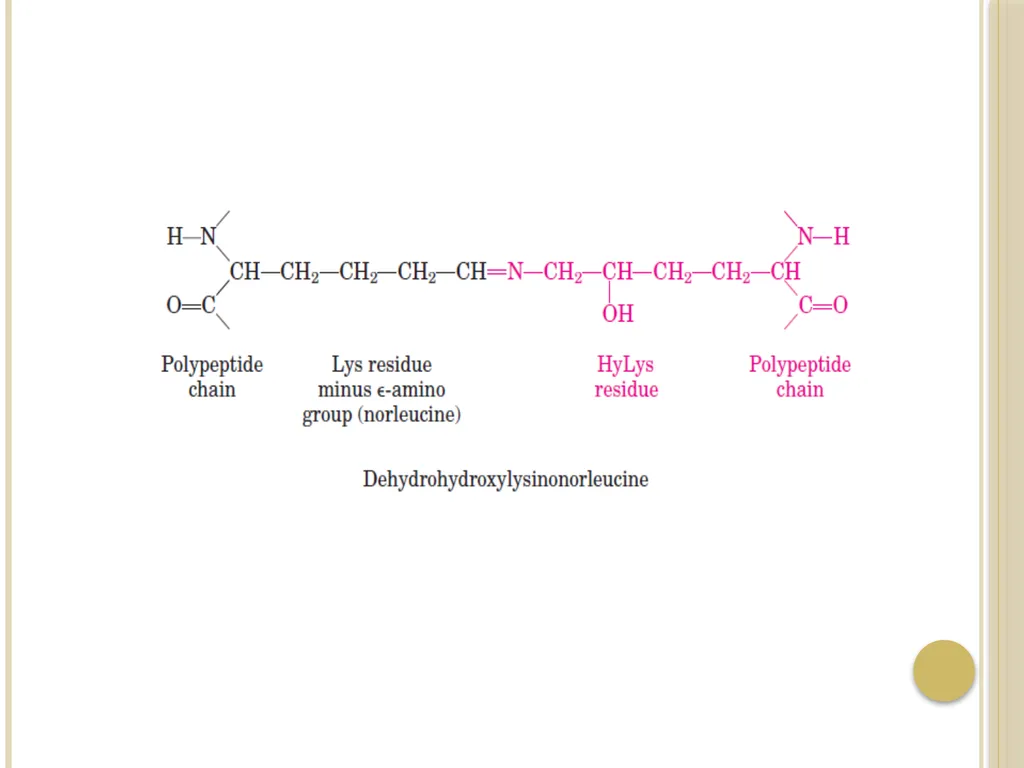
The polypeptide chains of tropocollagen are covalently cross linked by dehydrolysinonorleucine residues formed by an enzymatic reaction between two lysine residues of adjacent tropocollagen subunits.
The three polypeptide chains are held together by hydrogen bonds between chains.<br>
34
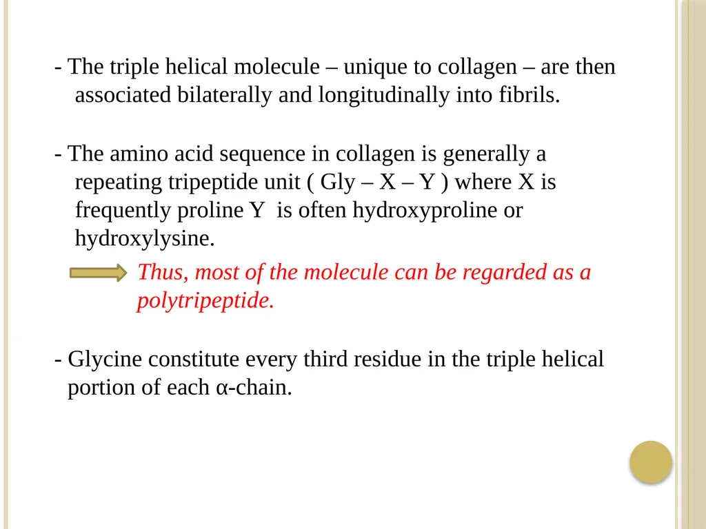
- The triple helical molecule – unique to collagen – are then associated bilaterally and longitudinally into fibrils.
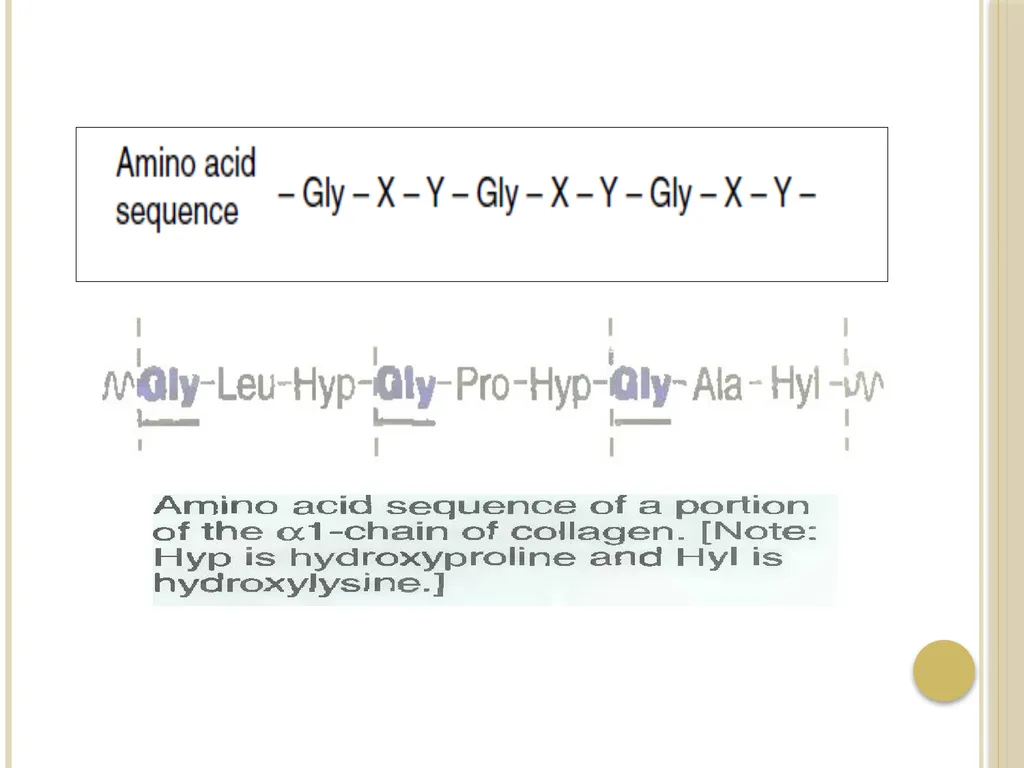
- The amino acid sequence in collagen is generally a repeating tripeptide unit ( Gly – X – Y ) where X is frequently proline Y is often hydroxyproline or hydroxylysine.
Thus, most of the molecule can be regarded as a polytripeptide.
- Glycine constitute every third residue in the triple helical portion of each α-chain.<br>
35
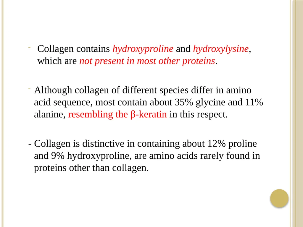
Collagen contains hydroxyproline and hydroxylysine, which are not present in most other proteins.
Although collagen of different species differ in amino acid sequence, most contain about 35% glycine and 11% alanine, resembling the β-keratin in this respect.
- Collagen is distinctive in containing about 12% proline and 9% hydroxyproline, are amino acids rarely found in proteins other than collagen.<br>
36
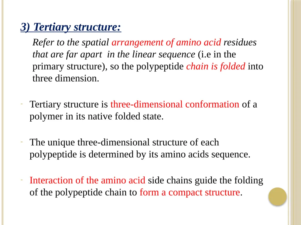
3) Tertiary structure:
Refer to the spatial arrangement of amino acid residues that are far apart in the linear sequence (i.e in the primary structure), so the polypeptide chain is folded into three dimension.
Tertiary structure is three-dimensional conformation of a polymer in its native folded state.
The unique three-dimensional structure of each polypeptide is determined by its amino acids sequence.
Interaction of the amino acid side chains guide the folding of the polypeptide chain to form a compact structure.<br>
37
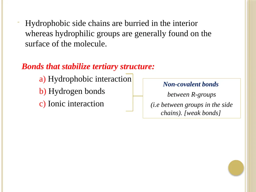
Bonds that stabilize tertiary structure:
a) Hydrophobic interaction
b) Hydrogen bonds
c) Ionic interaction Hydrophobic side chains are burried in the interior whereas hydrophilic groups are generally found on the surface of the molecule. Non-covalent bonds
between R-groups
(i.e between groups in the side chains). [weak bonds]<br>
a) Hydrophobic interaction
b) Hydrogen bonds
c) Ionic interaction Hydrophobic side chains are burried in the interior whereas hydrophilic groups are generally found on the surface of the molecule. Non-covalent bonds
between R-groups
(i.e between groups in the side chains). [weak bonds]<br>
38
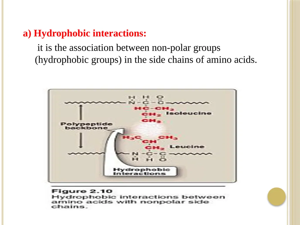
a) Hydrophobic interactions:
it is the association between non-polar groups (hydrophobic groups) in the side chains of amino acids.<br>
it is the association between non-polar groups (hydrophobic groups) in the side chains of amino acids.<br>
39
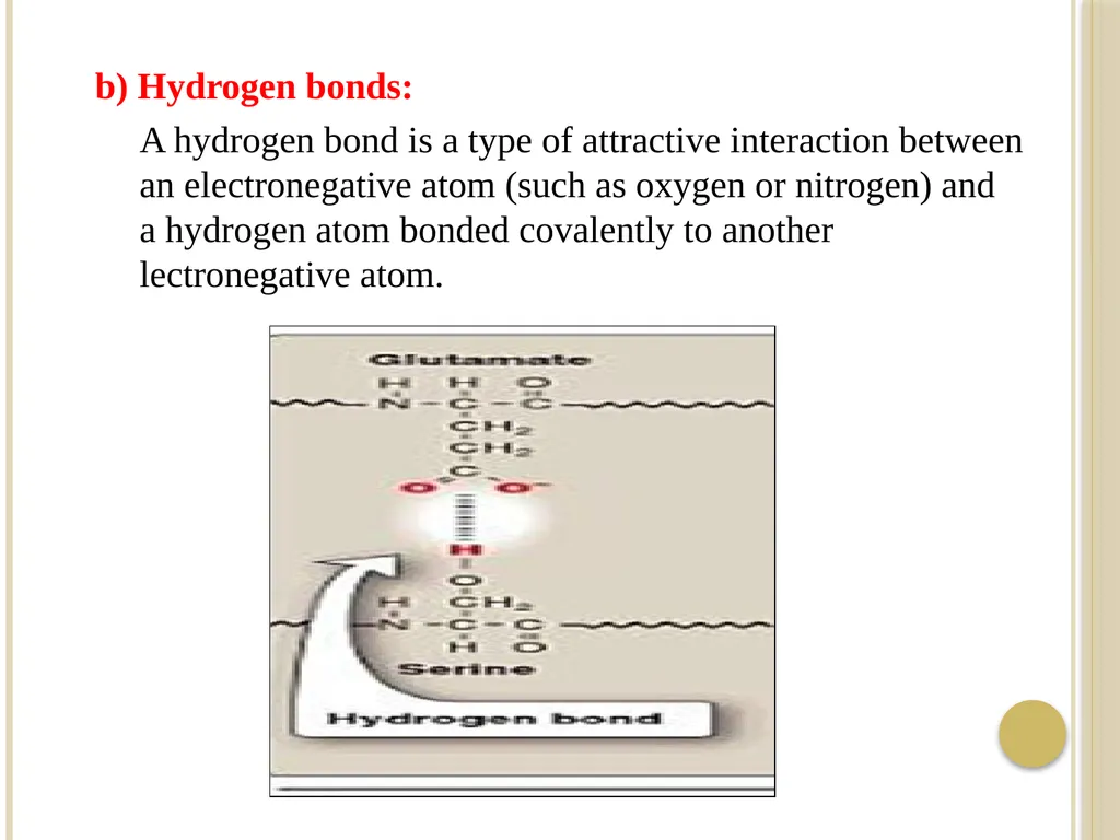
b) Hydrogen bonds:
A hydrogen bond is a type of attractive interaction between an electronegative atom (such as oxygen or nitrogen) and a hydrogen atom bonded covalently to another lectronegative atom.<br>
A hydrogen bond is a type of attractive interaction between an electronegative atom (such as oxygen or nitrogen) and a hydrogen atom bonded covalently to another lectronegative atom.<br>
40
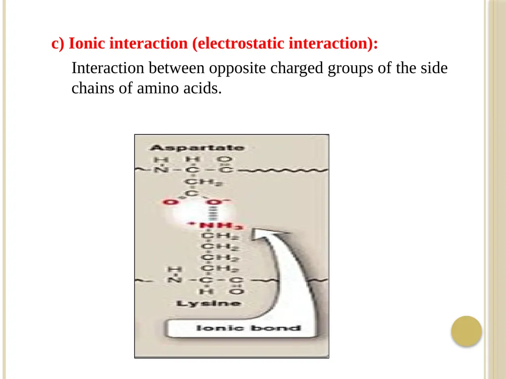
c) Ionic interaction (electrostatic interaction):
Interaction between opposite charged groups of the side chains of amino acids.<br>
Interaction between opposite charged groups of the side chains of amino acids.<br>
41
Example:
Myoglobin:
- It is small globular protein (hemoprotein), present in heart and skeletal muscle.
- It functions both as a reservoir for oxygen and as an oxygen carrier that increases the rate of transport of oxygen within the muscle cell.
- Myoglobin consists of a single polypeptide chain of 153 amino acid residues of known sequence and a single heme group.<br>
42
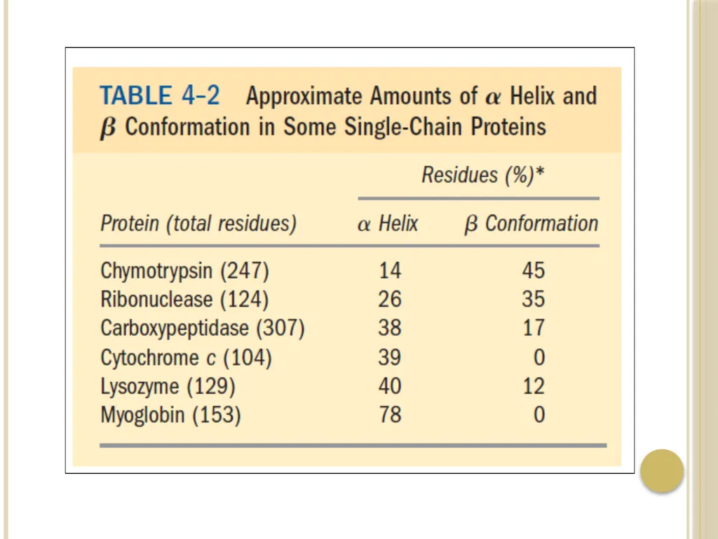
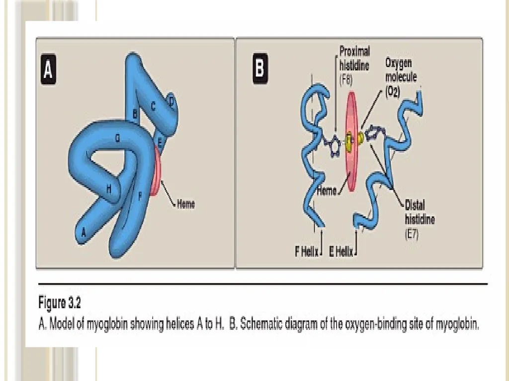
- Myoglobin is a compact molecule with approximately 80% of its polypeptide chain folded into eight stretches of α-helix. These α-helical regions, labeled A to H.
- The backbone of the myoglobin molecule is made up of 8 relatively straight segments of α-helix interrupted by bends.
- The longest α-helix has 23 amino acid residues and the
shortest only seven; all are right handed.
More than 70% of the amino acids in the myoglobin molecule are in these α-helical regions.<br>
43
- The eight α-helical regions, labeled A to H, are terminated either by the presence of proline, whose five-membered ring can not be accomedated in α-helix, or by β-bends and loops stabilize by hydrogen bonds and ionic bonds.<br>
44
Location of polar and non-polar amino acid residues:
-The interior of the myoglobin molecule is composed almost entirely of non-polar amino acids.
- They are packed closely together, forming a structure
stabilized by hydrophobic interactions among these clustered residues.
- In contrast, charged amino acids are located almost
exclusively on the surface, where they can form hydrogen bonds with water.<br>
45
Heme group:
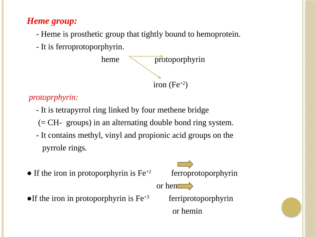
- Heme is prosthetic group that tightly bound to hemoprotein.
- It is ferroprotoporphyrin.
heme protoporphyrin
iron (Fe+2)
protoprphyrin:
- It is tetrapyrrol ring linked by four methene bridge
(= CH- groups) in an alternating double bond ring system.
- It contains methyl, vinyl and propionic acid groups on the
pyrrole rings.
● If the iron in protoporphyrin is Fe+2 ferroprotoporphyrin
or heme
●If the iron in protoporphyrin is Fe+3 ferriprotoporphyrin
or hemin<br>
46
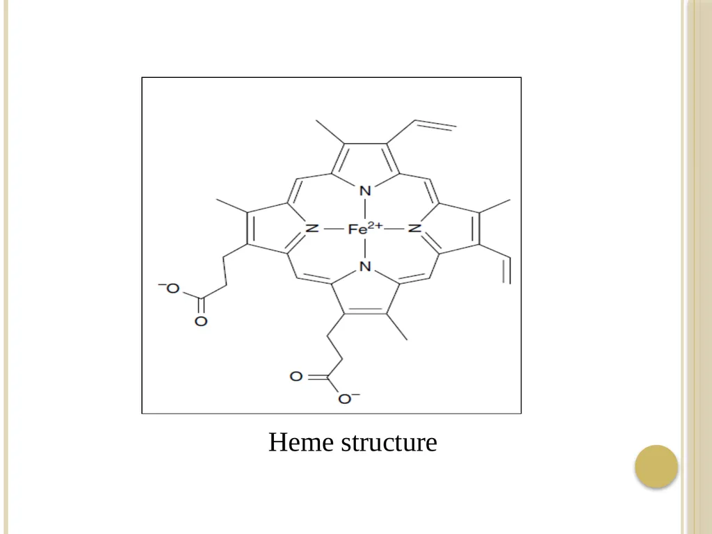
Heme structure<br>
47
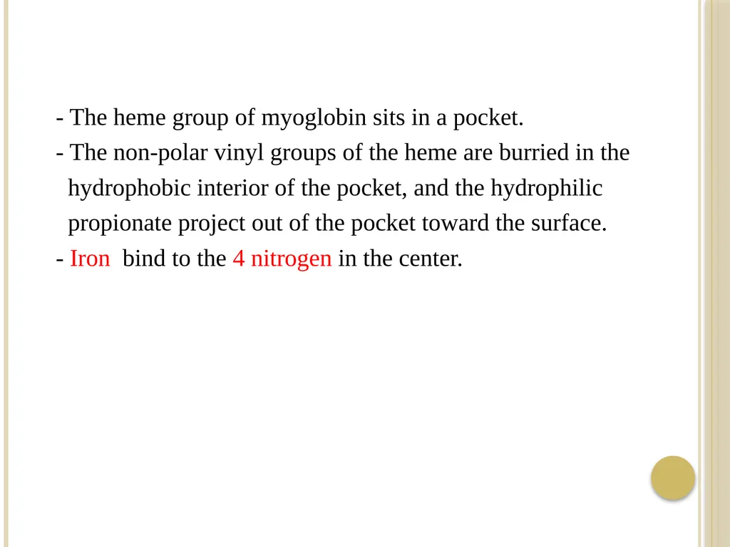
- The heme group of myoglobin sits in a pocket.
- The non-polar vinyl groups of the heme are burried in the
hydrophobic interior of the pocket, and the hydrophilic
propionate project out of the pocket toward the surface.
- Iron bind to the 4 nitrogen in the center.<br>
- The non-polar vinyl groups of the heme are burried in the
hydrophobic interior of the pocket, and the hydrophilic
propionate project out of the pocket toward the surface.
- Iron bind to the 4 nitrogen in the center.<br>
48
Binding of heme group:
- The heme group of myoglobin sits in a crevice in the
molecule, lined with non-polar amino acids.
- Notable exception are two histidine residues:
One, termed the proximal histidine (F8), binds directly to the iron of heme.
The second, or distal histidine (E7) does not directly interact with the heme group but helps stabilize the binding of oxygen to the ferrous iron.<br>
49
- Oxygen binding site is between iron and His E7 (distal His).
Myoglobin structure is stabilized by:
Non-covalent bonds.<br>
Myoglobin structure is stabilized by:
Non-covalent bonds.<br>
50
4) Quaternary structure:
- Proteins that are composed of more than one polypeptide chain (subunits) show a fourth level of protein structure which is the quaternary structure.
Quaternary structure is the three-dimensional structure of a multisubunit protein, particularly the manner in which the subunits fit together.
- Subunits may either function independently of each other or may work cooperatively, as in hemoglobin.<br>
51
Bonds that stabilize quaternary structure:
a) Hydrophobic bonds.
b) Hydrogen bonds.
c) Ionic bonds (electrostatic bonds). - The interaction between subunits are stabilized by the same forces that stabilize tertiary structure (non-covalent bonds). Non-covalent bonds between R-groups
(i.e between groups in the side chains).
[weak bonds]<br>
a) Hydrophobic bonds.
b) Hydrogen bonds.
c) Ionic bonds (electrostatic bonds). - The interaction between subunits are stabilized by the same forces that stabilize tertiary structure (non-covalent bonds). Non-covalent bonds between R-groups
(i.e between groups in the side chains).
[weak bonds]<br>
52
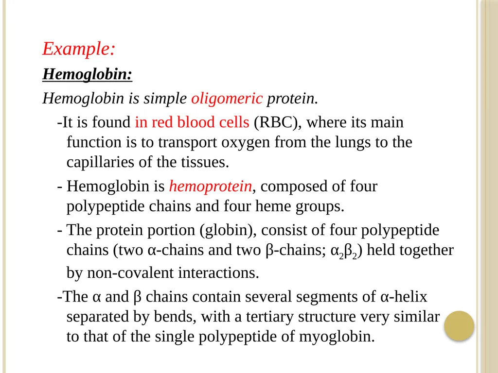
Example:
Hemoglobin:
Hemoglobin is simple oligomeric protein.
-It is found in red blood cells (RBC), where its main function is to transport oxygen from the lungs to the capillaries of the tissues.
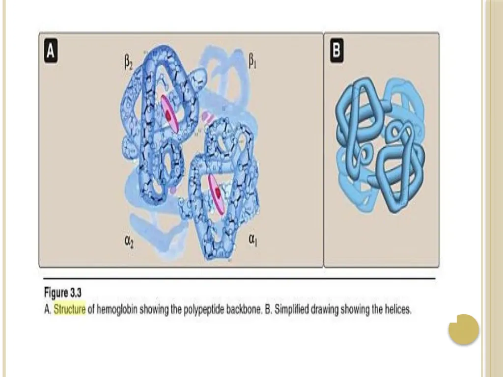
- Hemoglobin is hemoprotein, composed of four polypeptide chains and four heme groups.
- The protein portion (globin), consist of four polypeptide chains (two α-chains and two β-chains; α2β2) held together by non-covalent interactions.
-The α and β chains contain several segments of α-helix separated by bends, with a tertiary structure very similar to that of the single polypeptide of myoglobin.<br>
53
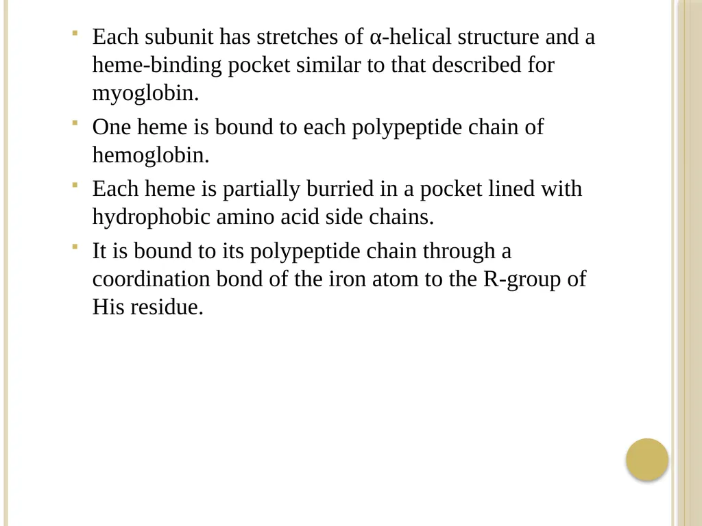
Each subunit has stretches of α-helical structure and a heme-binding pocket similar to that described for myoglobin.
One heme is bound to each polypeptide chain of hemoglobin.
Each heme is partially burried in a pocket lined with hydrophobic amino acid side chains.
It is bound to its polypeptide chain through a coordination bond of the iron atom to the R-group of His residue.<br>
One heme is bound to each polypeptide chain of hemoglobin.
Each heme is partially burried in a pocket lined with hydrophobic amino acid side chains.
It is bound to its polypeptide chain through a coordination bond of the iron atom to the R-group of His residue.<br>
54
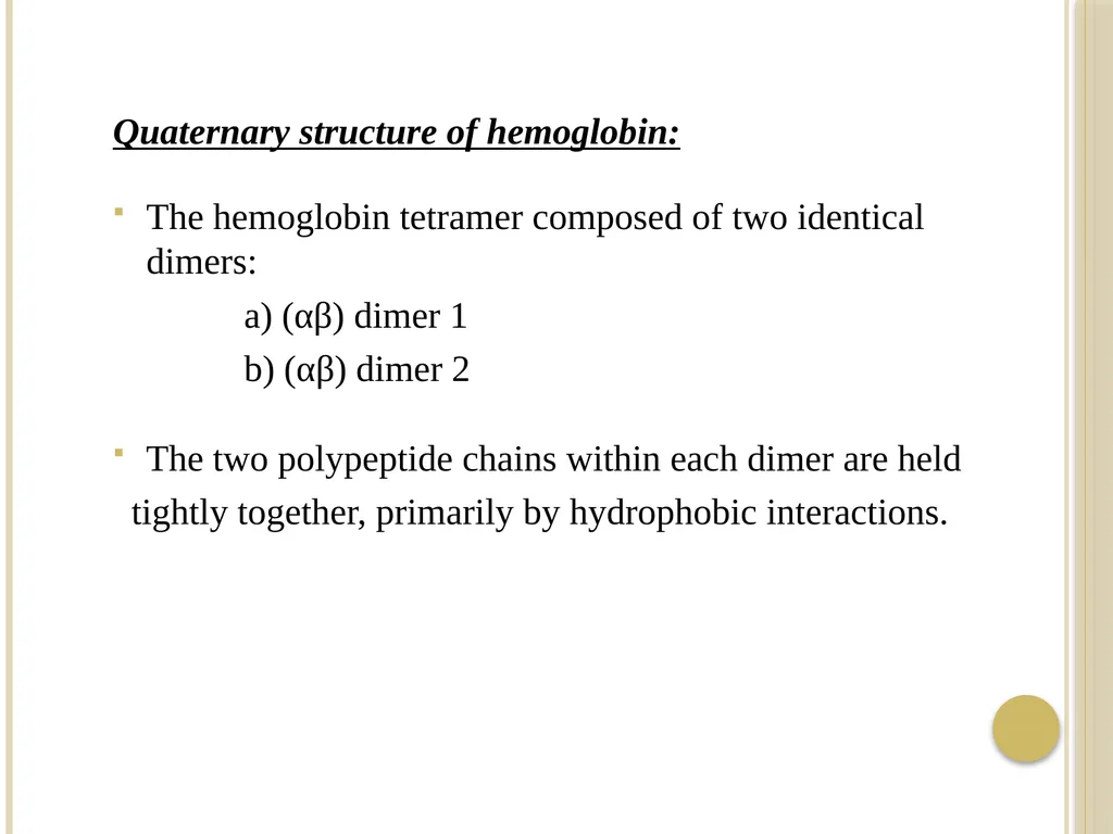
Quaternary structure of hemoglobin:
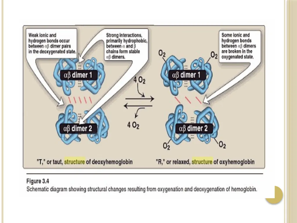
The hemoglobin tetramer composed of two identical dimers:
a) (αβ) dimer 1
b) (αβ) dimer 2
The two polypeptide chains within each dimer are held
tightly together, primarily by hydrophobic interactions.<br>
The hemoglobin tetramer composed of two identical dimers:
a) (αβ) dimer 1
b) (αβ) dimer 2
The two polypeptide chains within each dimer are held
tightly together, primarily by hydrophobic interactions.<br>
55
- Interchain hydrophobic interaction form association between α and β subunits in the dimers.
- Ionic and hydrogen bonds also occur between the members
of the dimer.
- The two dimers are held together primarily by polar bonds.
- The weaker interactions between these mobile dimers result
in the two dimers occupying different relative positions in
deoxyhemoglobin compared to oxyhemoglobin.
i) T form:
- The deoxy form of hemoglobin is called the “T” or taut (tense) form .<br>
56
- In the T form, the two αβ dimers interact through a network
or ionic bonds and hydrogen bonds that constrain the
movement of the polypeptide chains.
- The T form is the low oxygen-affinity form of hemoglobin.
ii) R form :
The binding of oxygen to hemoglobin causes rupture of some of the ionic bonds and hydrogen bonds between the αβ dimers.
this ds to a structure called the “R” or relaxed form, in which the polypeptide chains have more freedom of movement.
- The R form is the high oxygen affinity form of hemoglobin.<br>
57
Binding of oxygen to hemoglobin:
- Myoglobin can bind only one molecule of oxygen because it
contains only one heme group.
- In contrast, hemoglobin can bind one oxygen molecule (O2)
at each of its four heme groups.
- The degree of saturation (Y) of these oxygen-binding sites on all myoglobin or hemoglobin molecules can vary between zero (all sites are empty) and 100% (all sites are full).
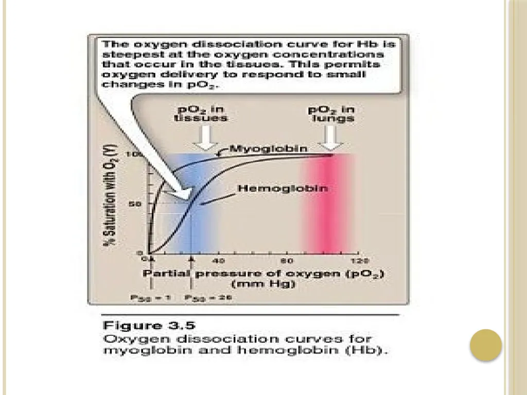
Oxygen dissociation curve:
- A plot of Y is measured at different partial pressures of oxygen (pO2) is called the oxygen dissociation curve.<br>
58
- This graph illustrates that myoglobin has a higher oxygen
affinity than does hemoglobin.
-The partial pressure of oxygen needed to achieve half-
saturation of the binding sites (P50) is approximately 1mm Hg for myoglobin and 26 mm Hg for hemoglobin.
[note: The higher the oxygen affinity (that is, the more tightly oxygen binds), the lower the P50 ].
a) myoglobin:
- The oxygen dissociation curve for myoglobin has a hyperbolic shape.
- This reflects the fact that myoglobin reversibly binds a single molecule of oxygen.<br>
59
b) Hemoglobin:
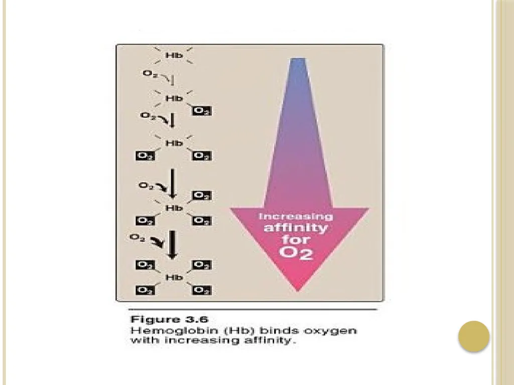
-The oxygen dissociation curve for hemoglobin is sigmoidal in shape, indicating that the subunits cooperate in binding oxygen.
- Cooperative binding of oxygen by the four subunits of hemoglobin means that the binding of an oxygen molecule at one heme group increases the oxygen affinity of the remaining heme groups in the same hemoglobin molecule.
-This effect is referred to heme – heme interaction.<br>
60
Denaturation of proteins:
- Protein denaturation results in unfolding and disorganization of the protein΄s structure, which are not accompanied by hydrolysis of peptide bonds.
Denaturing agents include:
● heat
● organic solvents
● mechanical mixing
● strong acids or bases
● detergents
● ions of heavy metals such as lead and mercury.
- Most proteins, once denatured, remain permanently disordered<br>
61
Steps in the determination of amino acid sequence
1) If the protein contains more than one polypeptide chain, the individual chains are first separated and purified.
2) All the disulfide groups are reduced and the resulting sulfhydryl
groups alkylated.
3) A sample of each polypeptide chain is subjected to total hydrolysis, and its amino acid composition is determined.
4) On another sample of the polypeptide chain the N-terminal and
C-terminal residues are identified.
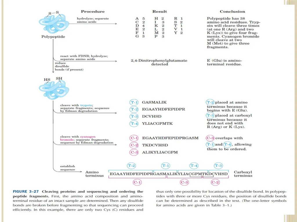
5) The intact polypeptide chain is cleaved into a series of smaller
peptides by enzymatic or chemical hydrolysis.
6) The peptide fragments resulting from step 5 are separated and their amino acid composition and sequence are determined.<br>
62
7) Another sample of the original polypeptide chain is partially
hydrolyzed by a second procedure to fragment the chain at points other than those cleaved by the first partial hydrolysis. The peptide fragments are separated and their amino acid composition and sequence determined.
8) By comparing the amino acid sequence of the two sets of peptide fragments, particularly where the fragments from the first partial hydrolysis overlap the cleavage points in the second, the peptide fragments can be placed in the proper order to yield the complete amino acid sequence.
9) The positions of the disulfide bonds and the amide groups in the original polypeptide chain are determined.<br>
63
1) Cleavage of disulfide bonds and separation of polypeptide chains:
- Before analyzing the amino acid sequence of a protein, we must determine whether the protein contains more than one polypeptide chain.
- If the polypeptide chains have no covalent cross-linkages, they can be separated by treating the protein with acid, base or high concentration of salt or denaturing agent.
- If the polypeptide chains are covalently cross linked by one or more disulfide bonds between half residues of cystine, these cross-linkages must be cleaved by appropriate chemical reaction.<br>
64
The commenest procedure:
a) is to reduce the disulfide bond to sulfhydryl groups with an excess of mercaptoethanol.
b) An alkylating agent like iodoacetate is then used to alkylate the sulfhydryl group of the cysteine residues to yield their S-carboxy-methyl derivatives.
-When the polypeptide chain is subsequently hydrolyzed, these residues appear as S-carboxymethyl cysteine, which is easily identified by the chromatographic procedure used for amino acid analysis.
- Alkylation of cysteine residues is desirable because the sulfhydryl group of cysteine is relatively unstable and tends to undergo oxidation.<br>
65
H2N
R1 - C - H
C = O
HN
SH - CH2 – CH
C = O HI
HN
R2 - C – H
COOH
Tripeptide containing
cysteine
+
ICH2COOH
Iodoacetate H2N
R1 - C - H
C = O
HN
COOH- CH2 - S - CH2 – CH
C = O
HN
R2 - C – H
COOH
S-carboxymethyl derivative of
cysteine-containing peptide<br>
R1 - C - H
C = O
HN
SH - CH2 – CH
C = O HI
HN
R2 - C – H
COOH
Tripeptide containing
cysteine
+
ICH2COOH
Iodoacetate H2N
R1 - C - H
C = O
HN
COOH- CH2 - S - CH2 – CH
C = O
HN
R2 - C – H
COOH
S-carboxymethyl derivative of
cysteine-containing peptide<br>
66
2) Complete hydrolysis of polypeptide chains and
determination of amino acid composition:
- Once the polypeptide chain to be examined has obtained with
no remaining disulfide cross-links or free sulfhydryl groups,
it is completely hydrolyzed and its amino acid composition
determined.
- Peptide bond are readily hydrolyzed by heating with either
acid or base.
a) Hydrolysis:
i) Acid hydrolysis:
Heating polypeptide with excess 6 N HCl at 100-120ͦ C for 10-24 hr , usually in an evacuated, sealed tube, is the usual procedure for complete hydrolysis.<br>
67
ii) Alkaline hydrolysis:
Polypeptide can hydrolyzed by boiling with strong NaOH solution.
b) Composition analysis:
The amino acid composition of hydrolyzate of polypeptide and protein is determined by automated ion exchange chromatography in an amino acid analyzer.
6M HCl Chromatography
Polypeptide Free amino acids amino acid
composition<br>
68
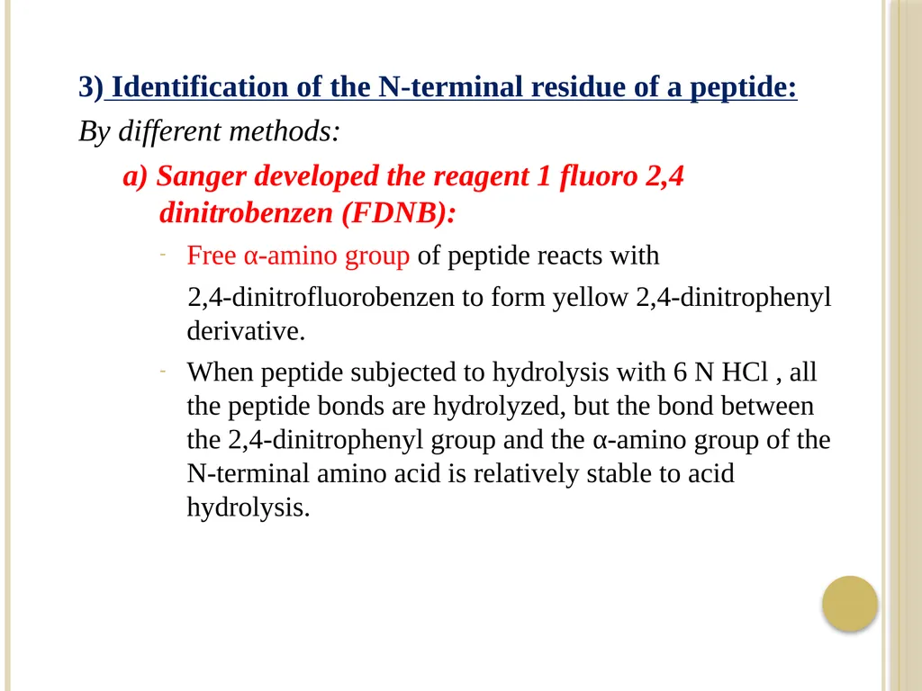
3) Identification of the N-terminal residue of a peptide:
By different methods:
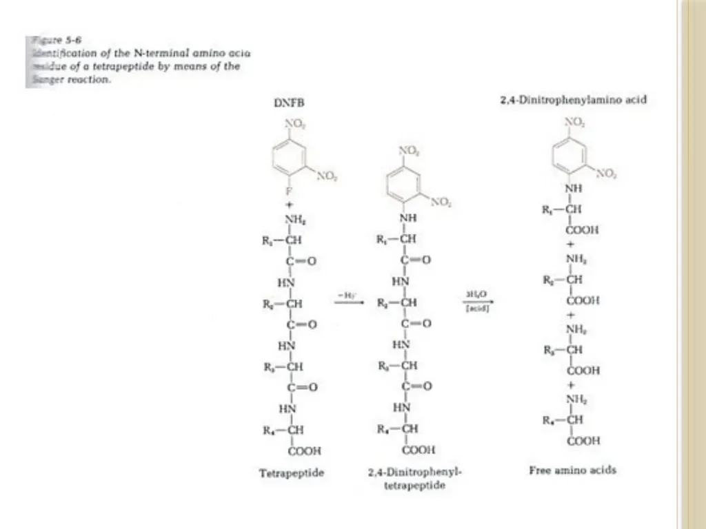
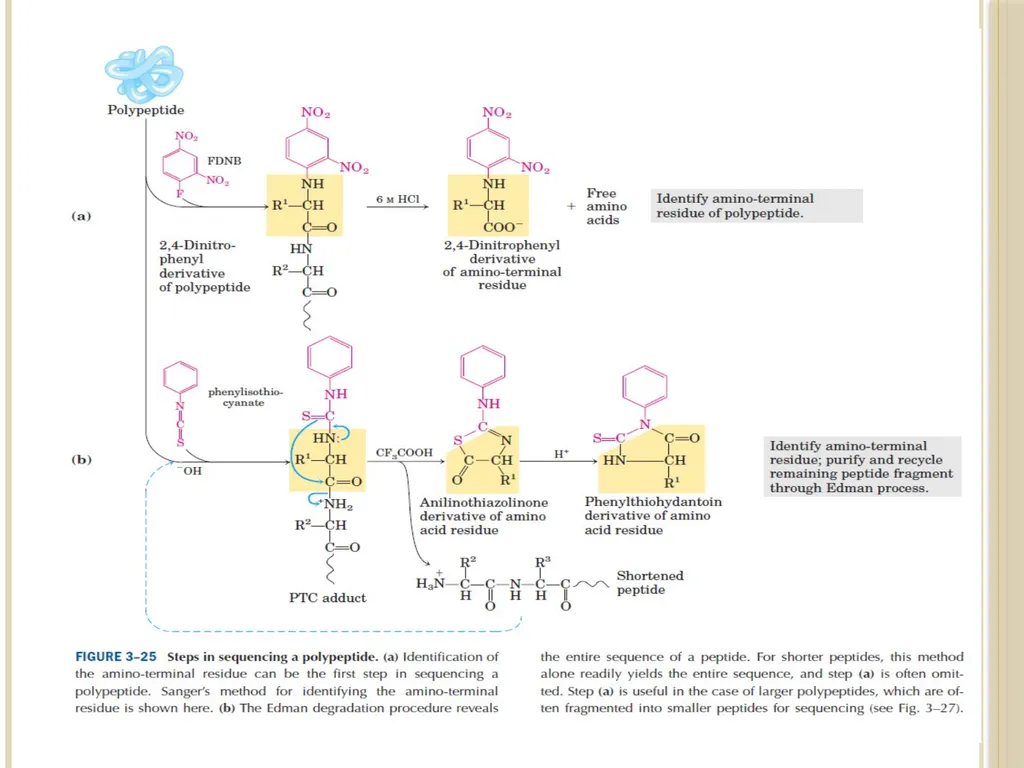
a) Sanger developed the reagent 1 fluoro 2,4 dinitrobenzen (FDNB):
Free α-amino group of peptide reacts with
2,4-dinitrofluorobenzen to form yellow 2,4-dinitrophenyl derivative.
When peptide subjected to hydrolysis with 6 N HCl , all the peptide bonds are hydrolyzed, but the bond between the 2,4-dinitrophenyl group and the α-amino group of the N-terminal amino acid is relatively stable to acid hydrolysis.<br>
69
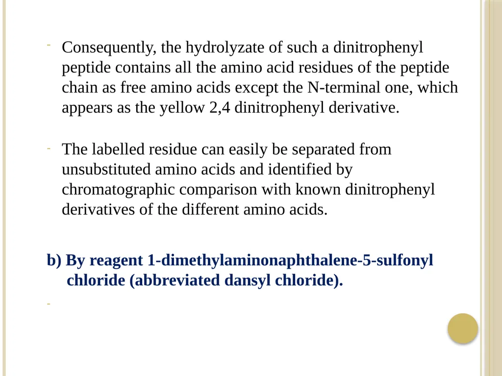
Consequently, the hydrolyzate of such a dinitrophenyl peptide contains all the amino acid residues of the peptide chain as free amino acids except the N-terminal one, which appears as the yellow 2,4 dinitrophenyl derivative.
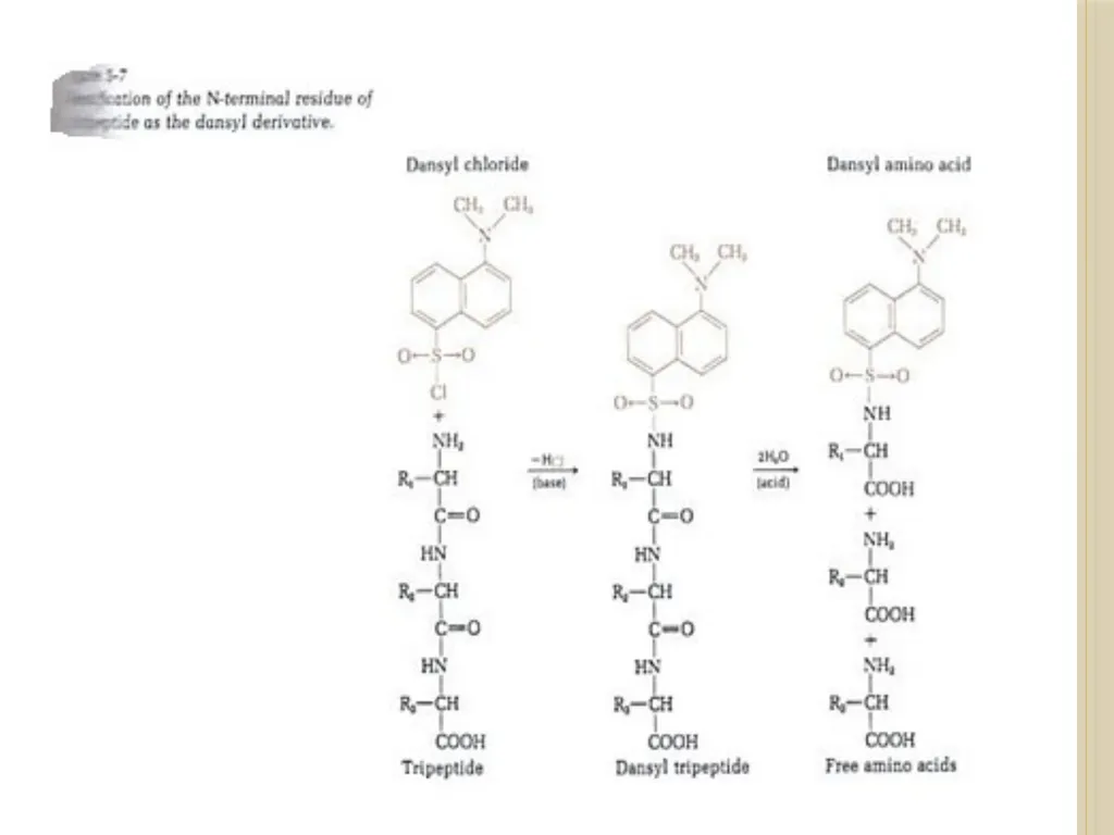
The labelled residue can easily be separated from unsubstituted amino acids and identified by chromatographic comparison with known dinitrophenyl derivatives of the different amino acids. b) By reagent 1-dimethylaminonaphthalene-5-sulfonyl chloride (abbreviated dansyl chloride).<br>
The labelled residue can easily be separated from unsubstituted amino acids and identified by chromatographic comparison with known dinitrophenyl derivatives of the different amino acids. b) By reagent 1-dimethylaminonaphthalene-5-sulfonyl chloride (abbreviated dansyl chloride).<br>
70
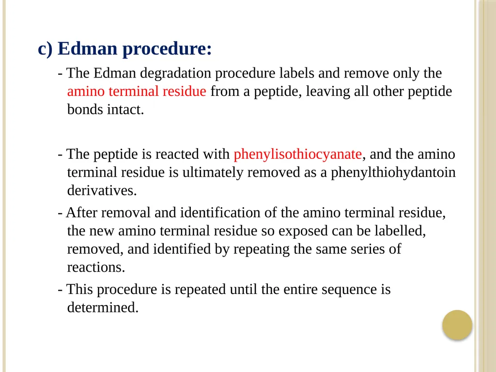
c) Edman procedure:
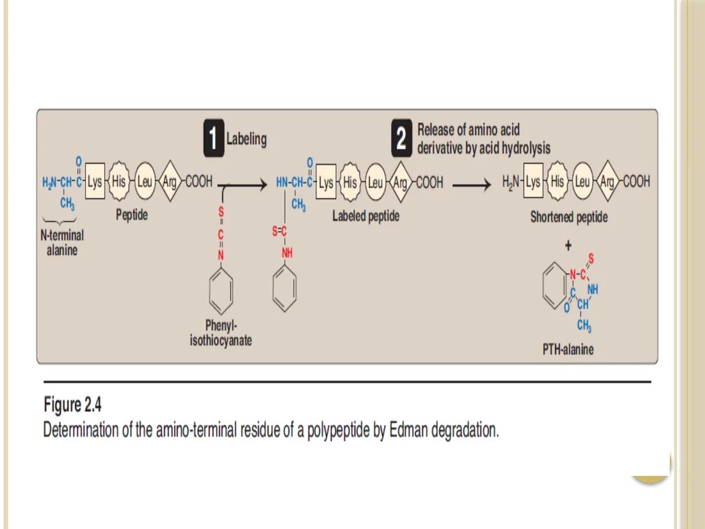
- The Edman degradation procedure labels and remove only the amino terminal residue from a peptide, leaving all other peptide bonds intact.
- The peptide is reacted with phenylisothiocyanate, and the amino terminal residue is ultimately removed as a phenylthiohydantoin derivatives.
- After removal and identification of the amino terminal residue, the new amino terminal residue so exposed can be labelled, removed, and identified by repeating the same series of reactions.
- This procedure is repeated until the entire sequence is determined.<br>
71
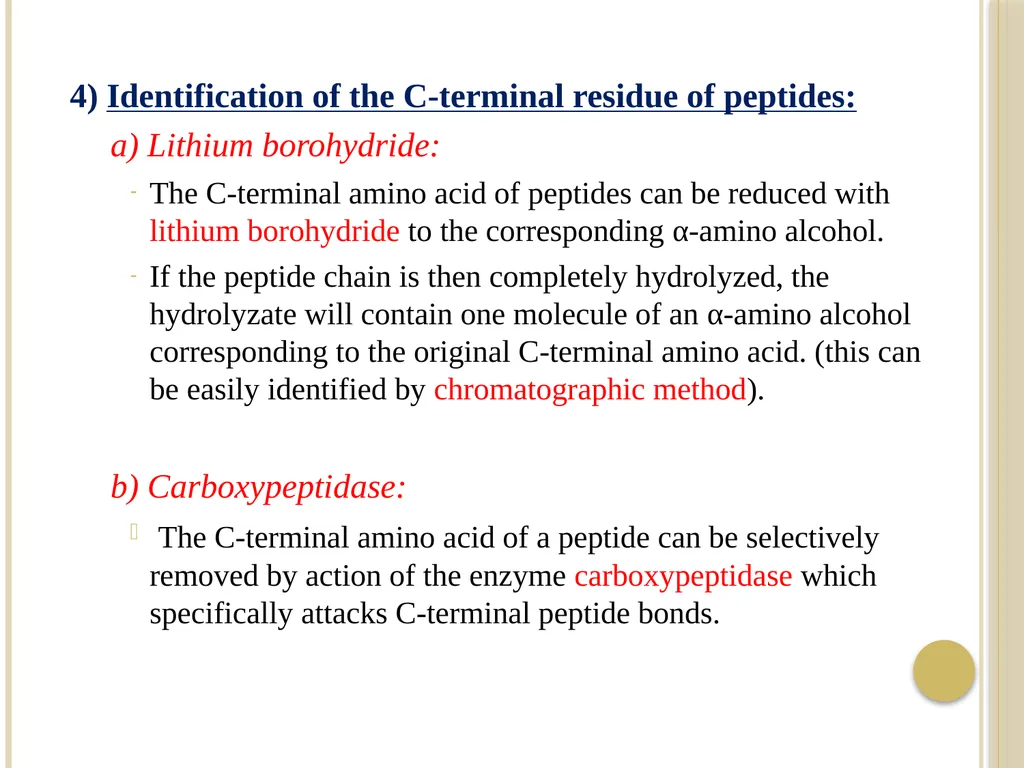
4) Identification of the C-terminal residue of peptides:
a) Lithium borohydride:
The C-terminal amino acid of peptides can be reduced with lithium borohydride to the corresponding α-amino alcohol.
If the peptide chain is then completely hydrolyzed, the hydrolyzate will contain one molecule of an α-amino alcohol corresponding to the original C-terminal amino acid. (this can be easily identified by chromatographic method).
b) Carboxypeptidase:
The C-terminal amino acid of a peptide can be selectively removed by action of the enzyme carboxypeptidase which specifically attacks C-terminal peptide bonds.<br>
72
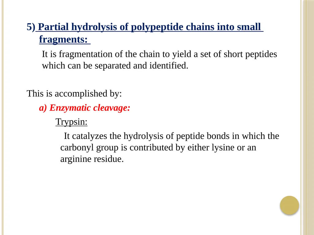
5) Partial hydrolysis of polypeptide chains into small fragments:
It is fragmentation of the chain to yield a set of short peptides which can be separated and identified.
This is accomplished by:
a) Enzymatic cleavage:
Trypsin:
It catalyzes the hydrolysis of peptide bonds in which the carbonyl group is contributed by either lysine or an arginine residue.<br>
It is fragmentation of the chain to yield a set of short peptides which can be separated and identified.
This is accomplished by:
a) Enzymatic cleavage:
Trypsin:
It catalyzes the hydrolysis of peptide bonds in which the carbonyl group is contributed by either lysine or an arginine residue.<br>
73
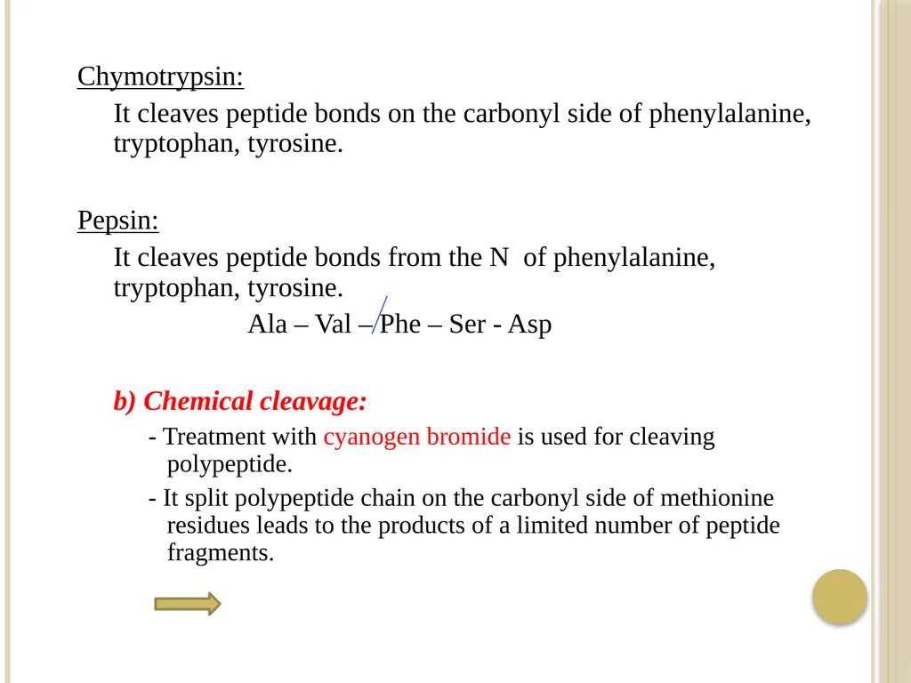
Chymotrypsin:
It cleaves peptide bonds on the carbonyl side of phenylalanine, tryptophan, tyrosine.
Pepsin:
It cleaves peptide bonds from the N of phenylalanine, tryptophan, tyrosine.
Ala – Val – Phe – Ser - Asp
b) Chemical cleavage:
- Treatment with cyanogen bromide is used for cleaving polypeptide.
- It split polypeptide chain on the carbonyl side of methionine residues leads to the products of a limited number of peptide fragments.<br>
74
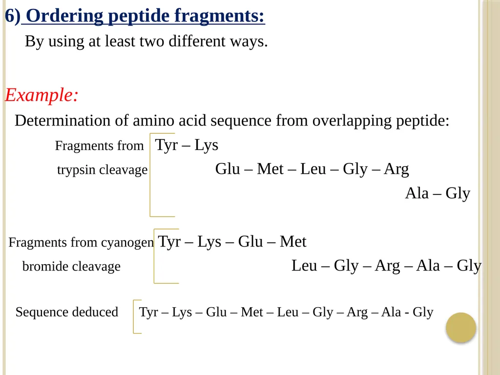
6) Ordering peptide fragments:
By using at least two different ways.
Example:
Determination of amino acid sequence from overlapping peptide:
Fragments from Tyr – Lys
trypsin cleavage Glu – Met – Leu – Gly – Arg
Ala – Gly
Fragments from cyanogen Tyr – Lys – Glu – Met
bromide cleavage Leu – Gly – Arg – Ala – Gly
Sequence deduced Tyr – Lys – Glu – Met – Leu – Gly – Arg – Ala - Gly<br>
75
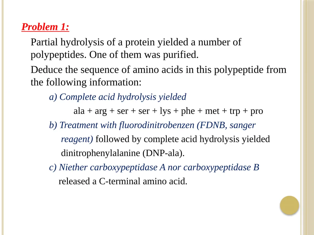
Problem 1:
Partial hydrolysis of a protein yielded a number of polypeptides. One of them was purified.
Deduce the sequence of amino acids in this polypeptide from the following information:
a) Complete acid hydrolysis yielded
ala + arg + ser + ser + lys + phe + met + trp + pro
b) Treatment with fluorodinitrobenzen (FDNB, sanger
reagent) followed by complete acid hydrolysis yielded
dinitrophenylalanine (DNP-ala).
c) Niether carboxypeptidase A nor carboxypeptidase B
released a C-terminal amino acid.<br>
76
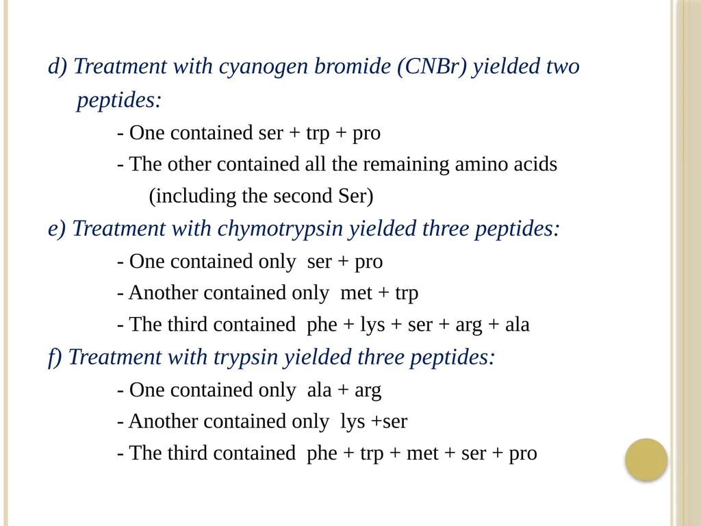
d) Treatment with cyanogen bromide (CNBr) yielded two
peptides:
- One contained ser + trp + pro
- The other contained all the remaining amino acids
(including the second Ser)
e) Treatment with chymotrypsin yielded three peptides:
- One contained only ser + pro
- Another contained only met + trp
- The third contained phe + lys + ser + arg + ala
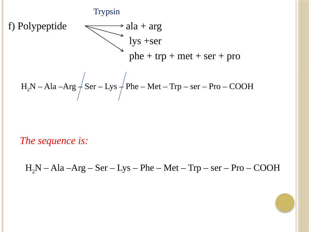
f) Treatment with trypsin yielded three peptides:
- One contained only ala + arg
- Another contained only lys +ser
- The third contained phe + trp + met + ser + pro<br>
77
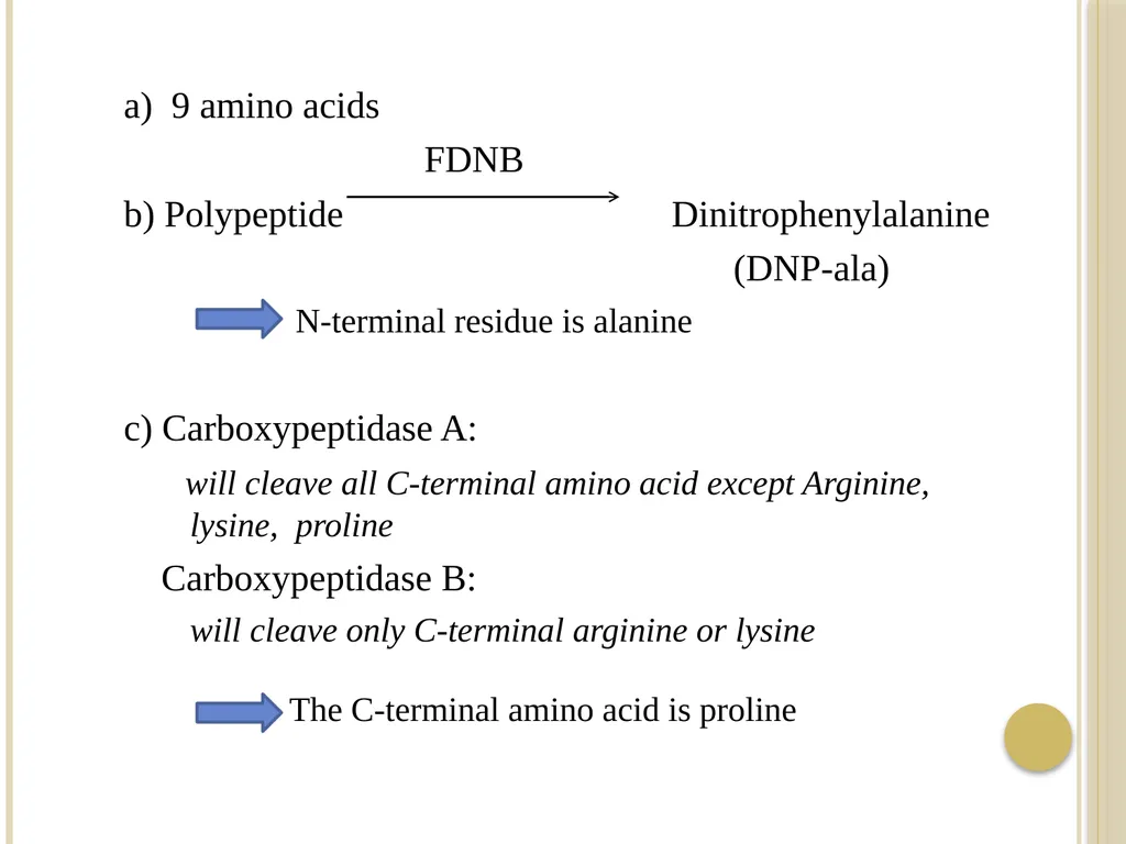
a) 9 amino acids
FDNB
b) Polypeptide Dinitrophenylalanine
(DNP-ala)
N-terminal residue is alanine
c) Carboxypeptidase A:
will cleave all C-terminal amino acid except Arginine, lysine, proline
Carboxypeptidase B:
will cleave only C-terminal arginine or lysine
The C-terminal amino acid is proline<br>
FDNB
b) Polypeptide Dinitrophenylalanine
(DNP-ala)
N-terminal residue is alanine
c) Carboxypeptidase A:
will cleave all C-terminal amino acid except Arginine, lysine, proline
Carboxypeptidase B:
will cleave only C-terminal arginine or lysine
The C-terminal amino acid is proline<br>
78
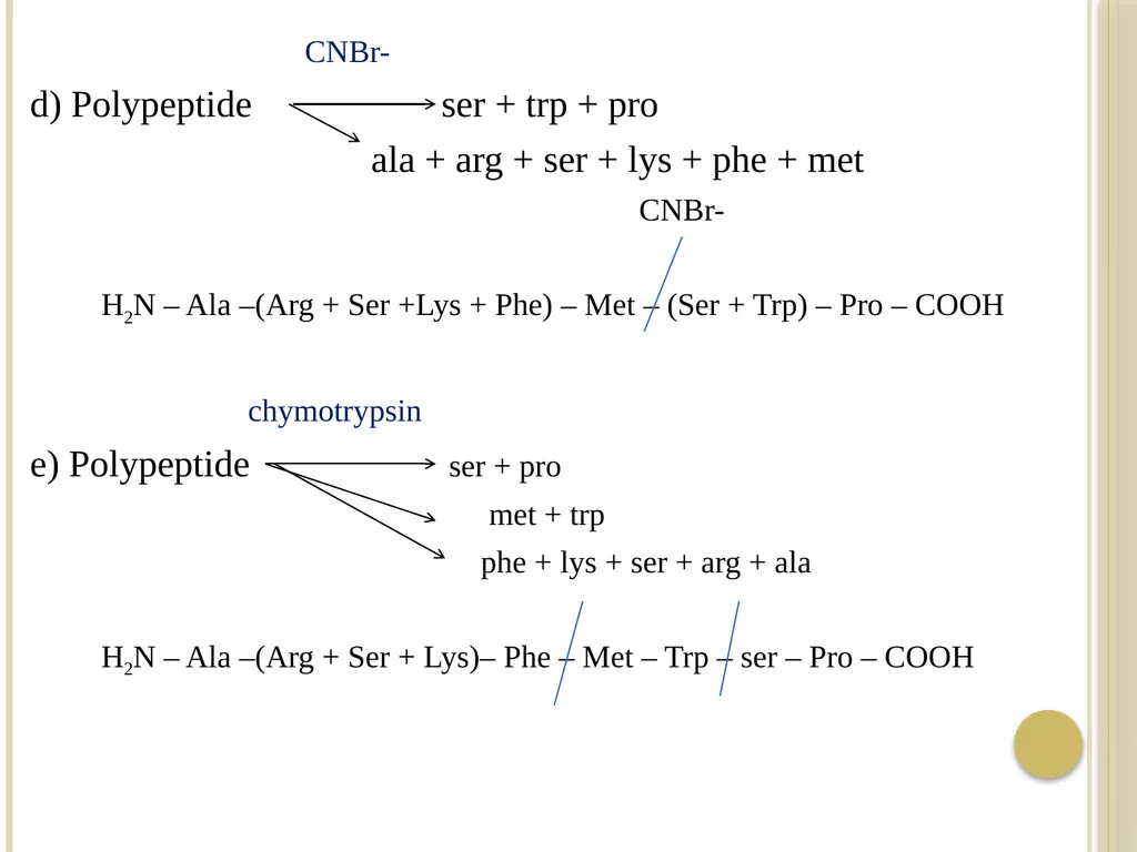
CNBr-
d) Polypeptide ser + trp + pro
ala + arg + ser + lys + phe + met
CNBr-
H2N – Ala –(Arg + Ser +Lys + Phe) – Met – (Ser + Trp) – Pro – COOH
chymotrypsin
e) Polypeptide ser + pro
met + trp
phe + lys + ser + arg + ala
H2N – Ala –(Arg + Ser + Lys)– Phe – Met – Trp – ser – Pro – COOH<br>
d) Polypeptide ser + trp + pro
ala + arg + ser + lys + phe + met
CNBr-
H2N – Ala –(Arg + Ser +Lys + Phe) – Met – (Ser + Trp) – Pro – COOH
chymotrypsin
e) Polypeptide ser + pro
met + trp
phe + lys + ser + arg + ala
H2N – Ala –(Arg + Ser + Lys)– Phe – Met – Trp – ser – Pro – COOH<br>
79
Trypsin
f) Polypeptide ala + arg
lys +ser
phe + trp + met + ser + pro
H2N – Ala –Arg – Ser – Lys – Phe – Met – Trp – ser – Pro – COOH
The sequence is:
H2N – Ala –Arg – Ser – Lys – Phe – Met – Trp – ser – Pro – COOH<br>
f) Polypeptide ala + arg
lys +ser
phe + trp + met + ser + pro
H2N – Ala –Arg – Ser – Lys – Phe – Met – Trp – ser – Pro – COOH
The sequence is:
H2N – Ala –Arg – Ser – Lys – Phe – Met – Trp – ser – Pro – COOH<br>
80
Problem 2:
- Reaction of a tetrapeptide with 2,4 dinitrofluorobenzen, followed by hydrolysis in 6 N HCl , yielded the 2,4- dinitrophenyl derivative of valine and three other amino acids.
- Hydrolysis of another sample of the tetrapeptide with trypsin
give two fragments: one was reduced with LiBH4 and then
hydrolyzed. In the hydrolyzate, the amino alcohol corresponding to glycine was detected, together with an amino acid forming a yellow reaction product with ninhydrin.
- Give a possible amino acid sequence for the tetrapeptide.<br>
81
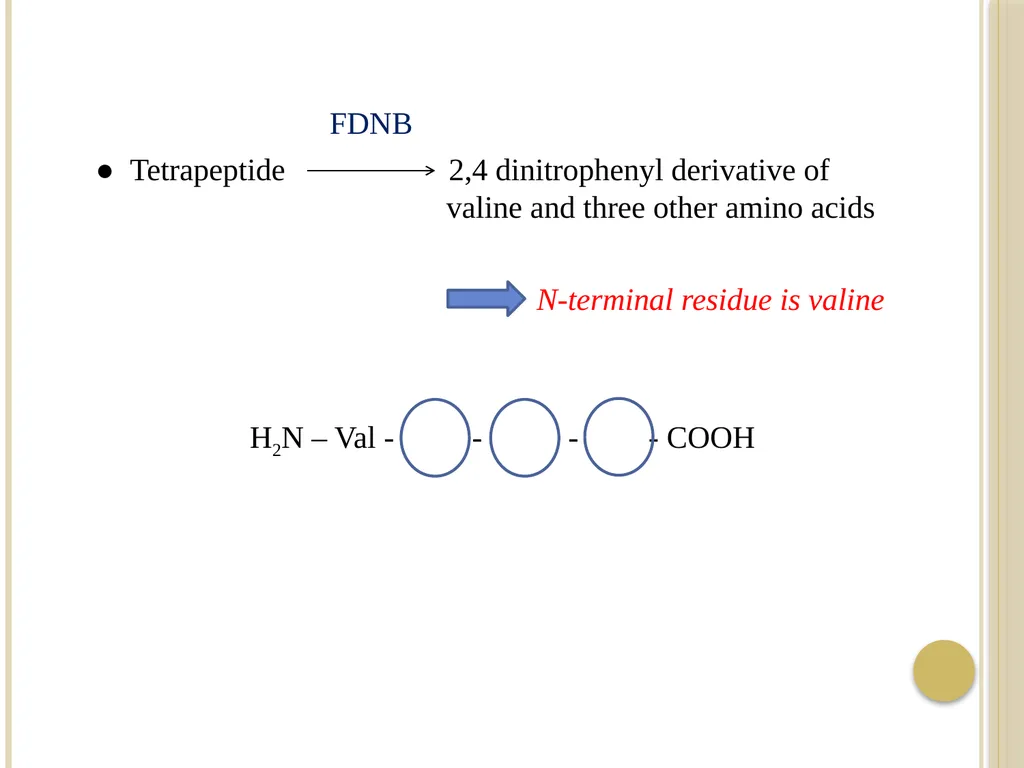
FDNB
● Tetrapeptide 2,4 dinitrophenyl derivative of valine and three other amino acids
N-terminal residue is valine
H2N – Val - - - - COOH<br>
● Tetrapeptide 2,4 dinitrophenyl derivative of valine and three other amino acids
N-terminal residue is valine
H2N – Val - - - - COOH<br>
82
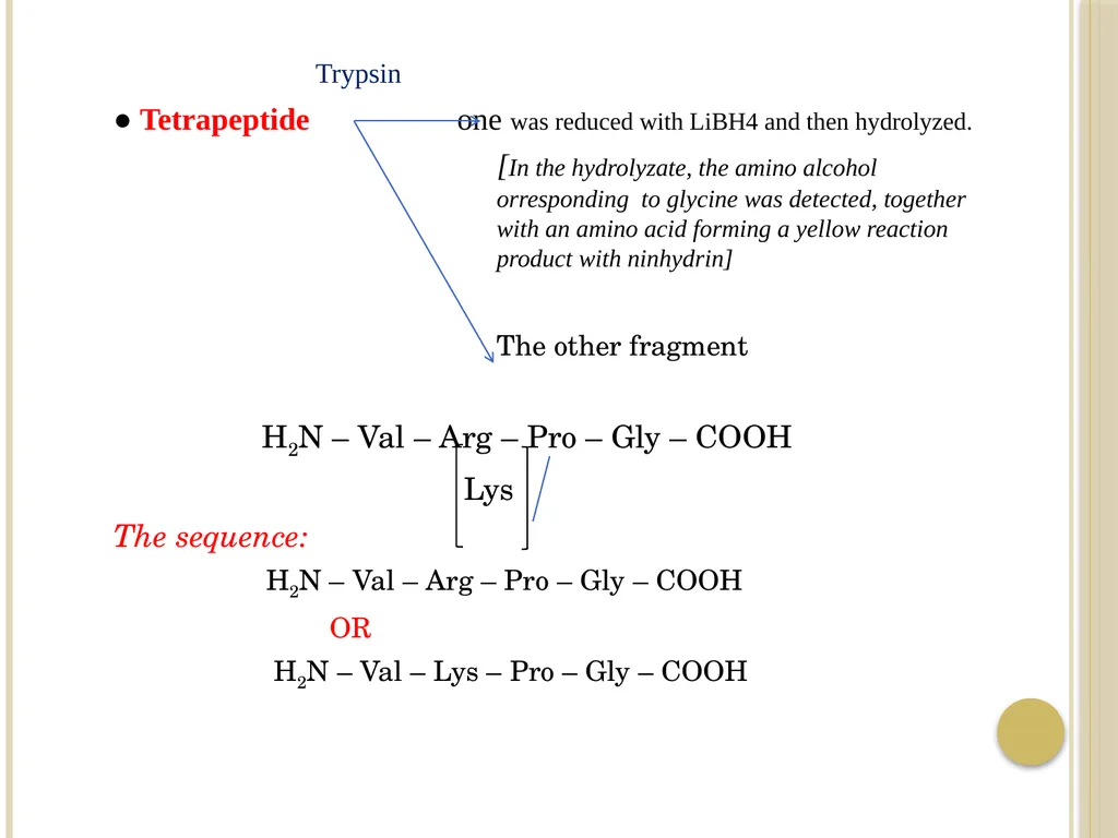
Trypsin
● Tetrapeptide one was reduced with LiBH4 and then hydrolyzed.
[In the hydrolyzate, the amino alcohol orresponding to glycine was detected, together with an amino acid forming a yellow reaction product with ninhydrin]
The other fragment
H2N – Val – Arg – Pro – Gly – COOH
Lys
The sequence:
H2N – Val – Arg – Pro – Gly – COOH
OR
H2N – Val – Lys – Pro – Gly – COOH<br>
83
References Lehninger, A.L., Nelson,D.L., Cox,M.M, Principles of Biochemistry,Worth publishers,Inc.,NewYork .<br>