
CHAPTER 34: Focused Lecture Environmental



Description: CHAPTER 34: Focused Lecture Environmental Emergencies National EMS Education Standard Competencies (1 of 4) Trauma Applies fundamental knowledge to provide basic and selected advanced emergency care and transportation based on assessment
Related Topics
Download Presentation
"CHAPTER 34: Focused Lecture Environmental" is the property of its rightful owner. Permission is granted to download and print the materials on this website for personal, non-commercial use only, and to display it on your personal computer provided you do not modify the materials and that you retain all copyright notices contained in the materials. By downloading content from our website, you accept the terms of this agreement.
Presentation Transcript
slide1. CHAPTER 34: Focused Lecture Environmental Emergencies<br>
slide2. National EMS Education Standard Competencies (1 of 4) Trauma
Applies fundamental knowledge to provide basic and selected advanced emergency care and transportation based on assessment findings for an acutely injured patient.<br>
slide3. National EMS Education Standard Competencies (2 of 4) Environmental Emergencies
Recognition and management of:
Submersion incidents
Temperature-related illness<br>
slide4. National EMS Education Standard Competencies (3 of 4) Pathophysiology, assessment, and management of:
Near drowning
Temperature-related illness
Bites and envenomations<br>
slide5. National EMS Education Standard Competencies (4 of 4) Pathophysiology, assessment, and management of:
Dysbarism
High altitude
Diving injuries
Electrical injury
Radiation exposure<br>
slide6. Introduction Medical condition caused or exacerbated by weather, terrain, or atmospheric pressure
Medical emergencies can result, particularly in:
Children
Older people
People with chronic illnesses
Young adults who overexert themselves<br>
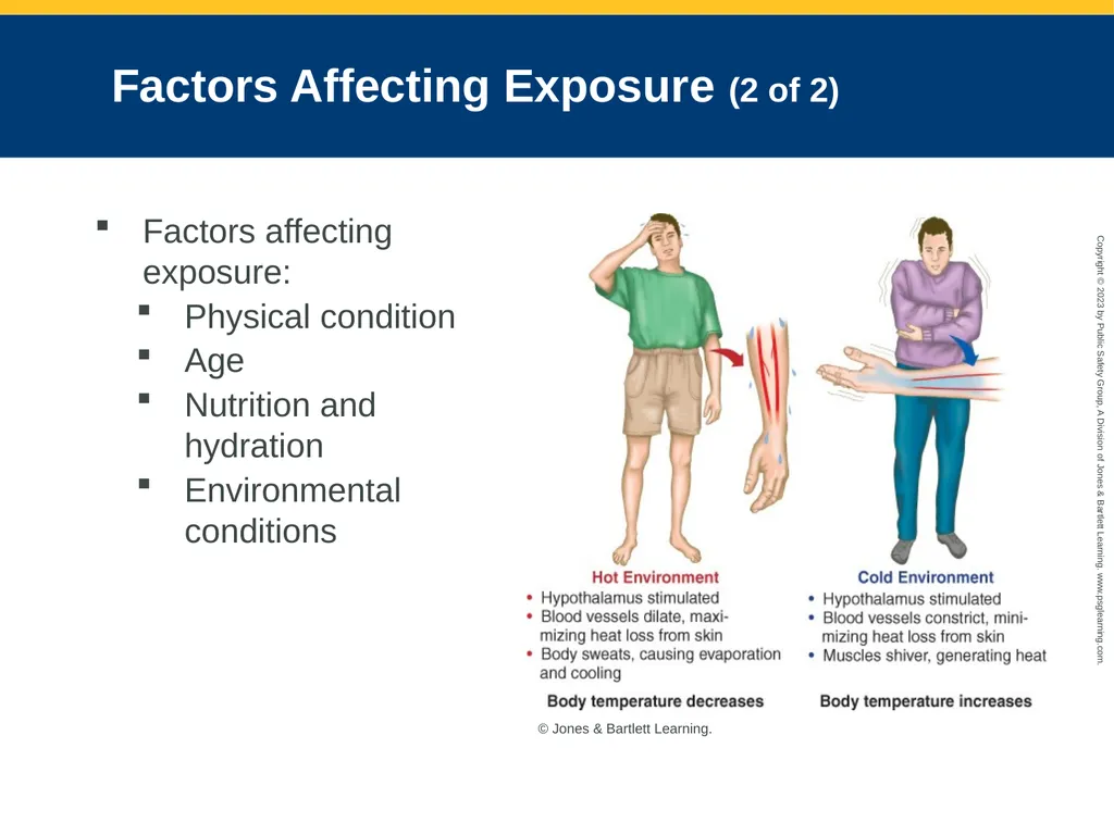
slide7. Factors Affecting Exposure (1 of 2) Thermoregulation: Balance between heat production and heat excretion
Rise in core body temperature elicits responses that increase heat loss and shut off normal heat production pathways (thermogenesis).
Decrease in core body temperature prompts heat production and conservation and turns off normal heat-liberating pathways (thermolysis).<br>
slide8. Factors Affecting Exposure (2 of 2) Factors affecting exposure:
Physical condition
Age
Nutrition and hydration
Environmental conditions © Jones & Bartlett Learning.<br>
slide9. Cold Exposure (1 of 9) Normal body temperature must be maintained within a narrow range for the body’s chemistry to work efficiently.
Hypothermia: Core body temperature falls because of inadequate internal heat production, excess cold stress, or a combination of both<br>
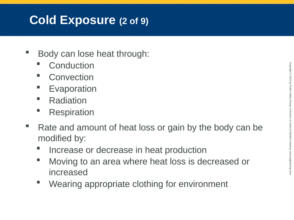
slide10. Cold Exposure (2 of 9) Body can lose heat through:
Conduction
Convection
Evaporation
Radiation
Respiration
Rate and amount of heat loss or gain by the body can be modified by:
Increase or decrease in heat production
Moving to an area where heat loss is decreased or increased
Wearing appropriate clothing for environment<br>
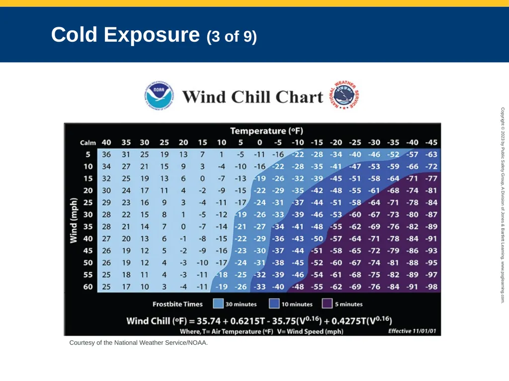
slide11. Cold Exposure (3 of 9) Courtesy of the National Weather Service/NOAA.<br>
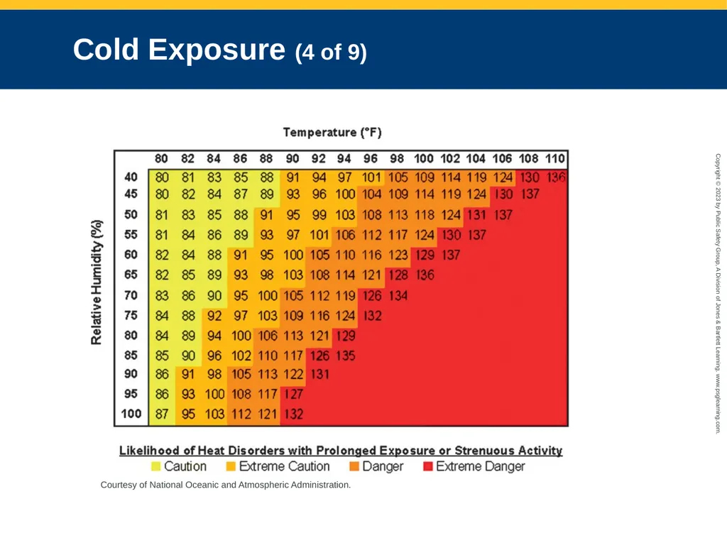
slide12. Cold Exposure (4 of 9) Courtesy of National Oceanic and Atmospheric Administration.<br>
slide13. Cold Exposure (5 of 9) Pathophysiology
Hypothermia
Occurs when core body temperature falls below 95°F
Body cannot regulate body temperature and generate body heat.
Can develop quickly or gradually
Impaired thermoregulation can occur with therapeutic use or overdoses of sedative medications, tricyclic antidepressants, and phenothiazines.<br>
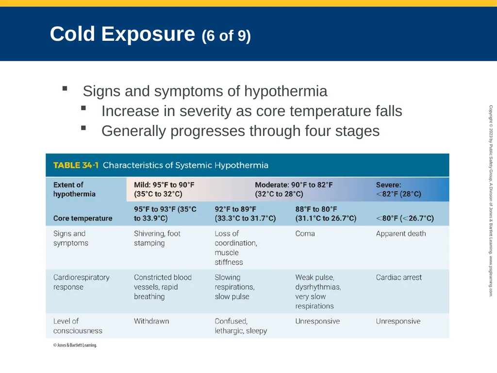
slide14. Cold Exposure (6 of 9) Signs and symptoms of hypothermia
Increase in severity as core temperature falls
Generally progresses through four stages<br>
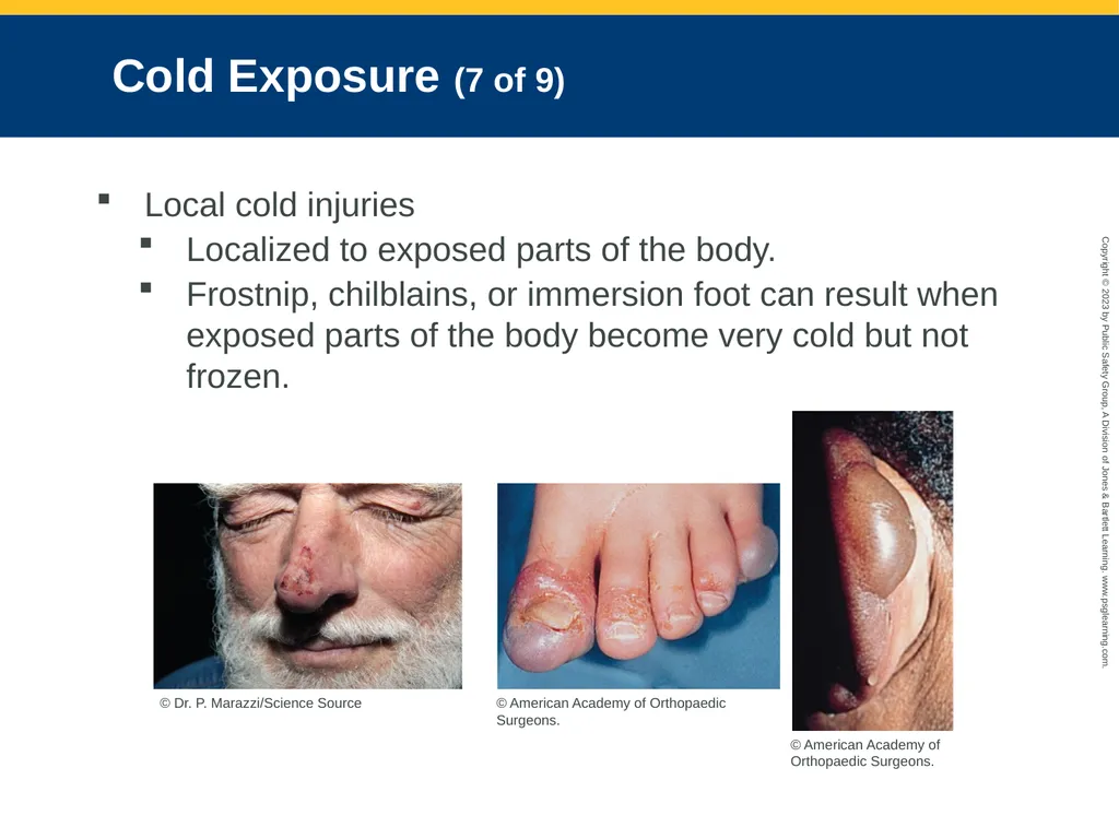
slide15. Cold Exposure (7 of 9) Local cold injuries
Localized to exposed parts of the body.
Frostnip, chilblains, or immersion foot can result when exposed parts of the body become very cold but not frozen. © Dr. P. Marazzi/Science Source © American Academy of Orthopaedic Surgeons. © American Academy of Orthopaedic Surgeons.<br>
slide16. Cold Exposure (8 of 9) Frostnip and immersion foot
Frostnip affects the ears, nose, and fingers and is not painful.
Immersion foot occurs after prolonged exposure to cold water.<br>
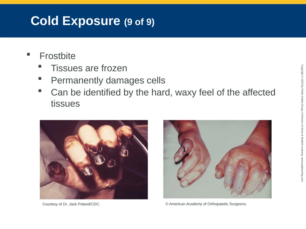
slide17. Cold Exposure (9 of 9) Frostbite
Tissues are frozen
Permanently damages cells
Can be identified by the hard, waxy feel of the affected tissues Courtesy of Dr. Jack Poland/CDC. © American Academy of Orthopaedic Surgeons.<br>
slide18. Assessment of Cold Injuries (1 of 9) Management: Stabilizing ABCs and preventing further heat loss
All patients who are injured are at risk for hypothermia.
Scene size-up
Important aspects: Air temperature, wind chill, whether it is wet or dry<br>
slide19. Assessment of Cold Injuries (2 of 9) Ensure that the scene is safe
Identify potential safety hazards, such as wet grass, mud, snow, or icy streets.
Consider special hazards.
Use appropriate standard precautions.
Consider the number of patients.
Summon additional help as quickly as possible.
Look for indicators of MOI.<br>
slide20. Assessment of Cold Injuries (3 of 9) Primary survey
Form a general impression.
Perform a rapid full-body scan to determine whether a life threat exists.
If chief complaint is simply being cold, quickly assess core temperature.
Evaluate mental status using AVPU.
Take into account physiologic changes that occur as a result of hypothermia.<br>
slide21. Assessment of Cold Injuries (4 of 9) Airway and breathing
If you believe the patient is in cardiac arrest, provide high-quality chest compressions.
If you cannot feel a radial pulse, gently palpate for a carotid pulse, and wait for up to 60 seconds before you decide that the patient is pulseless.
Assume shock is present and treat it.
Ensure adequate airway and breathing.
All patients with hypothermia require immediate transport for evaluation and treatment.<br>
slide22. Assessment of Cold Injuries (5 of 9) History taking
Investigate the chief complaint.
Obtain a medical history.
Be alert for injury-specific signs, symptoms, and pertinent negatives.
If possible, determine the duration and temperature of the exposure, and wind velocity.<br>
slide23. Assessment of Cold Injuries (6 of 9) SAMPLE history
Last oral intake and activity prior to the exposure will help to determine severity of the cold injury.
Investigate underlying factors:
Exposure to wet conditions
Inadequate insulation from cold or wind
Restricted circulation
Inadequate nutrition, alcohol or drug abuse
Diabetes, cardiovascular disease<br>
slide24. Assessment of Cold Injuries (7 of 9) Secondary assessment
Focus your physical examination on the severity of hypothermia.
Determine the degree and extent of cold injury.
Vital signs may be altered by the effects of hypothermia.
Respirations may be slow and shallow.<br>
slide25. Assessment of Cold Injuries (8 of 9) Low blood pressure and a slow pulse indicate moderate to severe hypothermia.
Determine core body temperature using a hypothermia thermometer based on local protocol.
Pulse oximetry will often be inaccurate because of the lack of perfusion in the extremities.<br>
slide26. Assessment of Cold Injuries (9 of 9) Reassessment
Repeat the primary survey.
Reassess vital signs and chief complaint.
Review all treatments.
Monitor level of consciousness and vital signs.
Communicate all information gathered to the receiving facility.<br>
slide27. Emergency Medical Care for Cold Emergencies (1 of 10) General steps to prevent further cold injury:
Remove wet clothing, and keep the patient dry.
Prevent conduction heat loss.
Insulate all exposed body parts.
Prevent convection heat loss.
Remove the patient from the cold environment.<br>
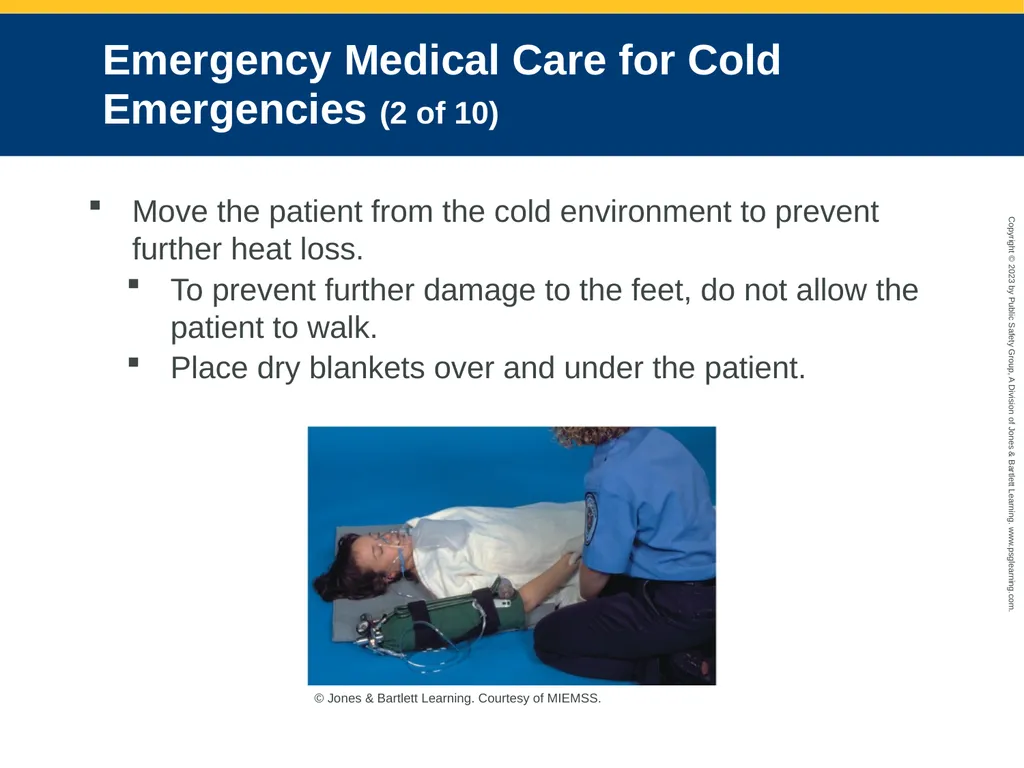
slide28. Emergency Medical Care for Cold Emergencies (2 of 10) Move the patient from the cold environment to prevent further heat loss.
To prevent further damage to the feet, do not allow the patient to walk.
Place dry blankets over and under the patient. © Jones & Bartlett Learning. Courtesy of MIEMSS.<br>
slide29. Emergency Medical Care for Cold Emergencies (3 of 10) If available, give the patient warm, humidified oxygen.
Obtain IV access, but follow local protocols.
When possible, administer fluid that has been warmed.<br>
slide30. Emergency Medical Care for Cold Emergencies (4 of 10) Handle the patient gently to avoid pain or further injury to the skin.
Mild hypothermia
Signs and symptoms: alertness, shivering, core body temperature between 90°F and 95°F
Treatment: Begin passive rewarming slowly, turn the heat up high in the patient compartment, give warm fluids<br>
slide31. Emergency Medical Care for Cold Emergencies (5 of 10) Moderate or severe hypothermia
Active rewarming is best accomplished in the ED.
Rewarming extremities too rapidly without rewarming the core can cause vasodilation.
Move the patient out of the wind and away from contact with any object that will conduct heat away from the body.<br>
slide32. Emergency Medical Care for Cold Emergencies (6 of 10) Withholding and cessation of resuscitative efforts
If submersion preceded the arrest, successful resuscitation is unlikely.
Trauma, alcohol overdose, and drug overdose could have resulted in hypothermia.
Fixed, dilated pupils, apparent rigor mortis, and dependent lividity are not contraindications for resuscitation of severely hypothermic patients.<br>
slide33. Emergency Medical Care for Cold Emergencies (7 of 10) Emergency treatment of local cold injuries
Remove the patient from further exposure to the cold.
Handle the injured part gently, and protect it from further injury.
Administer oxygen, if not already done.
Remove any wet or restrictive clothing over the injured part.<br>
slide34. Emergency Medical Care for Cold Emergencies (8 of 10) If there is no chance of reinjury or if transport to the ED will be delayed, consider active rewarming.
With frostnip, contact with a warm object may be all that is needed.
With immersion foot, remove wet shoes, boots, and socks.
Rewarm the foot gradually
Splint the extremity, and cover it loosely with a dry, sterile dressing.
Never rub injured tissues.
Do not re-expose injury to cold.<br>
slide35. Emergency Medical Care for Cold Emergencies (9 of 10) With a late or deep cold injury, remove any jewelry from the injured part.
Cover injury loosely with a dry, sterile dressing.
Do not apply heat or rewarm the part.
Do not allow the patient to stand or walk on a frostbitten foot.
Evaluate for signs or symptoms of systemic hypothermia.
Support vital functions and transport promptly.<br>
slide36. Emergency Medical Care for Cold Emergencies (10 of 10) Rewarming in the field
Use a warm-water bath.
Immerse the frostbitten part in water with a temperature of between 102°F and 104°F.
Dress the area with dry, sterile dressings.
Expect the patient to report severe pain.
Cover frostbitten part with soft, padded, sterile cotton dressings.
If blisters have formed, do not break them.<br>
slide37. Cold Exposure and You You are also at risk for hypothermia if you work in a cold environment.
Be aware of existing and potential weather conditions.
Wear proper clothing whenever appropriate.
Properly maintain and equip your vehicle.<br>
slide38. Heat Exposure (1 of 8) Normal body temperature is 98.6°F (37°C).
Thermolysis: getting rid of excess heat
Sweating (and evaporation of the sweat)
Dilation of peripheral blood vessels
Hyperthermia is a high core temperature, usually 101°F or higher.<br>
slide39. Heat Exposure (2 of 8) Pathophysiology
When mechanisms to decrease body heat are overwhelmed, illness develops.
High air temperature
High humidity
Vigorous exercise
Heat exposure can cause heat cramps, heat exhaustion, and heatstroke.<br>
slide40. Heat Exposure (3 of 8) Persons at greatest risk for heat illnesses:
Children
Geriatric patients
Patients with heart disease, COPD, diabetes, dehydration, and obesity
Patients with limited mobility
Contributing factors:
Length of exposure
Intensity of exposure
Enviroment<br>
slide41. Heat Exposure (4 of 8) Preventive measures:
Maintain an adequate fluid intake.
Acclimatize to the environment.
Limit exposure.
Drink water continually.<br>
slide42. Heat Exposure (5 of 8) Heat cramps
Painful muscle spasms that occur after vigorous exercise
Sweat causes a change in electrolyte balance.
Dehydration may also have a role in development.<br>
slide43. Heat Exposure (6 of 8) Heat exhaustion
Most common illness caused by heat
Result of the body losing too much water and electrolytes through very heavy sweating
Signs and symptoms
Cold, clammy skin with ashen pallor
Dry tongue and thirst
Dizziness, weakness, or faintness
Normal vital signs
Normal or slightly elevated body temperature<br>
slide44. Heat Exposure (7 of 8) Heatstroke
Least common but most serious illness caused by heat exposure
Body is subjected to more heat than it can effectively remove.
Hot, dry, flushed skin
First sign is a change in behavior.
A patient can have a heatstroke even if still sweating.<br>
slide45. Heat Exposure (8 of 8) Classic heatstroke commonly presents in people with chronic illnesses.
Exertional heatstroke commonly presents in people who are in good general health but have an increased core body temperature because of overwhelming heat stress.
Recovery depends on the speed with which treatment is administered.<br>
slide46. Assessment of Heat Injuries (1 of 5) Scene size-up
Perform an environmental assessment.
Use appropriate standard precautions.
Look for indicators of MOI.
Heat emergencies commonly occur during athletic events and practices.
Overcooling can result in shivering, which generates more heat.
Consider calling medical control for paramedic backup.<br>
slide47. Assessment of Heat Injuries (2 of 5) Primary survey
Observe how the patient interacts with you and the environment.
Perform a rapid full-body scan.
Assess mental status using AVPU.
Assess ABCs.
Treat aggressively for shock.
For signs of heatstroke, transport without delay.<br>
slide48. Assessment of Heat Injuries (3 of 5) History taking
Investigate the chief complaint.
Obtain medical history.
Be alert for specific signs and symptoms.
Obtain a SAMPLE history.
Be thorough in your questioning.
Determine patient’s exposure to heat and humidity and activities prior to the onset of symptoms.<br>
slide49. Assessment of Heat Injuries (4 of 5) Secondary assessment
If the patient is alert, perform a focused assessment.
Assess for muscle cramps or confusion.
Examine patient’s mental status, and obtain the patient’s vital signs.
Pay special attention to the skin.
Perform a neurologic examination.
Patients with hyperthermia will be tachycardic and tachypneic.
Check patient’s temperature.<br>
slide50. Assessment of Heat Injuries (5 of 5) Reassessment
Watch your patient’s condition carefully for deterioration.
Evaluate effectiveness of your interventions.
Be careful not to cause shivering when cooling down a patient.
Document the weather conditions and the activities the patient was performing prior to the emergency.<br>
slide51. Emergency Medical Care for Heat Emergencies (1 of 4) Heat cramps
Treatment:
Remove patient from hot environment.
Administer high-flow oxygen if indicated.
Rest the cramping muscles.
Replace fluids by mouth.
If the cramps do not go away, initiate an IV line and transport the patient to the hospital.<br>
slide52. Emergency Medical Care for Heat Emergencies (2 of 4) Heat exhaustion
Remove the patient from exposure to heat and urgently reversing the increase in body temperature.
Provide cold-water immersion to the patient if allowed.<br>
slide53. Emergency Medical Care for Heat Emergencies (3 of 4) Prepare to transport the patient if:
Symptoms do not clear up promptly.
Level of consciousness decreases.
Body temperature remains elevated.
Person is very young, older, or has any underlying medical condition.<br>
slide54. Emergency Medical Care for Heat Emergencies (4 of 4) Heatstroke
When treating a patient with heatstroke:
Move patient out of the hot environment.
Remove the patient’s clothing.
Cover the patient with wet towels or sheet.
Administer oxygen if indicated.
Obtain IV access and administer 20-mL/kg bolus of isotonic crystalloid solution.
Transport immediately.<br>
slide55. Water Emergencies (1 of 12) Drowning
Respiratory impairment from submersion or immersion in liquid
Patients with submersion injury can die of secondary complications that occur beyond 24 hours.
Alcohol consumption, preexisting seizure disorders, geriatric patients with cardiovascular disease, and unsupervised access to water are among the major risk factors.<br>
slide56. Water Emergencies (2 of 12) Inhaling very small amounts of fresh or salt water can severely irritate the larynx resulting to laryngospasm.
Hypothermia is a major concern.
The body’s diving reflex may slow metabolism to the point of protecting vital organs.
Hypoxia is always the first concern.<br>
slide57. Water Emergencies (3 of 12) Diving emergencies
Most serious water-related injuries are associated with dives.
Physical principles of pressure effects
Most SCUBA diving is done at depths between 30 and 60 fsw (2 to 3 ATA).
Liquids such as water are not compressible.<br>
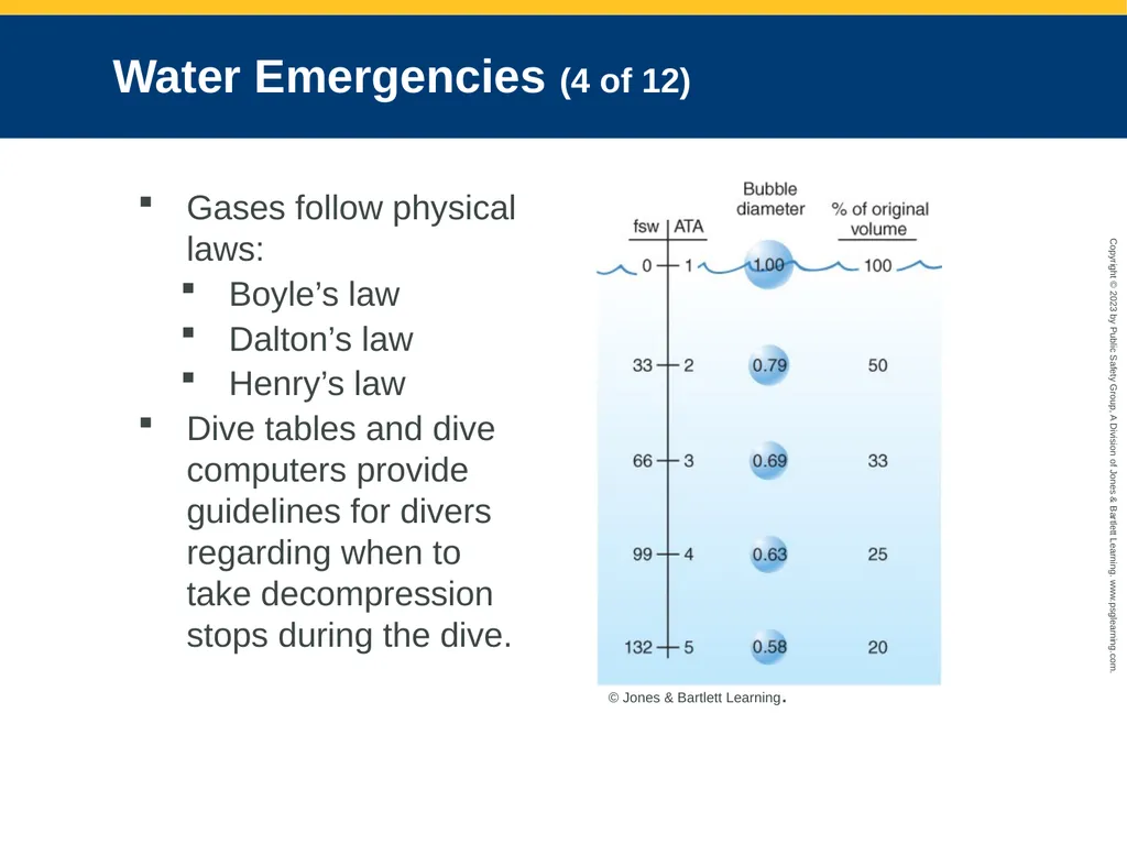
slide58. Water Emergencies (4 of 12) Gases follow physical laws:
Boyle’s law
Dalton’s law
Henry’s law
Dive tables and dive computers provide guidelines for divers regarding when to take decompression stops during the dive. © Jones & Bartlett Learning.<br>
slide59. Water Emergencies (5 of 12) General assessment: Diving history
Obtain as many details as you can.
Record:
When did symptoms start?
What type of diving was done, and what type of equipment was used?
Where is the diving site, and what was the water temperature?
Were safety stops used?
Were there any dive complications?<br>
slide60. Water Emergencies (6 of 12) Descent emergencies
Major problem during descent is barotrauma.
Can result from compression of gases within body spaces during descent or expansion of gases within those spaces during ascent
A person with a perforated tympanic membrane may develop a special problem while diving.
Nitrogen narcosis: A state of altered mental status caused by breathing compressed nitrogen-containing air at depth<br>
slide61. Water Emergencies (7 of 12) Ascent emergencies
Usually require aggressive resuscitation
Pulmonary overpressurization syndrome (POPS)
Can occur when divers fail to exhale during an ascent
Can cause pneumothorax, mediastinal and subcutaneous emphysema, alveolar hemorrhage, and lethal air embolism<br>
slide62. Water Emergencies (8 of 12) POPS (cont’d)
Physical examination may reveal palpable subcutaneous air above the clavicles.
Always look for unequal breath sounds, low pulse oximetry values, and hyperresonance on the affected side of the chest.
Pneumothorax may require needle decompression or a chest tube.
Provide 100% oxygen by nonrebreathing mask.<br>
slide63. Water Emergencies (9 of 12) Air embolism
Bubbles of air in blood vessels
Air inside the lungs expands rapidly.
Air released from this rupture can enter:
Pleural space
Mediastinum
Bloodstream
Pneumothorax and pneumomediastinum both result in pain and severe dyspnea.<br>
slide64. Water Emergencies (10 of 12) Potential signs and symptoms of air embolism:
Blotching
Froth at the nose and mouth
Severe pain in muscles, joints, or abdomen
Dyspnea, localized pleuritic chest pain
Dizziness, nausea, and vomiting
Dysphasia
Cough
Cyanosis
Difficulty with vision
Paralysis and/or coma
Irregular pulse or cardiac arrest<br>
slide65. Water Emergencies (11 of 12) Decompression sickness
Bubbles of gas obstruct blood vessels.
Results from too rapid an ascent from a dive, too long of a dive at too deep a depth, or repeated dives within a short period
Most striking symptom is abdominal/joint pain.<br>
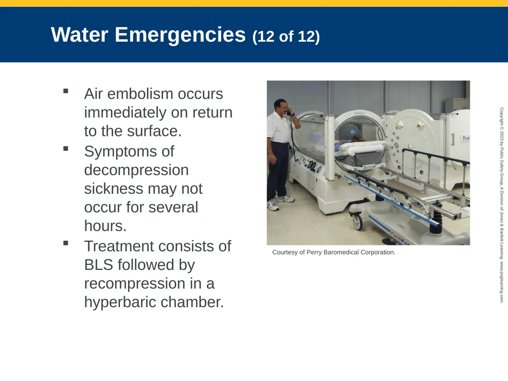
slide66. Water Emergencies (12 of 12) Air embolism occurs immediately on return to the surface.
Symptoms of decompression sickness may not occur for several hours.
Treatment consists of BLS followed by recompression in a hyperbaric chamber. Courtesy of Perry Baromedical Corporation.<br>
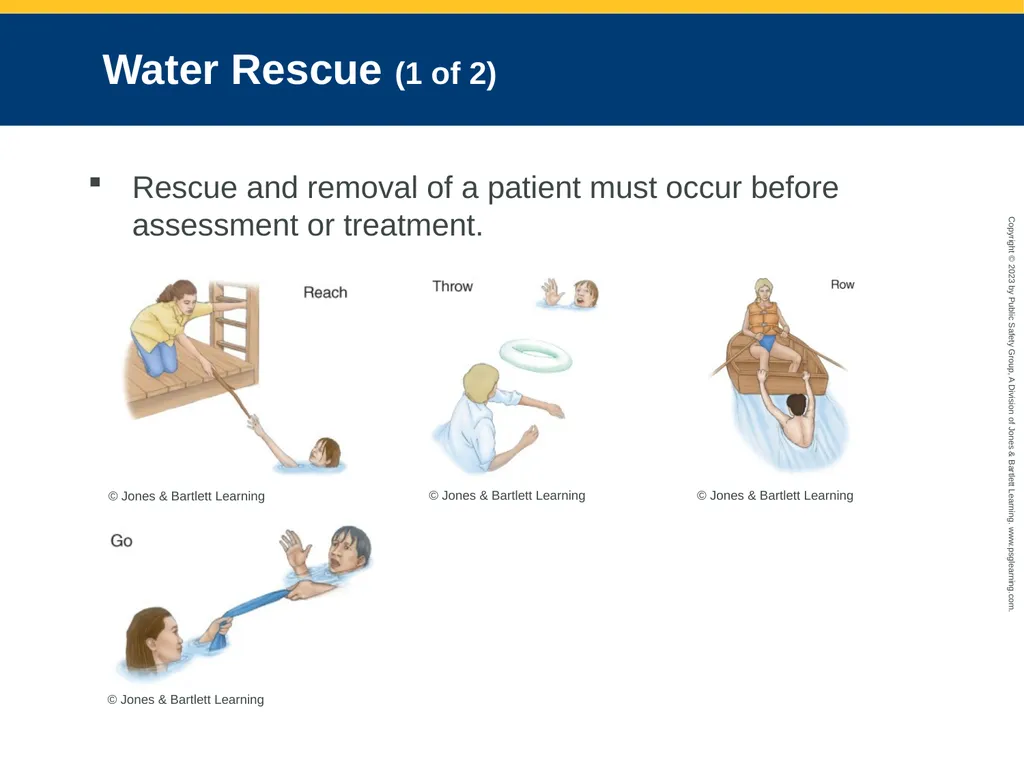
slide67. Water Rescue (1 of 2) Rescue and removal of a patient must occur before assessment or treatment. © Jones & Bartlett Learning © Jones & Bartlett Learning © Jones & Bartlett Learning © Jones & Bartlett Learning<br>
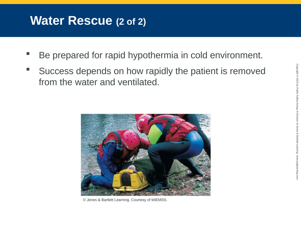
slide68. Water Rescue (2 of 2) Be prepared for rapid hypothermia in cold environment.
Success depends on how rapidly the patient is removed from the water and ventilated. © Jones & Bartlett Learning. Courtesy of MIEMSS.<br>
slide69. Spinal Injuries in Submersion Incidents Submersion incidents may be complicated by spinal fractures and spinal cord injuries.
Assume spinal injury exists when:
Submersion has resulted from a diving mishap or fall from a substantial height.
Patient is unresponsive.
Patient is responsive but reports weakness, paralysis, numbness, or tingling in the arms or legs.<br>
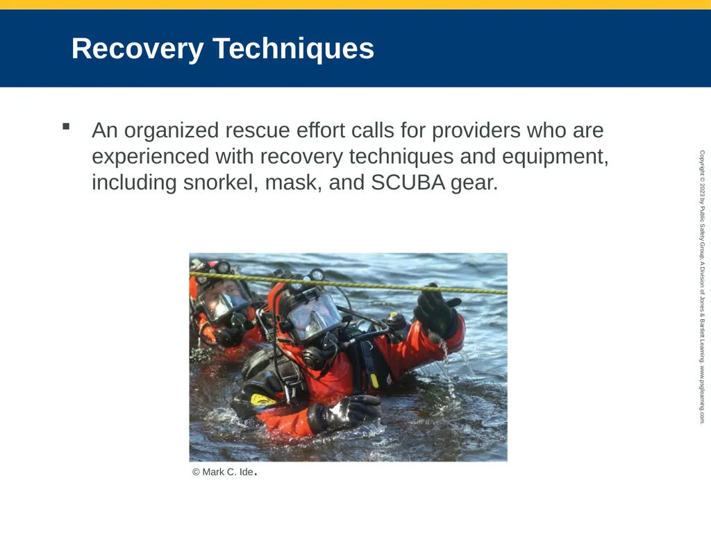
slide70. Recovery Techniques An organized rescue effort calls for providers who are experienced with recovery techniques and equipment, including snorkel, mask, and SCUBA gear. © Mark C. Ide.<br>
slide71. Patient Assessment of Drowning and Diving Emergencies (1 of 8) Scene size-up
Check for hazards.
Never drive through moving water.
Never attempt a water rescue without proper training and equipment.
If the patient is still in water, look for the best, safest means of removal.
Call for additional resources early.
Look for indicators of the MOI.<br>
slide72. Patient Assessment of Drowning and Diving Emergencies (2 of 8) Primary survey
Form a general impression.
Assess life threats and determine whether spinal stabilization is necessary.
Pay attention to chest pain, dyspnea, and complaints of sensory changes when a diving emergency is suspected.
Determine level of consciousness using the AVPU scale.
Be suspicious of drug and alcohol use and its effects.<br>
slide73. Patient Assessment of Drowning and Diving Emergencies (3 of 8) Airway and breathing
Open the airway and assess breathing in unresponsive patients.
Suction if the patient has vomited or if pink, frothy secretions are found in the airway.
Provide ventilations with a bag-mask device for inadequate breathing.
If the patient is responsive, provide high-flow oxygen with a nonrebreathing mask.
Obtain and continually monitor breath sounds.<br>
slide74. Patient Assessment of Drowning and Diving Emergencies (4 of 8) Circulation
Check for a pulse.
Never give up on resuscitating a cold-water drowning victim.
Exposure to cold water will occasionally activate certain primitive reflexes.
Evaluate the patient for adequate perfusion.<br>
slide75. Patient Assessment of Drowning and Diving Emergencies (5 of 8) Transport decision
Always transport patients to the hospital.
ARDS or renal failure may occur after resuscitation.
Inhalation of any amount of fluid can lead to delayed complications lasting for days or weeks.
Decompression sickness and air embolism must be treated in a recompression chamber.
Perform all interventions en route.<br>
slide76. Patient Assessment of Drowning and Diving Emergencies (6 of 8) History taking
Investigate the chief complaint.
Obtain a medical history.
Be alert for injury-specific signs and symptoms.
Obtain a SAMPLE history with special attention to the dive parameters.
Note any physical activity, alcohol or drug consumption, and other medical conditions.<br>
slide77. Patient Assessment of Drowning and Diving Emergencies (7 of 8) Secondary assessment
If patient is responsive, focus your physical examination on the basis of the chief complaint.
Begin with a rapid full-body scan.
Assess and ensure ABCs
Check for signs of hypothermia.
Obtain a baseline Glasgow Coma Scale score.
Assess for peripheral pulses, skin color and discoloration, itching, pain, and paresthesia.<br>
slide78. Patient Assessment of Drowning and Diving Emergencies (8 of 8) Reassessment
Repeat primary survey.
Reassess vital signs and chief complaint.
Recheck patient interventions.
Assess your patient’s mental status constantly, and assess vital signs at least every 5 minutes.
Pay attention to respirations and breath sounds.
Document the circumstances of the drowning and extrication.<br>
slide79. Emergency Care for Drowning or Diving Emergencies (1 of 7) Patients should be kept warm.
Provide blankets and protection from the environment as needed.
Obtain IV access.
Do not delay transport by attempting IV access.
Recognize that an emergency exists and treat as soon as possible.<br>
slide80. Emergency Care for Drowning or Diving Emergencies (2 of 7) Treating responsive patients who are suspected of having air embolism or decompression sickness
Administer oxygen via nonrebreathing mask or bag-mask device assist.
Place the patient in a left lateral recumbent position with the head down.
Consider the possibility of pneumothorax and monitor the patient’s breath sounds.
Provide prompt transport.<br>
slide81. Emergency Care for Drowning or Diving Emergencies (3 of 7) Submerged vehicle incidents
The safest time to escape from a submerging vehicle is immediately after it enters the water.
Attempt to remove the passengers from the vehicle as quickly as possible.<br>
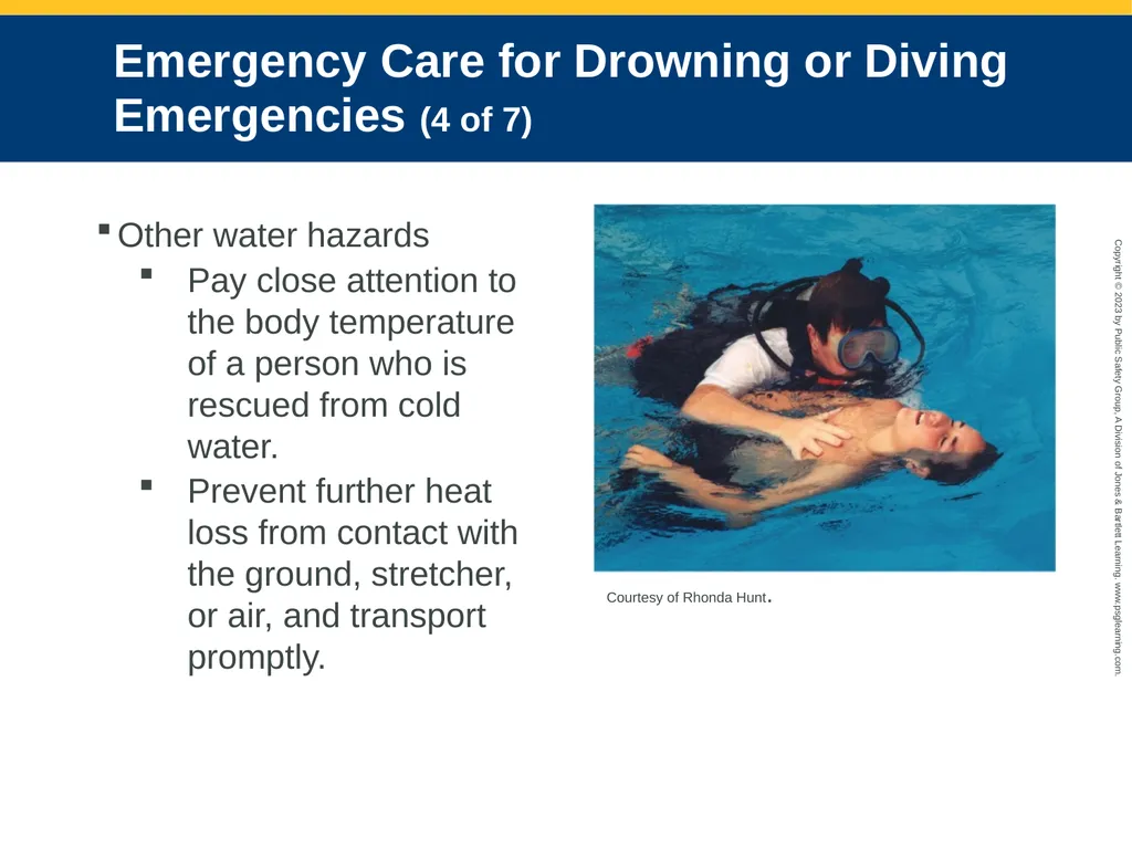
slide82. Emergency Care for Drowning or Diving Emergencies (4 of 7) Other water hazards
Pay close attention to the body temperature of a person who is rescued from cold water.
Prevent further heat loss from contact with the ground, stretcher, or air, and transport promptly. Courtesy of Rhonda Hunt.<br>
slide83. Emergency Care for Drowning or Diving Emergencies (5 of 7) Breath-holding syncope
Swimmers breathe in and out rapidly and deeply before entering the water
Hyperventilation lowers the carbon dioxide level.
Injuries caused by water hazards may be complicated by immersion in cold water.
Injuries from boat propellers, sharp rocks, water skis, dangerous marine life<br>
slide84. Emergency Care for Drowning or Diving Emergencies (6 of 7) Treatment for injuries:
Remove the patient from the water.
Protect the spine.
Administer oxygen.
Apply dressings and splints if indicated.
Monitor closely for signs of immersion or cold injury.
A child who is involved in a drowning or submersion may be the victim of child abuse<br>
slide85. Emergency Care for Drowning or Diving Emergencies (7 of 7) Prevention
All pools should be surrounded by a fence at least 6 feet high with slats no farther apart than 3 inches and self-closing, self-locking gates.
Most common problem is lack of adult supervision.
Be involved with public education to make people aware of hazards.<br>
slide86. High Altitude (1 of 6) Dysbarism injuries
Signs and symptoms caused by the difference between the surrounding atmospheric pressure and the total gas pressure in various tissues, fluids, and cavities of the body<br>
slide87. High Altitude (2 of 6) Altitude illness
Occurs when an unacclimated person is exposed to diminished oxygen pressure in the air at high altitudes
Affects the central nervous system and pulmonary system
Range from the common acute mountain sickness to rare deaths from HACE and HAPE<br>
slide88. High Altitude (3 of 6) Acute mountain sickness
Caused by diminished oxygen pressure in the air at altitudes above 5,000 feet
Results in hypoxia
Signs and symptoms: Headache, lightheadedness, fatigue, loss of appetite, nausea, difficulty sleeping, shortness of breath during physical exertion, and swollen face<br>
slide89. High Altitude (4 of 6) High-altitude pulmonary edema (HAPE)
Can occur at altitudes of 8,000 feet or greater
Fluid collects in the lungs, hindering passage of oxygen into the bloodstream.
Signs and symptoms
Shortness of breath
Cough with pink sputum
Cyanosis
Rapid pulse<br>
slide90. High Altitude (5 of 6) High-altitude cerebral edema (HACE)
Occurs in climbers who climb above 12,000 feet
May accompany HAPE and can quickly become life threatening
Symptoms of HACE and HAPE may overlap.
Severe constant throbbing headache
Ataxia
Extreme fatigue
Vomiting
Loss of consciousness<br>
slide91. High Altitude (6 of 6) Treatment
Provide oxygen.
Descend from the height.
Transport the patient.
If local protocols allow, CPAP may help.<br>
slide92. Lightning Injuries (1 of 4) Approximately 49 people die from lightning strikes in the United States each year.
Most deaths and injuries caused by lightning occur during the summer months.
Lightning strikes when a massive discharge of electricity occurs between two bodies that have different charges.<br>
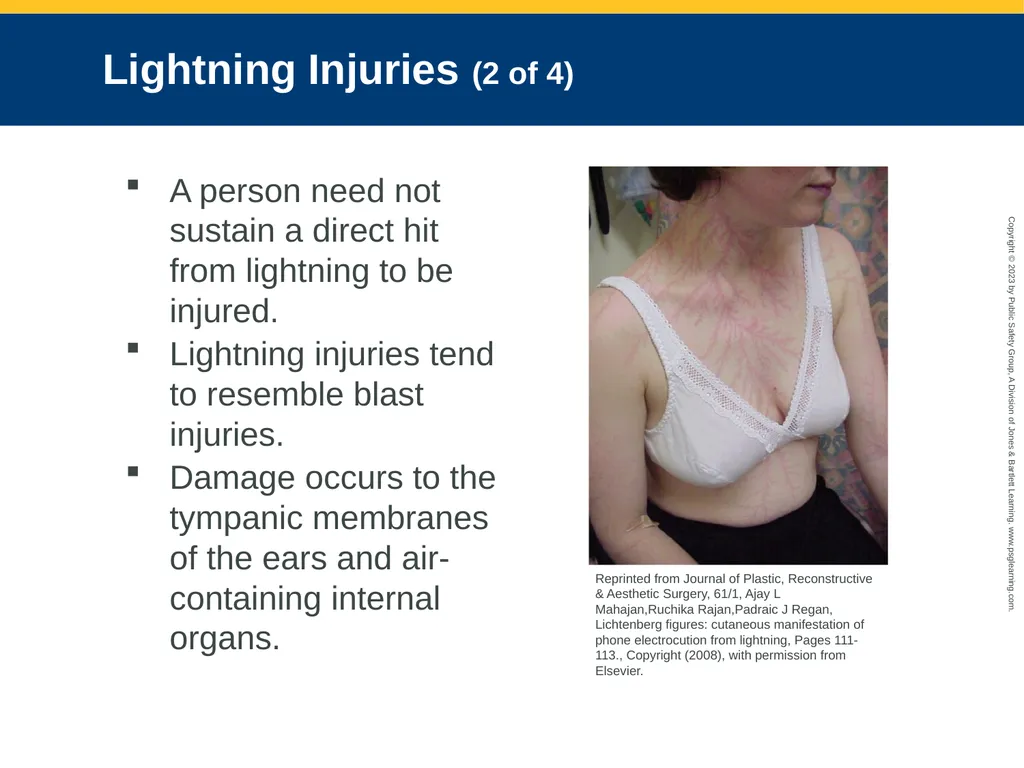
slide93. Lightning Injuries (2 of 4) A person need not sustain a direct hit from lightning to be injured.
Lightning injuries tend to resemble blast injuries.
Damage occurs to the tympanic membranes of the ears and air-containing internal organs. Reprinted from Journal of Plastic, Reconstructive & Aesthetic Surgery, 61/1, Ajay L Mahajan,Ruchika Rajan,Padraic J Regan, Lichtenberg figures: cutaneous manifestation of phone electrocution from lightning, Pages 111-113., Copyright (2008), with permission from Elsevier.<br>
slide94. Lightning Injuries (3 of 4) For the cardiovascular system, lightning acts as a cosmic defibrillator.
Someone regaining a pulse after a lightning strike and having respiratory arrest is known to result in a secondary cardiac arrest.
Central nervous system is almost invariably affected by a lightning strike.
Look for the entrance and exit wound.<br>
slide95. Lightning Injuries (4 of 4) Skin burns are usually superficial; full-thickness burns are rare.
Lightning injuries are categorized as being mild, moderate, or severe.
Immediate threats to life:
Airway obstruction
Respiratory arrest
Cardiac arrest<br>
slide96. Emergency Medical Care for Lightning Injuries (1 of 3) First priority is to get any patients and rescuers to a safe place.
If you suddenly feel a tingling sensation or your hair stands on end, the area around you has become charged.
Curl up in a ball and squat; make yourself as small a target as possible.<br>
slide97. Emergency Medical Care for Lightning Injuries (2 of 3) Determine the number of patients.
Reverse triage: Focus your efforts on those who are in respiratory or cardiac arrest.
Start CPR when necessary.
Manually stabilize the head in a neutral inline position.
If patient is in respiratory arrest with a pulse, begin immediate bag-mask ventilations with 100% oxygen.<br>
slide98. Emergency Medical Care for Lightning Injuries (3 of 3) Treatment of lightning injuries:
Make sure the scene is safe.
Priority for treatment goes to patients who are not breathing.
Perform CPR as needed. Establish an airway, and take cervical spine precautions.
Administer supplemental oxygen.
Insert a large-bore IV catheter and deliver an isotonic crystalloid solution wide open to keep the kidneys flushed out.<br>
slide99. Spider Bites (1 of 7) Spiders are numerous and widespread in the United States.
Female black widow spider and brown recluse spider deliver serious, even life-threatening venom.
Your safety is of paramount importance.<br>
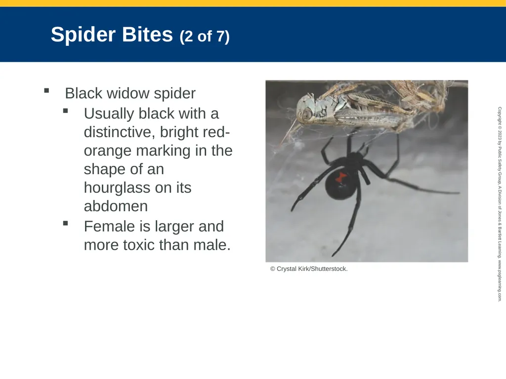
slide100. Spider Bites (2 of 7) Black widow spider
Usually black with a distinctive, bright red-orange marking in the shape of an hourglass on its abdomen
Female is larger and more toxic than male. © Crystal Kirk/Shutterstock.<br>
slide101. Spider Bites (3 of 7) The bite is sometimes overlooked.
If the site becomes numb right away, the patient may not even recall being bitten.
Most black widow spider bites cause localized pain and symptoms.
A bite on the abdomen may cause muscle spasms so severe that they resemble an acute abdomen condition.
Venom is neurotoxic.<br>
slide102. Spider Bites (4 of 7) Other systemic symptoms: Dizziness, sweating, nausea, vomiting, rashes
Tightness in the chest, difficulty breathing, and severe cramps typically develop in 24 hours.
Signs and symptoms subside over 48 hours.
A physician can administer a specific antivenin.<br>
slide103. Spider Bites (5 of 7) Severe muscle spasms are usually treated in the hospital with IV benzodiazepines such as diazepam (Valium) or lorazepam (Ativan).
Emergency treatment consists of BLS for the patient in respiratory distress, cleaning the bite with soap and water, and applying an ice pack.
Transport as soon as possible.<br>
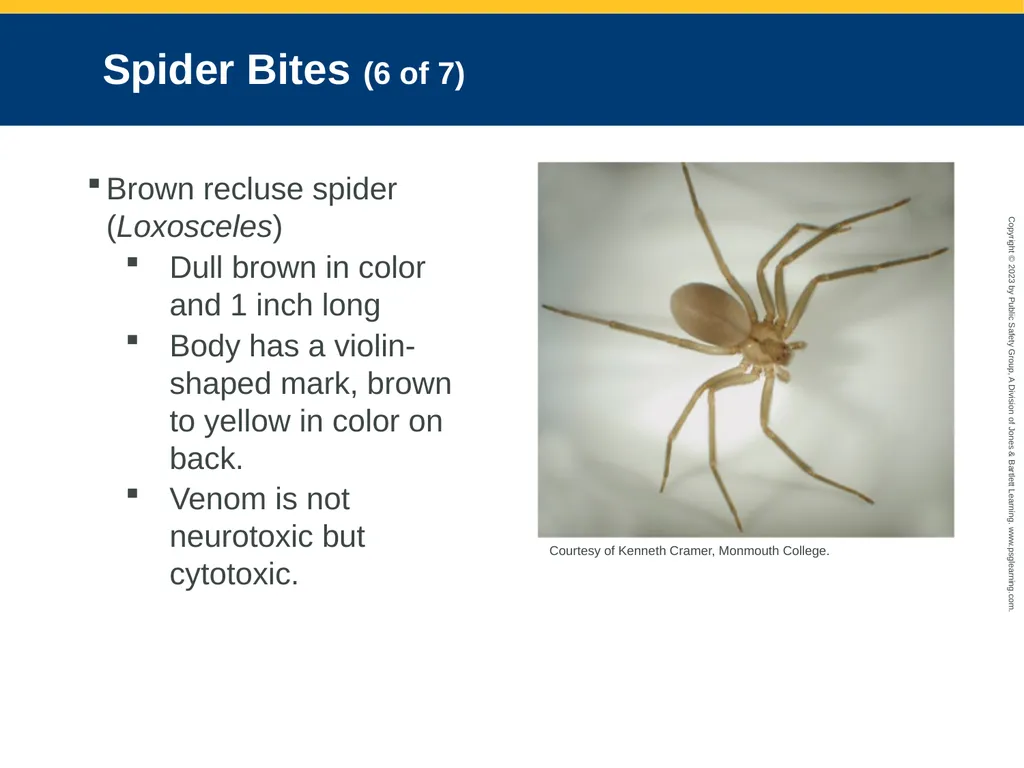
slide104. Spider Bites (6 of 7) Brown recluse spider (Loxosceles)
Dull brown in color and 1 inch long
Body has a violin-shaped mark, brown to yellow in color on back.
Venom is not neurotoxic but cytotoxic. Courtesy of Kenneth Cramer, Monmouth College. Courtesy of Kenneth Cramer, MonmCourtesy of Kenneth Cramer, Monmouth College.outh College. Courtesy of Kenneth Cramer, Monmouth College.<br>
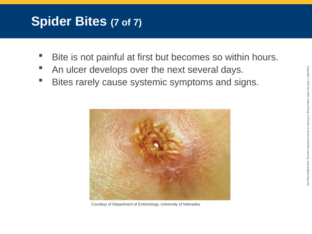
slide105. Spider Bites (7 of 7) Bite is not painful at first but becomes so within hours.
An ulcer develops over the next several days.
Bites rarely cause systemic symptoms and signs. Courtesy of Department of Entomology, University of Nebraska.<br>
slide106. Hymenoptera Stings (1 of 2) Painful but are not a medical emergency
Remove the stinger and, if still present, the venom sac.
Best done by using a firm-edged item
Clean thoroughly with soap and water or an antiseptic solution.<br>
slide107. Hymenoptera Stings (2 of 2) Anaphylaxis may occur.
Signs and symptoms are flushed skin, low blood pressure, and difficulty breathing.
Be prepared to administer epinephrine and support airway and breathing.<br>
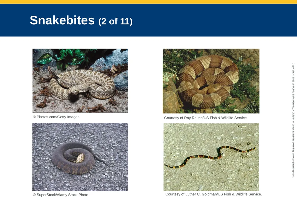
slide108. Snakebites (1 of 11) Only 19 species of snakes in the United States are venomous.
Rattlesnake (Crotalus)
Copperhead (Agkistrodon contortrix)
Cottonmouth, or water moccasin (Agkistrodon piscivorus)
Coral snakes (Micrurus fulvius and Micruroides euryxanthus)<br>
slide109. Snakebites (2 of 11) © Photos.com/Getty Images Courtesy of Ray Rauch/US Fish & Wildlife Service © SuperStock/Alamy Stock Photo Courtesy of Luther C. Goldman/US Fish & Wildlife Service.<br>
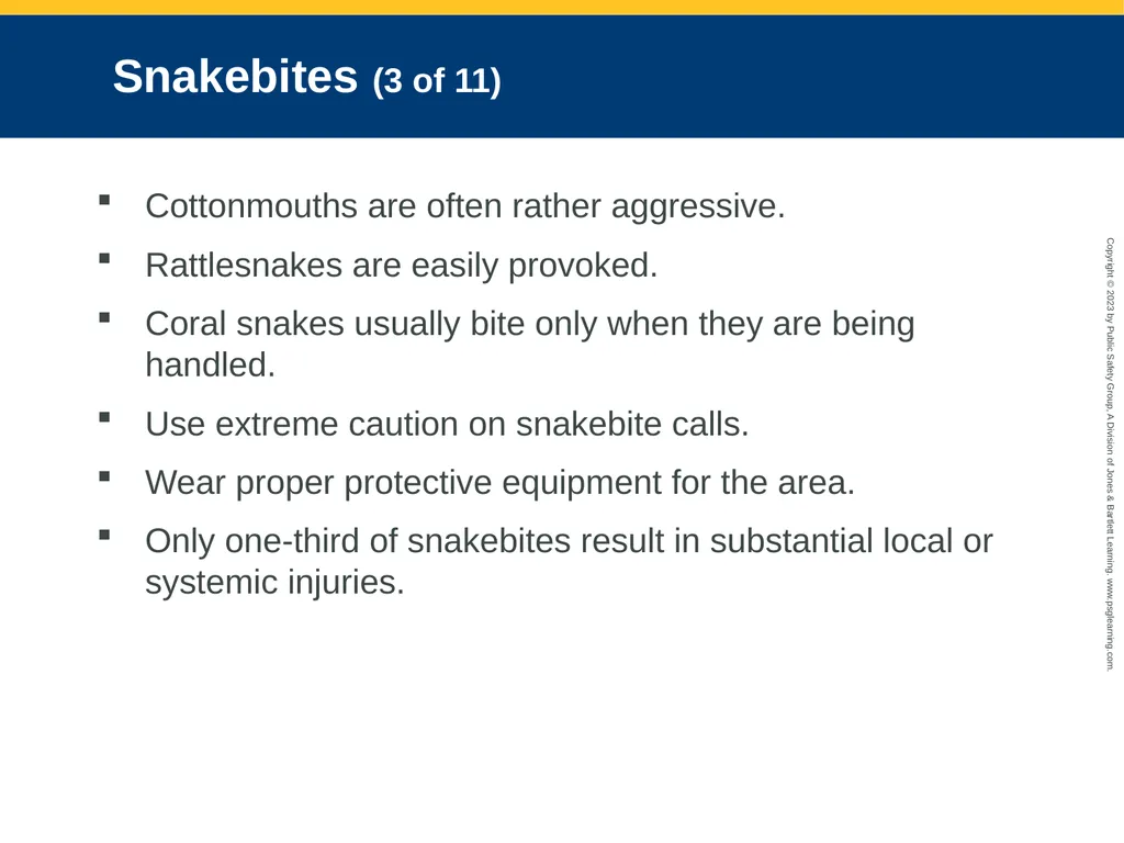
slide110. Snakebites (3 of 11) Cottonmouths are often rather aggressive.
Rattlesnakes are easily provoked.
Coral snakes usually bite only when they are being handled.
Use extreme caution on snakebite calls.
Wear proper protective equipment for the area.
Only one-third of snakebites result in substantial local or systemic injuries.<br>
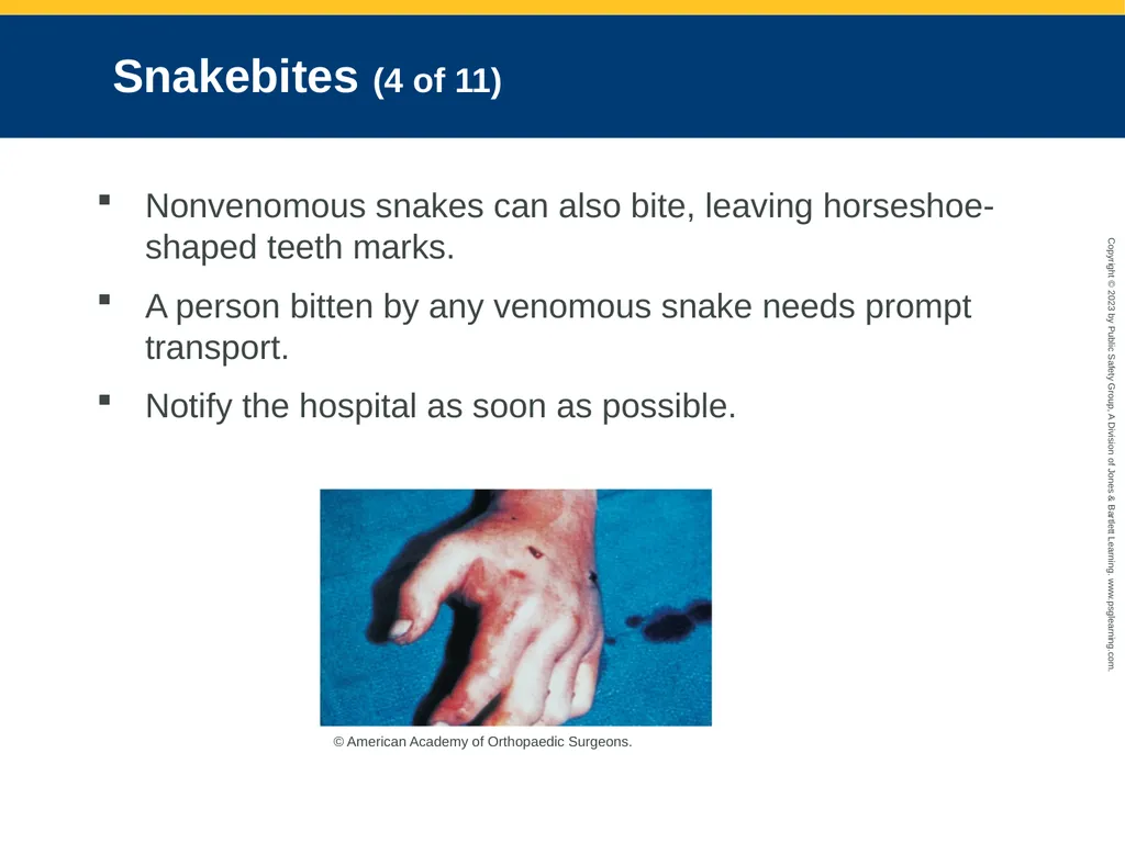
slide111. Snakebites (4 of 11) Nonvenomous snakes can also bite, leaving horseshoe-shaped teeth marks.
A person bitten by any venomous snake needs prompt transport.
Notify the hospital as soon as possible. © American Academy of Orthopaedic Surgeons.<br>
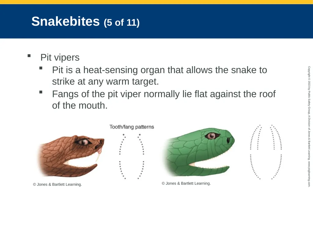
slide112. Snakebites (5 of 11) Pit vipers
Pit is a heat-sensing organ that allows the snake to strike at any warm target.
Fangs of the pit viper normally lie flat against the roof of the mouth. © Jones & Bartlett Learning. © Jones & Bartlett Learning.<br>
slide113. Snakebites (6 of 11) Signs of envenomation by a pit viper:
Burning pain at the site of the injury
Swelling
Bluish discoloration in light-skinned people
Venom can interfere with body’s blood-clotting mechanisms.
The smaller the patient, the more severe the symptoms.
If swelling has occurred, use a marker to mark its edges on the skin.<br>
slide114. Snakebites (7 of 11) Coral snakes
Small reptile with a series of bright red, yellow, and black bands completely encircling the body
Have tiny fangs and inject the venom with its teeth by a chewing motion
Leaves one or more puncture or scratchlike wounds
Coral snake venom causes paralysis of the nervous system.
Often limited or no local symptoms.<br>
slide115. Snakebites (8 of 11) Treatment of snakebites
Calm the patient.
Have the patient lie supine.
Determine the approximate time of the bite and document your time en route to a receiving facility.
Clean the bite area gently with soap and water or a mild antiseptic.
Do not apply ice.<br>
slide116. Snakebites (9 of 11) If the bite occurred on an arm or leg, use a preformed pressure immobilization bandage and place the affected extremity below the level of the heart.
Be alert for an anaphylactic reaction.
Do not give anything by mouth, and be alert for vomiting.
If the patient was bitten on the trunk, keep the patient supine and calm and transport as quickly as possible.<br>
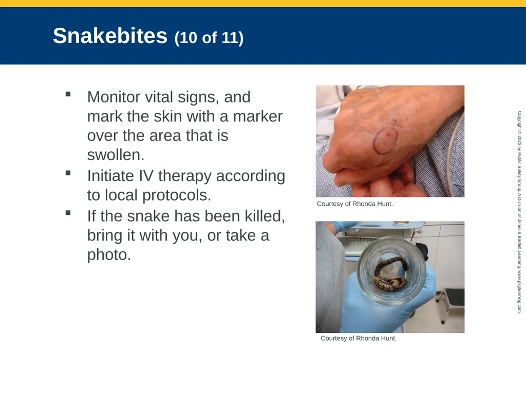
slide117. Snakebites (10 of 11) Monitor vital signs, and mark the skin with a marker over the area that is swollen.
Initiate IV therapy according to local protocols.
If the snake has been killed, bring it with you, or take a photo. Courtesy of Rhonda Hunt. Courtesy of Rhonda Hunt.<br>
slide118. Snakebites (11 of 11) If the patient shows no sign of envenomation:
Provide BLS as needed.
Place a sterile dressing over the suspected bite area.
Immobilize the injury site.
All patients with a suspected snakebite should be taken to the ED.<br>
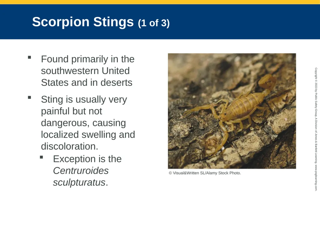
slide119. Scorpion Stings (1 of 3) Found primarily in the southwestern United States and in deserts
Sting is usually very painful but not dangerous, causing localized swelling and discoloration.
Exception is the Centruroides sculpturatus. © Visual&Written SL/Alamy Stock Photo.<br>
slide120. Scorpion Stings (2 of 3) Venom may produce a severe systemic reaction causing:
Circulatory collapse
Severe muscle contractions
Excessive salivation
Hypertension
Seizures
Cardiac failure
Antivenin is available.<br>
slide121. Scorpion Stings (3 of 3) Notify medical control as soon as possible.
Administer BLS and provide rapid transport to the ED.<br>
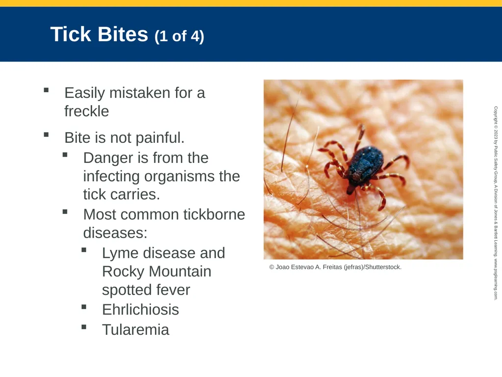
slide122. Tick Bites (1 of 4) Easily mistaken for a freckle
Bite is not painful.
Danger is from the infecting organisms the tick carries.
Most common tickborne diseases:
Lyme disease and Rocky Mountain spotted fever
Ehrlichiosis
Tularemia © Joao Estevao A. Freitas (jefras)/Shutterstock.<br>
slide123. Tick Bites (2 of 4) The longer a tick stays embedded, the greater the chance that a disease will be transmitted.
Rocky Mountain spotted fever
Occurs within 7 to 10 days after bite
Symptoms: Nausea, vomiting, headache, weakness, paralysis, and possibly cardiorespiratory collapse<br>
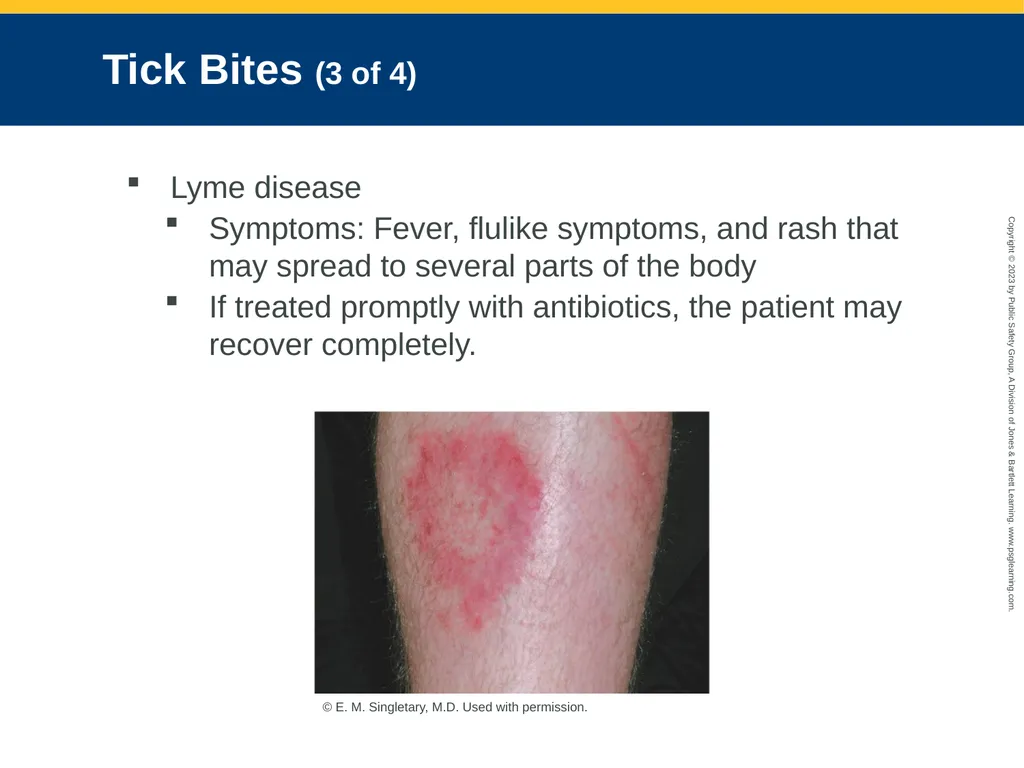
slide124. Tick Bites (3 of 4) Lyme disease
Symptoms: Fever, flulike symptoms, and rash that may spread to several parts of the body
If treated promptly with antibiotics, the patient may recover completely. © E. M. Singletary, M.D. Used with permission.<br>
slide125. Tick Bites (4 of 4) Transmission of infection from tick to person takes at least 12 hours.
To remove a tick:
Using fine tweezers, grasp the tick by the head and pull it gently but firmly straight up.
Cleanse the area with antiseptic.
Save the tick so that it can be identified.
Do not handle tick with your fingers.<br>
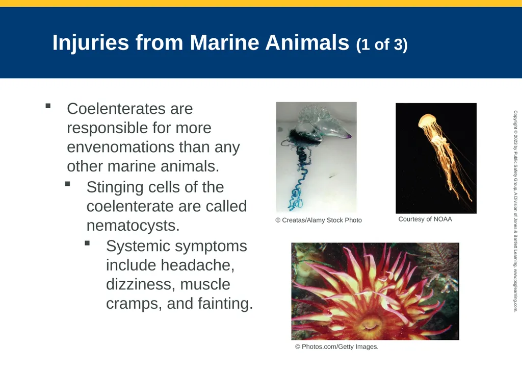
slide126. Injuries from Marine Animals (1 of 3) Coelenterates are responsible for more envenomations than any other marine animals.
Stinging cells of the coelenterate are called nematocysts.
Systemic symptoms include headache, dizziness, muscle cramps, and fainting. © Creatas/Alamy Stock Photo Courtesy of NOAA © Photos.com/Getty Images.<br>
slide127. Injuries from Marine Animals (2 of 3) To treat a jellyfish sting:
Remove the patient from the water and remove the tentacles by scraping them off with the edge of a stiff object.
Wash the affected area with vinegar immediately if available for at least 30 seconds.
Limit further discharge of nematocysts by avoiding fresh water, wet sand, showers, or careless manipulation of tentacles.
Reduce motion of the affected extremity.<br>
slide128. Injuries from Marine Animals (3 of 3) A patient may have a systemic allergic reaction to the sting.
Toxins from the spines of urchins, stingrays, and certain spiny fish are heat sensitive.
The best treatment is to immobilize the affected area and soak it in hot water for 30 minutes.<br>
slide2. National EMS Education Standard Competencies (1 of 4) Trauma
Applies fundamental knowledge to provide basic and selected advanced emergency care and transportation based on assessment findings for an acutely injured patient.<br>
slide3. National EMS Education Standard Competencies (2 of 4) Environmental Emergencies
Recognition and management of:
Submersion incidents
Temperature-related illness<br>
slide4. National EMS Education Standard Competencies (3 of 4) Pathophysiology, assessment, and management of:
Near drowning
Temperature-related illness
Bites and envenomations<br>
slide5. National EMS Education Standard Competencies (4 of 4) Pathophysiology, assessment, and management of:
Dysbarism
High altitude
Diving injuries
Electrical injury
Radiation exposure<br>
slide6. Introduction Medical condition caused or exacerbated by weather, terrain, or atmospheric pressure
Medical emergencies can result, particularly in:
Children
Older people
People with chronic illnesses
Young adults who overexert themselves<br>
slide7. Factors Affecting Exposure (1 of 2) Thermoregulation: Balance between heat production and heat excretion
Rise in core body temperature elicits responses that increase heat loss and shut off normal heat production pathways (thermogenesis).
Decrease in core body temperature prompts heat production and conservation and turns off normal heat-liberating pathways (thermolysis).<br>
slide8. Factors Affecting Exposure (2 of 2) Factors affecting exposure:
Physical condition
Age
Nutrition and hydration
Environmental conditions © Jones & Bartlett Learning.<br>
slide9. Cold Exposure (1 of 9) Normal body temperature must be maintained within a narrow range for the body’s chemistry to work efficiently.
Hypothermia: Core body temperature falls because of inadequate internal heat production, excess cold stress, or a combination of both<br>
slide10. Cold Exposure (2 of 9) Body can lose heat through:
Conduction
Convection
Evaporation
Radiation
Respiration
Rate and amount of heat loss or gain by the body can be modified by:
Increase or decrease in heat production
Moving to an area where heat loss is decreased or increased
Wearing appropriate clothing for environment<br>
slide11. Cold Exposure (3 of 9) Courtesy of the National Weather Service/NOAA.<br>
slide12. Cold Exposure (4 of 9) Courtesy of National Oceanic and Atmospheric Administration.<br>
slide13. Cold Exposure (5 of 9) Pathophysiology
Hypothermia
Occurs when core body temperature falls below 95°F
Body cannot regulate body temperature and generate body heat.
Can develop quickly or gradually
Impaired thermoregulation can occur with therapeutic use or overdoses of sedative medications, tricyclic antidepressants, and phenothiazines.<br>
slide14. Cold Exposure (6 of 9) Signs and symptoms of hypothermia
Increase in severity as core temperature falls
Generally progresses through four stages<br>
slide15. Cold Exposure (7 of 9) Local cold injuries
Localized to exposed parts of the body.
Frostnip, chilblains, or immersion foot can result when exposed parts of the body become very cold but not frozen. © Dr. P. Marazzi/Science Source © American Academy of Orthopaedic Surgeons. © American Academy of Orthopaedic Surgeons.<br>
slide16. Cold Exposure (8 of 9) Frostnip and immersion foot
Frostnip affects the ears, nose, and fingers and is not painful.
Immersion foot occurs after prolonged exposure to cold water.<br>
slide17. Cold Exposure (9 of 9) Frostbite
Tissues are frozen
Permanently damages cells
Can be identified by the hard, waxy feel of the affected tissues Courtesy of Dr. Jack Poland/CDC. © American Academy of Orthopaedic Surgeons.<br>
slide18. Assessment of Cold Injuries (1 of 9) Management: Stabilizing ABCs and preventing further heat loss
All patients who are injured are at risk for hypothermia.
Scene size-up
Important aspects: Air temperature, wind chill, whether it is wet or dry<br>
slide19. Assessment of Cold Injuries (2 of 9) Ensure that the scene is safe
Identify potential safety hazards, such as wet grass, mud, snow, or icy streets.
Consider special hazards.
Use appropriate standard precautions.
Consider the number of patients.
Summon additional help as quickly as possible.
Look for indicators of MOI.<br>
slide20. Assessment of Cold Injuries (3 of 9) Primary survey
Form a general impression.
Perform a rapid full-body scan to determine whether a life threat exists.
If chief complaint is simply being cold, quickly assess core temperature.
Evaluate mental status using AVPU.
Take into account physiologic changes that occur as a result of hypothermia.<br>
slide21. Assessment of Cold Injuries (4 of 9) Airway and breathing
If you believe the patient is in cardiac arrest, provide high-quality chest compressions.
If you cannot feel a radial pulse, gently palpate for a carotid pulse, and wait for up to 60 seconds before you decide that the patient is pulseless.
Assume shock is present and treat it.
Ensure adequate airway and breathing.
All patients with hypothermia require immediate transport for evaluation and treatment.<br>
slide22. Assessment of Cold Injuries (5 of 9) History taking
Investigate the chief complaint.
Obtain a medical history.
Be alert for injury-specific signs, symptoms, and pertinent negatives.
If possible, determine the duration and temperature of the exposure, and wind velocity.<br>
slide23. Assessment of Cold Injuries (6 of 9) SAMPLE history
Last oral intake and activity prior to the exposure will help to determine severity of the cold injury.
Investigate underlying factors:
Exposure to wet conditions
Inadequate insulation from cold or wind
Restricted circulation
Inadequate nutrition, alcohol or drug abuse
Diabetes, cardiovascular disease<br>
slide24. Assessment of Cold Injuries (7 of 9) Secondary assessment
Focus your physical examination on the severity of hypothermia.
Determine the degree and extent of cold injury.
Vital signs may be altered by the effects of hypothermia.
Respirations may be slow and shallow.<br>
slide25. Assessment of Cold Injuries (8 of 9) Low blood pressure and a slow pulse indicate moderate to severe hypothermia.
Determine core body temperature using a hypothermia thermometer based on local protocol.
Pulse oximetry will often be inaccurate because of the lack of perfusion in the extremities.<br>
slide26. Assessment of Cold Injuries (9 of 9) Reassessment
Repeat the primary survey.
Reassess vital signs and chief complaint.
Review all treatments.
Monitor level of consciousness and vital signs.
Communicate all information gathered to the receiving facility.<br>
slide27. Emergency Medical Care for Cold Emergencies (1 of 10) General steps to prevent further cold injury:
Remove wet clothing, and keep the patient dry.
Prevent conduction heat loss.
Insulate all exposed body parts.
Prevent convection heat loss.
Remove the patient from the cold environment.<br>
slide28. Emergency Medical Care for Cold Emergencies (2 of 10) Move the patient from the cold environment to prevent further heat loss.
To prevent further damage to the feet, do not allow the patient to walk.
Place dry blankets over and under the patient. © Jones & Bartlett Learning. Courtesy of MIEMSS.<br>
slide29. Emergency Medical Care for Cold Emergencies (3 of 10) If available, give the patient warm, humidified oxygen.
Obtain IV access, but follow local protocols.
When possible, administer fluid that has been warmed.<br>
slide30. Emergency Medical Care for Cold Emergencies (4 of 10) Handle the patient gently to avoid pain or further injury to the skin.
Mild hypothermia
Signs and symptoms: alertness, shivering, core body temperature between 90°F and 95°F
Treatment: Begin passive rewarming slowly, turn the heat up high in the patient compartment, give warm fluids<br>
slide31. Emergency Medical Care for Cold Emergencies (5 of 10) Moderate or severe hypothermia
Active rewarming is best accomplished in the ED.
Rewarming extremities too rapidly without rewarming the core can cause vasodilation.
Move the patient out of the wind and away from contact with any object that will conduct heat away from the body.<br>
slide32. Emergency Medical Care for Cold Emergencies (6 of 10) Withholding and cessation of resuscitative efforts
If submersion preceded the arrest, successful resuscitation is unlikely.
Trauma, alcohol overdose, and drug overdose could have resulted in hypothermia.
Fixed, dilated pupils, apparent rigor mortis, and dependent lividity are not contraindications for resuscitation of severely hypothermic patients.<br>
slide33. Emergency Medical Care for Cold Emergencies (7 of 10) Emergency treatment of local cold injuries
Remove the patient from further exposure to the cold.
Handle the injured part gently, and protect it from further injury.
Administer oxygen, if not already done.
Remove any wet or restrictive clothing over the injured part.<br>
slide34. Emergency Medical Care for Cold Emergencies (8 of 10) If there is no chance of reinjury or if transport to the ED will be delayed, consider active rewarming.
With frostnip, contact with a warm object may be all that is needed.
With immersion foot, remove wet shoes, boots, and socks.
Rewarm the foot gradually
Splint the extremity, and cover it loosely with a dry, sterile dressing.
Never rub injured tissues.
Do not re-expose injury to cold.<br>
slide35. Emergency Medical Care for Cold Emergencies (9 of 10) With a late or deep cold injury, remove any jewelry from the injured part.
Cover injury loosely with a dry, sterile dressing.
Do not apply heat or rewarm the part.
Do not allow the patient to stand or walk on a frostbitten foot.
Evaluate for signs or symptoms of systemic hypothermia.
Support vital functions and transport promptly.<br>
slide36. Emergency Medical Care for Cold Emergencies (10 of 10) Rewarming in the field
Use a warm-water bath.
Immerse the frostbitten part in water with a temperature of between 102°F and 104°F.
Dress the area with dry, sterile dressings.
Expect the patient to report severe pain.
Cover frostbitten part with soft, padded, sterile cotton dressings.
If blisters have formed, do not break them.<br>
slide37. Cold Exposure and You You are also at risk for hypothermia if you work in a cold environment.
Be aware of existing and potential weather conditions.
Wear proper clothing whenever appropriate.
Properly maintain and equip your vehicle.<br>
slide38. Heat Exposure (1 of 8) Normal body temperature is 98.6°F (37°C).
Thermolysis: getting rid of excess heat
Sweating (and evaporation of the sweat)
Dilation of peripheral blood vessels
Hyperthermia is a high core temperature, usually 101°F or higher.<br>
slide39. Heat Exposure (2 of 8) Pathophysiology
When mechanisms to decrease body heat are overwhelmed, illness develops.
High air temperature
High humidity
Vigorous exercise
Heat exposure can cause heat cramps, heat exhaustion, and heatstroke.<br>
slide40. Heat Exposure (3 of 8) Persons at greatest risk for heat illnesses:
Children
Geriatric patients
Patients with heart disease, COPD, diabetes, dehydration, and obesity
Patients with limited mobility
Contributing factors:
Length of exposure
Intensity of exposure
Enviroment<br>
slide41. Heat Exposure (4 of 8) Preventive measures:
Maintain an adequate fluid intake.
Acclimatize to the environment.
Limit exposure.
Drink water continually.<br>
slide42. Heat Exposure (5 of 8) Heat cramps
Painful muscle spasms that occur after vigorous exercise
Sweat causes a change in electrolyte balance.
Dehydration may also have a role in development.<br>
slide43. Heat Exposure (6 of 8) Heat exhaustion
Most common illness caused by heat
Result of the body losing too much water and electrolytes through very heavy sweating
Signs and symptoms
Cold, clammy skin with ashen pallor
Dry tongue and thirst
Dizziness, weakness, or faintness
Normal vital signs
Normal or slightly elevated body temperature<br>
slide44. Heat Exposure (7 of 8) Heatstroke
Least common but most serious illness caused by heat exposure
Body is subjected to more heat than it can effectively remove.
Hot, dry, flushed skin
First sign is a change in behavior.
A patient can have a heatstroke even if still sweating.<br>
slide45. Heat Exposure (8 of 8) Classic heatstroke commonly presents in people with chronic illnesses.
Exertional heatstroke commonly presents in people who are in good general health but have an increased core body temperature because of overwhelming heat stress.
Recovery depends on the speed with which treatment is administered.<br>
slide46. Assessment of Heat Injuries (1 of 5) Scene size-up
Perform an environmental assessment.
Use appropriate standard precautions.
Look for indicators of MOI.
Heat emergencies commonly occur during athletic events and practices.
Overcooling can result in shivering, which generates more heat.
Consider calling medical control for paramedic backup.<br>
slide47. Assessment of Heat Injuries (2 of 5) Primary survey
Observe how the patient interacts with you and the environment.
Perform a rapid full-body scan.
Assess mental status using AVPU.
Assess ABCs.
Treat aggressively for shock.
For signs of heatstroke, transport without delay.<br>
slide48. Assessment of Heat Injuries (3 of 5) History taking
Investigate the chief complaint.
Obtain medical history.
Be alert for specific signs and symptoms.
Obtain a SAMPLE history.
Be thorough in your questioning.
Determine patient’s exposure to heat and humidity and activities prior to the onset of symptoms.<br>
slide49. Assessment of Heat Injuries (4 of 5) Secondary assessment
If the patient is alert, perform a focused assessment.
Assess for muscle cramps or confusion.
Examine patient’s mental status, and obtain the patient’s vital signs.
Pay special attention to the skin.
Perform a neurologic examination.
Patients with hyperthermia will be tachycardic and tachypneic.
Check patient’s temperature.<br>
slide50. Assessment of Heat Injuries (5 of 5) Reassessment
Watch your patient’s condition carefully for deterioration.
Evaluate effectiveness of your interventions.
Be careful not to cause shivering when cooling down a patient.
Document the weather conditions and the activities the patient was performing prior to the emergency.<br>
slide51. Emergency Medical Care for Heat Emergencies (1 of 4) Heat cramps
Treatment:
Remove patient from hot environment.
Administer high-flow oxygen if indicated.
Rest the cramping muscles.
Replace fluids by mouth.
If the cramps do not go away, initiate an IV line and transport the patient to the hospital.<br>
slide52. Emergency Medical Care for Heat Emergencies (2 of 4) Heat exhaustion
Remove the patient from exposure to heat and urgently reversing the increase in body temperature.
Provide cold-water immersion to the patient if allowed.<br>
slide53. Emergency Medical Care for Heat Emergencies (3 of 4) Prepare to transport the patient if:
Symptoms do not clear up promptly.
Level of consciousness decreases.
Body temperature remains elevated.
Person is very young, older, or has any underlying medical condition.<br>
slide54. Emergency Medical Care for Heat Emergencies (4 of 4) Heatstroke
When treating a patient with heatstroke:
Move patient out of the hot environment.
Remove the patient’s clothing.
Cover the patient with wet towels or sheet.
Administer oxygen if indicated.
Obtain IV access and administer 20-mL/kg bolus of isotonic crystalloid solution.
Transport immediately.<br>
slide55. Water Emergencies (1 of 12) Drowning
Respiratory impairment from submersion or immersion in liquid
Patients with submersion injury can die of secondary complications that occur beyond 24 hours.
Alcohol consumption, preexisting seizure disorders, geriatric patients with cardiovascular disease, and unsupervised access to water are among the major risk factors.<br>
slide56. Water Emergencies (2 of 12) Inhaling very small amounts of fresh or salt water can severely irritate the larynx resulting to laryngospasm.
Hypothermia is a major concern.
The body’s diving reflex may slow metabolism to the point of protecting vital organs.
Hypoxia is always the first concern.<br>
slide57. Water Emergencies (3 of 12) Diving emergencies
Most serious water-related injuries are associated with dives.
Physical principles of pressure effects
Most SCUBA diving is done at depths between 30 and 60 fsw (2 to 3 ATA).
Liquids such as water are not compressible.<br>
slide58. Water Emergencies (4 of 12) Gases follow physical laws:
Boyle’s law
Dalton’s law
Henry’s law
Dive tables and dive computers provide guidelines for divers regarding when to take decompression stops during the dive. © Jones & Bartlett Learning.<br>
slide59. Water Emergencies (5 of 12) General assessment: Diving history
Obtain as many details as you can.
Record:
When did symptoms start?
What type of diving was done, and what type of equipment was used?
Where is the diving site, and what was the water temperature?
Were safety stops used?
Were there any dive complications?<br>
slide60. Water Emergencies (6 of 12) Descent emergencies
Major problem during descent is barotrauma.
Can result from compression of gases within body spaces during descent or expansion of gases within those spaces during ascent
A person with a perforated tympanic membrane may develop a special problem while diving.
Nitrogen narcosis: A state of altered mental status caused by breathing compressed nitrogen-containing air at depth<br>
slide61. Water Emergencies (7 of 12) Ascent emergencies
Usually require aggressive resuscitation
Pulmonary overpressurization syndrome (POPS)
Can occur when divers fail to exhale during an ascent
Can cause pneumothorax, mediastinal and subcutaneous emphysema, alveolar hemorrhage, and lethal air embolism<br>
slide62. Water Emergencies (8 of 12) POPS (cont’d)
Physical examination may reveal palpable subcutaneous air above the clavicles.
Always look for unequal breath sounds, low pulse oximetry values, and hyperresonance on the affected side of the chest.
Pneumothorax may require needle decompression or a chest tube.
Provide 100% oxygen by nonrebreathing mask.<br>
slide63. Water Emergencies (9 of 12) Air embolism
Bubbles of air in blood vessels
Air inside the lungs expands rapidly.
Air released from this rupture can enter:
Pleural space
Mediastinum
Bloodstream
Pneumothorax and pneumomediastinum both result in pain and severe dyspnea.<br>
slide64. Water Emergencies (10 of 12) Potential signs and symptoms of air embolism:
Blotching
Froth at the nose and mouth
Severe pain in muscles, joints, or abdomen
Dyspnea, localized pleuritic chest pain
Dizziness, nausea, and vomiting
Dysphasia
Cough
Cyanosis
Difficulty with vision
Paralysis and/or coma
Irregular pulse or cardiac arrest<br>
slide65. Water Emergencies (11 of 12) Decompression sickness
Bubbles of gas obstruct blood vessels.
Results from too rapid an ascent from a dive, too long of a dive at too deep a depth, or repeated dives within a short period
Most striking symptom is abdominal/joint pain.<br>
slide66. Water Emergencies (12 of 12) Air embolism occurs immediately on return to the surface.
Symptoms of decompression sickness may not occur for several hours.
Treatment consists of BLS followed by recompression in a hyperbaric chamber. Courtesy of Perry Baromedical Corporation.<br>
slide67. Water Rescue (1 of 2) Rescue and removal of a patient must occur before assessment or treatment. © Jones & Bartlett Learning © Jones & Bartlett Learning © Jones & Bartlett Learning © Jones & Bartlett Learning<br>
slide68. Water Rescue (2 of 2) Be prepared for rapid hypothermia in cold environment.
Success depends on how rapidly the patient is removed from the water and ventilated. © Jones & Bartlett Learning. Courtesy of MIEMSS.<br>
slide69. Spinal Injuries in Submersion Incidents Submersion incidents may be complicated by spinal fractures and spinal cord injuries.
Assume spinal injury exists when:
Submersion has resulted from a diving mishap or fall from a substantial height.
Patient is unresponsive.
Patient is responsive but reports weakness, paralysis, numbness, or tingling in the arms or legs.<br>
slide70. Recovery Techniques An organized rescue effort calls for providers who are experienced with recovery techniques and equipment, including snorkel, mask, and SCUBA gear. © Mark C. Ide.<br>
slide71. Patient Assessment of Drowning and Diving Emergencies (1 of 8) Scene size-up
Check for hazards.
Never drive through moving water.
Never attempt a water rescue without proper training and equipment.
If the patient is still in water, look for the best, safest means of removal.
Call for additional resources early.
Look for indicators of the MOI.<br>
slide72. Patient Assessment of Drowning and Diving Emergencies (2 of 8) Primary survey
Form a general impression.
Assess life threats and determine whether spinal stabilization is necessary.
Pay attention to chest pain, dyspnea, and complaints of sensory changes when a diving emergency is suspected.
Determine level of consciousness using the AVPU scale.
Be suspicious of drug and alcohol use and its effects.<br>
slide73. Patient Assessment of Drowning and Diving Emergencies (3 of 8) Airway and breathing
Open the airway and assess breathing in unresponsive patients.
Suction if the patient has vomited or if pink, frothy secretions are found in the airway.
Provide ventilations with a bag-mask device for inadequate breathing.
If the patient is responsive, provide high-flow oxygen with a nonrebreathing mask.
Obtain and continually monitor breath sounds.<br>
slide74. Patient Assessment of Drowning and Diving Emergencies (4 of 8) Circulation
Check for a pulse.
Never give up on resuscitating a cold-water drowning victim.
Exposure to cold water will occasionally activate certain primitive reflexes.
Evaluate the patient for adequate perfusion.<br>
slide75. Patient Assessment of Drowning and Diving Emergencies (5 of 8) Transport decision
Always transport patients to the hospital.
ARDS or renal failure may occur after resuscitation.
Inhalation of any amount of fluid can lead to delayed complications lasting for days or weeks.
Decompression sickness and air embolism must be treated in a recompression chamber.
Perform all interventions en route.<br>
slide76. Patient Assessment of Drowning and Diving Emergencies (6 of 8) History taking
Investigate the chief complaint.
Obtain a medical history.
Be alert for injury-specific signs and symptoms.
Obtain a SAMPLE history with special attention to the dive parameters.
Note any physical activity, alcohol or drug consumption, and other medical conditions.<br>
slide77. Patient Assessment of Drowning and Diving Emergencies (7 of 8) Secondary assessment
If patient is responsive, focus your physical examination on the basis of the chief complaint.
Begin with a rapid full-body scan.
Assess and ensure ABCs
Check for signs of hypothermia.
Obtain a baseline Glasgow Coma Scale score.
Assess for peripheral pulses, skin color and discoloration, itching, pain, and paresthesia.<br>
slide78. Patient Assessment of Drowning and Diving Emergencies (8 of 8) Reassessment
Repeat primary survey.
Reassess vital signs and chief complaint.
Recheck patient interventions.
Assess your patient’s mental status constantly, and assess vital signs at least every 5 minutes.
Pay attention to respirations and breath sounds.
Document the circumstances of the drowning and extrication.<br>
slide79. Emergency Care for Drowning or Diving Emergencies (1 of 7) Patients should be kept warm.
Provide blankets and protection from the environment as needed.
Obtain IV access.
Do not delay transport by attempting IV access.
Recognize that an emergency exists and treat as soon as possible.<br>
slide80. Emergency Care for Drowning or Diving Emergencies (2 of 7) Treating responsive patients who are suspected of having air embolism or decompression sickness
Administer oxygen via nonrebreathing mask or bag-mask device assist.
Place the patient in a left lateral recumbent position with the head down.
Consider the possibility of pneumothorax and monitor the patient’s breath sounds.
Provide prompt transport.<br>
slide81. Emergency Care for Drowning or Diving Emergencies (3 of 7) Submerged vehicle incidents
The safest time to escape from a submerging vehicle is immediately after it enters the water.
Attempt to remove the passengers from the vehicle as quickly as possible.<br>
slide82. Emergency Care for Drowning or Diving Emergencies (4 of 7) Other water hazards
Pay close attention to the body temperature of a person who is rescued from cold water.
Prevent further heat loss from contact with the ground, stretcher, or air, and transport promptly. Courtesy of Rhonda Hunt.<br>
slide83. Emergency Care for Drowning or Diving Emergencies (5 of 7) Breath-holding syncope
Swimmers breathe in and out rapidly and deeply before entering the water
Hyperventilation lowers the carbon dioxide level.
Injuries caused by water hazards may be complicated by immersion in cold water.
Injuries from boat propellers, sharp rocks, water skis, dangerous marine life<br>
slide84. Emergency Care for Drowning or Diving Emergencies (6 of 7) Treatment for injuries:
Remove the patient from the water.
Protect the spine.
Administer oxygen.
Apply dressings and splints if indicated.
Monitor closely for signs of immersion or cold injury.
A child who is involved in a drowning or submersion may be the victim of child abuse<br>
slide85. Emergency Care for Drowning or Diving Emergencies (7 of 7) Prevention
All pools should be surrounded by a fence at least 6 feet high with slats no farther apart than 3 inches and self-closing, self-locking gates.
Most common problem is lack of adult supervision.
Be involved with public education to make people aware of hazards.<br>
slide86. High Altitude (1 of 6) Dysbarism injuries
Signs and symptoms caused by the difference between the surrounding atmospheric pressure and the total gas pressure in various tissues, fluids, and cavities of the body<br>
slide87. High Altitude (2 of 6) Altitude illness
Occurs when an unacclimated person is exposed to diminished oxygen pressure in the air at high altitudes
Affects the central nervous system and pulmonary system
Range from the common acute mountain sickness to rare deaths from HACE and HAPE<br>
slide88. High Altitude (3 of 6) Acute mountain sickness
Caused by diminished oxygen pressure in the air at altitudes above 5,000 feet
Results in hypoxia
Signs and symptoms: Headache, lightheadedness, fatigue, loss of appetite, nausea, difficulty sleeping, shortness of breath during physical exertion, and swollen face<br>
slide89. High Altitude (4 of 6) High-altitude pulmonary edema (HAPE)
Can occur at altitudes of 8,000 feet or greater
Fluid collects in the lungs, hindering passage of oxygen into the bloodstream.
Signs and symptoms
Shortness of breath
Cough with pink sputum
Cyanosis
Rapid pulse<br>
slide90. High Altitude (5 of 6) High-altitude cerebral edema (HACE)
Occurs in climbers who climb above 12,000 feet
May accompany HAPE and can quickly become life threatening
Symptoms of HACE and HAPE may overlap.
Severe constant throbbing headache
Ataxia
Extreme fatigue
Vomiting
Loss of consciousness<br>
slide91. High Altitude (6 of 6) Treatment
Provide oxygen.
Descend from the height.
Transport the patient.
If local protocols allow, CPAP may help.<br>
slide92. Lightning Injuries (1 of 4) Approximately 49 people die from lightning strikes in the United States each year.
Most deaths and injuries caused by lightning occur during the summer months.
Lightning strikes when a massive discharge of electricity occurs between two bodies that have different charges.<br>
slide93. Lightning Injuries (2 of 4) A person need not sustain a direct hit from lightning to be injured.
Lightning injuries tend to resemble blast injuries.
Damage occurs to the tympanic membranes of the ears and air-containing internal organs. Reprinted from Journal of Plastic, Reconstructive & Aesthetic Surgery, 61/1, Ajay L Mahajan,Ruchika Rajan,Padraic J Regan, Lichtenberg figures: cutaneous manifestation of phone electrocution from lightning, Pages 111-113., Copyright (2008), with permission from Elsevier.<br>
slide94. Lightning Injuries (3 of 4) For the cardiovascular system, lightning acts as a cosmic defibrillator.
Someone regaining a pulse after a lightning strike and having respiratory arrest is known to result in a secondary cardiac arrest.
Central nervous system is almost invariably affected by a lightning strike.
Look for the entrance and exit wound.<br>
slide95. Lightning Injuries (4 of 4) Skin burns are usually superficial; full-thickness burns are rare.
Lightning injuries are categorized as being mild, moderate, or severe.
Immediate threats to life:
Airway obstruction
Respiratory arrest
Cardiac arrest<br>
slide96. Emergency Medical Care for Lightning Injuries (1 of 3) First priority is to get any patients and rescuers to a safe place.
If you suddenly feel a tingling sensation or your hair stands on end, the area around you has become charged.
Curl up in a ball and squat; make yourself as small a target as possible.<br>
slide97. Emergency Medical Care for Lightning Injuries (2 of 3) Determine the number of patients.
Reverse triage: Focus your efforts on those who are in respiratory or cardiac arrest.
Start CPR when necessary.
Manually stabilize the head in a neutral inline position.
If patient is in respiratory arrest with a pulse, begin immediate bag-mask ventilations with 100% oxygen.<br>
slide98. Emergency Medical Care for Lightning Injuries (3 of 3) Treatment of lightning injuries:
Make sure the scene is safe.
Priority for treatment goes to patients who are not breathing.
Perform CPR as needed. Establish an airway, and take cervical spine precautions.
Administer supplemental oxygen.
Insert a large-bore IV catheter and deliver an isotonic crystalloid solution wide open to keep the kidneys flushed out.<br>
slide99. Spider Bites (1 of 7) Spiders are numerous and widespread in the United States.
Female black widow spider and brown recluse spider deliver serious, even life-threatening venom.
Your safety is of paramount importance.<br>
slide100. Spider Bites (2 of 7) Black widow spider
Usually black with a distinctive, bright red-orange marking in the shape of an hourglass on its abdomen
Female is larger and more toxic than male. © Crystal Kirk/Shutterstock.<br>
slide101. Spider Bites (3 of 7) The bite is sometimes overlooked.
If the site becomes numb right away, the patient may not even recall being bitten.
Most black widow spider bites cause localized pain and symptoms.
A bite on the abdomen may cause muscle spasms so severe that they resemble an acute abdomen condition.
Venom is neurotoxic.<br>
slide102. Spider Bites (4 of 7) Other systemic symptoms: Dizziness, sweating, nausea, vomiting, rashes
Tightness in the chest, difficulty breathing, and severe cramps typically develop in 24 hours.
Signs and symptoms subside over 48 hours.
A physician can administer a specific antivenin.<br>
slide103. Spider Bites (5 of 7) Severe muscle spasms are usually treated in the hospital with IV benzodiazepines such as diazepam (Valium) or lorazepam (Ativan).
Emergency treatment consists of BLS for the patient in respiratory distress, cleaning the bite with soap and water, and applying an ice pack.
Transport as soon as possible.<br>
slide104. Spider Bites (6 of 7) Brown recluse spider (Loxosceles)
Dull brown in color and 1 inch long
Body has a violin-shaped mark, brown to yellow in color on back.
Venom is not neurotoxic but cytotoxic. Courtesy of Kenneth Cramer, Monmouth College. Courtesy of Kenneth Cramer, MonmCourtesy of Kenneth Cramer, Monmouth College.outh College. Courtesy of Kenneth Cramer, Monmouth College.<br>
slide105. Spider Bites (7 of 7) Bite is not painful at first but becomes so within hours.
An ulcer develops over the next several days.
Bites rarely cause systemic symptoms and signs. Courtesy of Department of Entomology, University of Nebraska.<br>
slide106. Hymenoptera Stings (1 of 2) Painful but are not a medical emergency
Remove the stinger and, if still present, the venom sac.
Best done by using a firm-edged item
Clean thoroughly with soap and water or an antiseptic solution.<br>
slide107. Hymenoptera Stings (2 of 2) Anaphylaxis may occur.
Signs and symptoms are flushed skin, low blood pressure, and difficulty breathing.
Be prepared to administer epinephrine and support airway and breathing.<br>
slide108. Snakebites (1 of 11) Only 19 species of snakes in the United States are venomous.
Rattlesnake (Crotalus)
Copperhead (Agkistrodon contortrix)
Cottonmouth, or water moccasin (Agkistrodon piscivorus)
Coral snakes (Micrurus fulvius and Micruroides euryxanthus)<br>
slide109. Snakebites (2 of 11) © Photos.com/Getty Images Courtesy of Ray Rauch/US Fish & Wildlife Service © SuperStock/Alamy Stock Photo Courtesy of Luther C. Goldman/US Fish & Wildlife Service.<br>
slide110. Snakebites (3 of 11) Cottonmouths are often rather aggressive.
Rattlesnakes are easily provoked.
Coral snakes usually bite only when they are being handled.
Use extreme caution on snakebite calls.
Wear proper protective equipment for the area.
Only one-third of snakebites result in substantial local or systemic injuries.<br>
slide111. Snakebites (4 of 11) Nonvenomous snakes can also bite, leaving horseshoe-shaped teeth marks.
A person bitten by any venomous snake needs prompt transport.
Notify the hospital as soon as possible. © American Academy of Orthopaedic Surgeons.<br>
slide112. Snakebites (5 of 11) Pit vipers
Pit is a heat-sensing organ that allows the snake to strike at any warm target.
Fangs of the pit viper normally lie flat against the roof of the mouth. © Jones & Bartlett Learning. © Jones & Bartlett Learning.<br>
slide113. Snakebites (6 of 11) Signs of envenomation by a pit viper:
Burning pain at the site of the injury
Swelling
Bluish discoloration in light-skinned people
Venom can interfere with body’s blood-clotting mechanisms.
The smaller the patient, the more severe the symptoms.
If swelling has occurred, use a marker to mark its edges on the skin.<br>
slide114. Snakebites (7 of 11) Coral snakes
Small reptile with a series of bright red, yellow, and black bands completely encircling the body
Have tiny fangs and inject the venom with its teeth by a chewing motion
Leaves one or more puncture or scratchlike wounds
Coral snake venom causes paralysis of the nervous system.
Often limited or no local symptoms.<br>
slide115. Snakebites (8 of 11) Treatment of snakebites
Calm the patient.
Have the patient lie supine.
Determine the approximate time of the bite and document your time en route to a receiving facility.
Clean the bite area gently with soap and water or a mild antiseptic.
Do not apply ice.<br>
slide116. Snakebites (9 of 11) If the bite occurred on an arm or leg, use a preformed pressure immobilization bandage and place the affected extremity below the level of the heart.
Be alert for an anaphylactic reaction.
Do not give anything by mouth, and be alert for vomiting.
If the patient was bitten on the trunk, keep the patient supine and calm and transport as quickly as possible.<br>
slide117. Snakebites (10 of 11) Monitor vital signs, and mark the skin with a marker over the area that is swollen.
Initiate IV therapy according to local protocols.
If the snake has been killed, bring it with you, or take a photo. Courtesy of Rhonda Hunt. Courtesy of Rhonda Hunt.<br>
slide118. Snakebites (11 of 11) If the patient shows no sign of envenomation:
Provide BLS as needed.
Place a sterile dressing over the suspected bite area.
Immobilize the injury site.
All patients with a suspected snakebite should be taken to the ED.<br>
slide119. Scorpion Stings (1 of 3) Found primarily in the southwestern United States and in deserts
Sting is usually very painful but not dangerous, causing localized swelling and discoloration.
Exception is the Centruroides sculpturatus. © Visual&Written SL/Alamy Stock Photo.<br>
slide120. Scorpion Stings (2 of 3) Venom may produce a severe systemic reaction causing:
Circulatory collapse
Severe muscle contractions
Excessive salivation
Hypertension
Seizures
Cardiac failure
Antivenin is available.<br>
slide121. Scorpion Stings (3 of 3) Notify medical control as soon as possible.
Administer BLS and provide rapid transport to the ED.<br>
slide122. Tick Bites (1 of 4) Easily mistaken for a freckle
Bite is not painful.
Danger is from the infecting organisms the tick carries.
Most common tickborne diseases:
Lyme disease and Rocky Mountain spotted fever
Ehrlichiosis
Tularemia © Joao Estevao A. Freitas (jefras)/Shutterstock.<br>
slide123. Tick Bites (2 of 4) The longer a tick stays embedded, the greater the chance that a disease will be transmitted.
Rocky Mountain spotted fever
Occurs within 7 to 10 days after bite
Symptoms: Nausea, vomiting, headache, weakness, paralysis, and possibly cardiorespiratory collapse<br>
slide124. Tick Bites (3 of 4) Lyme disease
Symptoms: Fever, flulike symptoms, and rash that may spread to several parts of the body
If treated promptly with antibiotics, the patient may recover completely. © E. M. Singletary, M.D. Used with permission.<br>
slide125. Tick Bites (4 of 4) Transmission of infection from tick to person takes at least 12 hours.
To remove a tick:
Using fine tweezers, grasp the tick by the head and pull it gently but firmly straight up.
Cleanse the area with antiseptic.
Save the tick so that it can be identified.
Do not handle tick with your fingers.<br>
slide126. Injuries from Marine Animals (1 of 3) Coelenterates are responsible for more envenomations than any other marine animals.
Stinging cells of the coelenterate are called nematocysts.
Systemic symptoms include headache, dizziness, muscle cramps, and fainting. © Creatas/Alamy Stock Photo Courtesy of NOAA © Photos.com/Getty Images.<br>
slide127. Injuries from Marine Animals (2 of 3) To treat a jellyfish sting:
Remove the patient from the water and remove the tentacles by scraping them off with the edge of a stiff object.
Wash the affected area with vinegar immediately if available for at least 30 seconds.
Limit further discharge of nematocysts by avoiding fresh water, wet sand, showers, or careless manipulation of tentacles.
Reduce motion of the affected extremity.<br>
slide128. Injuries from Marine Animals (3 of 3) A patient may have a systemic allergic reaction to the sting.
Toxins from the spines of urchins, stingrays, and certain spiny fish are heat sensitive.
The best treatment is to immobilize the affected area and soak it in hot water for 30 minutes.<br>