
CIRCULATORY SYSTEM LESSON -7A BY JITENDER YADAV
MJ
Published · 70 slides · 0 views

1 / 1









Description
CIRCULATORY SYSTEM LESSON -7A BY JITENDER YADAV INSPPH OBJECTIVES Upon completion of this lesson you will be able to: To study the characteristics composition of Blood. To know about the functions of blood. To know about the mechanism of
Related Topics
Share
Embed code
Download this presentation From Below
"CIRCULATORY SYSTEM LESSON -7A BY JITENDER YADAV" is the property of its rightful owner. Permission is granted to download and print the materials on this website for personal, non-commercial use only, and to display it on your personal computer provided you do not modify the materials and that you retain all copyright notices contained in the materials. By downloading content from our website, you accept the terms of this agreement.
Presentation Transcript
01
CIRCULATORY SYSTEM LESSON -7A BY
JITENDER YADAV
INSP/PH<br>
JITENDER YADAV
INSP/PH<br>
02
OBJECTIVES Upon completion of this lesson you will be able to:
To study the characteristics & composition of Blood.
To know about the functions of blood.
To know about the mechanism of clotting.
To gain knowledge about the various types of blood groups.<br>
To study the characteristics & composition of Blood.
To know about the functions of blood.
To know about the mechanism of clotting.
To gain knowledge about the various types of blood groups.<br>
03
OBJECTIVES 5. To know about the structure of arteries,
Veins & Capillaries.
6. To know about the Nervous control of blood
vessels.
7. To know about the structure of Heart &
cardiac cycle .
8. To know about the blood pressure, Pulse &
circulation of blood.
9. To learn brief details about important blood
vessels<br>
Veins & Capillaries.
6. To know about the Nervous control of blood
vessels.
7. To know about the structure of Heart &
cardiac cycle .
8. To know about the blood pressure, Pulse &
circulation of blood.
9. To learn brief details about important blood
vessels<br>
04
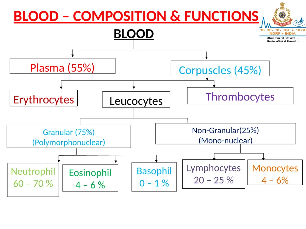
BLOOD – COMPOSITION & FUNCTIONS Plasma (55%) Corpuscles (45%) Erythrocytes Leucocytes Thrombocytes Granular (75%)
(Polymorphonuclear) Non-Granular(25%)
(Mono-nuclear) Neutrophil
60 – 70 % Eosinophil
4 – 6 % Basophil
0 – 1 % Lymphocytes
20 – 25 % Monocytes
4 – 6% BLOOD<br>
(Polymorphonuclear) Non-Granular(25%)
(Mono-nuclear) Neutrophil
60 – 70 % Eosinophil
4 – 6 % Basophil
0 – 1 % Lymphocytes
20 – 25 % Monocytes
4 – 6% BLOOD<br>
05
CHARACTERISTICS OF BLOOD:
Commonly described as a fluid connective tissue
Bright red when oxygenated (in arteries) & dark purplish-red when deoxygenated (in veins)
Slightly alkaline in reaction (pH – 7.4)
Total volume of blood in the body – 5 to 6 litres (approx. 8% of body weight
Specific gravity is 1.005<br>
Commonly described as a fluid connective tissue
Bright red when oxygenated (in arteries) & dark purplish-red when deoxygenated (in veins)
Slightly alkaline in reaction (pH – 7.4)
Total volume of blood in the body – 5 to 6 litres (approx. 8% of body weight
Specific gravity is 1.005<br>
06
COMPOSITION OF BLOOD:
PLASMA:
Fluid part of blood
Pale, yellowish fluid in which the corpuscles (solid part of blood) float
Contents:
Water – 90%<br>
PLASMA:
Fluid part of blood
Pale, yellowish fluid in which the corpuscles (solid part of blood) float
Contents:
Water – 90%<br>
07
Plasma proteins (7%) – Albumin; Globulin; Fibrinogen; Prothrombin; Heparin
Mineral Salts (o-9%) – Chlorides; Phosphates; Sodium-bi-carbonate & small amounts of potassium, magnesium, calcium, iron, copper & iodine
Nutrient material (from digested food) – Glucose; Amino-acid; Fatty acids; Vitamins<br>
Mineral Salts (o-9%) – Chlorides; Phosphates; Sodium-bi-carbonate & small amounts of potassium, magnesium, calcium, iron, copper & iodine
Nutrient material (from digested food) – Glucose; Amino-acid; Fatty acids; Vitamins<br>
08
Organic waste products – Urea; Uric acid; Creatinine
Anti-bodies & Anti-toxins
Hormones – from the endocrine (duct-less) glands
Enzymes – from the exocrine (ducted) glands
Gases – small amounts of dissolved Oxygen; CO2; Nitrogen<br>
Anti-bodies & Anti-toxins
Hormones – from the endocrine (duct-less) glands
Enzymes – from the exocrine (ducted) glands
Gases – small amounts of dissolved Oxygen; CO2; Nitrogen<br>
09
CELLULAR CONTENTS:
ERYTHROCYTES (Red Blood Corpuscles – RBCs):
Minute, disc shaped bodies with a concave surface
Non-nucleated
Approx. 7 microns in diameter
Normal counts:
5 – 5.5 million/cubic mm (men); 4.5 – 5 million/cubic mm (women)<br>
ERYTHROCYTES (Red Blood Corpuscles – RBCs):
Minute, disc shaped bodies with a concave surface
Non-nucleated
Approx. 7 microns in diameter
Normal counts:
5 – 5.5 million/cubic mm (men); 4.5 – 5 million/cubic mm (women)<br>
10
Formed in the bone-marrow
After passing through several stages of development, separate from the marrow & pass into circulating blood
Contain a special protein called Haemoglobin (Hb)
Development of RBCs is controlled by a feed-back system (same number are produced as they are destroyed)<br>
After passing through several stages of development, separate from the marrow & pass into circulating blood
Contain a special protein called Haemoglobin (Hb)
Development of RBCs is controlled by a feed-back system (same number are produced as they are destroyed)<br>
11
Destruction of RBCs (haemolysis) takes place in Spleen; iron is preserved for further use.
Average life-span is about 120 days.
Main function is to carry Oxygen by means of Hemoglobin to all tissues of the body.<br>
Average life-span is about 120 days.
Main function is to carry Oxygen by means of Hemoglobin to all tissues of the body.<br>
12
HAEMOGLOBIN
Complex protein containing a protein called Globin combined with an iron pigment called Haem.
Deficiency of Iron results in Anemia.
Has strong affinity for Oxygen.
Carries O2 by combining with it in the lungs to form oxyhaemoglobin.
Also has strong affinity for CO (forms carboxymethaemoglobin), in which case it is unable to carry O2, hence causing Anoxia.
Normal values: 14 -16 gms% (men), 12 -14 gms% (females).<br>
Complex protein containing a protein called Globin combined with an iron pigment called Haem.
Deficiency of Iron results in Anemia.
Has strong affinity for Oxygen.
Carries O2 by combining with it in the lungs to form oxyhaemoglobin.
Also has strong affinity for CO (forms carboxymethaemoglobin), in which case it is unable to carry O2, hence causing Anoxia.
Normal values: 14 -16 gms% (men), 12 -14 gms% (females).<br>
13
LEUCOCYTES (WHITE BLOOD CORPUSCLES – WBCs):
Colourless cells containing nuclei.
Slightly larger in size than RBCs, but less numerous.
Normal values: 7000 – 11000/ cubic mm.
Number increases during infections.<br>
Colourless cells containing nuclei.
Slightly larger in size than RBCs, but less numerous.
Normal values: 7000 – 11000/ cubic mm.
Number increases during infections.<br>
14
Two types of Leucocytes –
Granular
Non-granular
1. GRANULAR-
Develop in the bone-marrow from same parent cell (myeloblasts) as RBCs
As the cell develops, granules are formed in the cytoplasm. After passing through several stages of development, they pass into the circulating blood<br>
Granular
Non-granular
1. GRANULAR-
Develop in the bone-marrow from same parent cell (myeloblasts) as RBCs
As the cell develops, granules are formed in the cytoplasm. After passing through several stages of development, they pass into the circulating blood<br>
15
3 varieties – Neutrophils; Eosinophils; Basophils
Function –
To protect the body against invasion of bacteria by phagocytosis
To remove dead & injured tissue 2. NON-GRANULAR:
2 varieties – Lymphocytes & Monocytes
Lymphocytes develop in the lymphatic tissue
of the body (lymphatic glands & lymphatic
tissue of spleen, liver, bone-marrow)<br>
Function –
To protect the body against invasion of bacteria by phagocytosis
To remove dead & injured tissue 2. NON-GRANULAR:
2 varieties – Lymphocytes & Monocytes
Lymphocytes develop in the lymphatic tissue
of the body (lymphatic glands & lymphatic
tissue of spleen, liver, bone-marrow)<br>
16
Lymphocytes are concerned with production of anti-bodies & hence immunity against infections
Increase in lymphocytes is called lymphocytosis
Function of monocytes closely resembles that of neutrophils<br>
Increase in lymphocytes is called lymphocytosis
Function of monocytes closely resembles that of neutrophils<br>
17
THROMBOCYTES:
Extremely small cells
Non-nucleated
Contain granules in cytoplasm
Derived from Myeloblasts in the bone-marrow
Normal values: 3 – 4 lacs/ cubic mm
Function: Plays an important role in the blood clotting mechanism<br>
Extremely small cells
Non-nucleated
Contain granules in cytoplasm
Derived from Myeloblasts in the bone-marrow
Normal values: 3 – 4 lacs/ cubic mm
Function: Plays an important role in the blood clotting mechanism<br>
18
FUNCTIONS OF BLOOD
To carry oxygen to all tissues of the body.
To carry waste products to concerned excretory organs.
CO2 to lungs.
Urea, Uric acid to kidneys.
Water to lungs, kidneys & sweat glands.
To carry nutrient material to the tissues.
To carry hormones.<br>
To carry oxygen to all tissues of the body.
To carry waste products to concerned excretory organs.
CO2 to lungs.
Urea, Uric acid to kidneys.
Water to lungs, kidneys & sweat glands.
To carry nutrient material to the tissues.
To carry hormones.<br>
19
To carry anti-bodies against invading organisms.
To aid in defence of the body against pathogenic infections. BLOOD CLOTTING The process by which coagulation of blood takes place to prevent loss of blood when a blood vessel is damaged Following substances must be present before clotting can take place:<br>
To aid in defence of the body against pathogenic infections. BLOOD CLOTTING The process by which coagulation of blood takes place to prevent loss of blood when a blood vessel is damaged Following substances must be present before clotting can take place:<br>
20
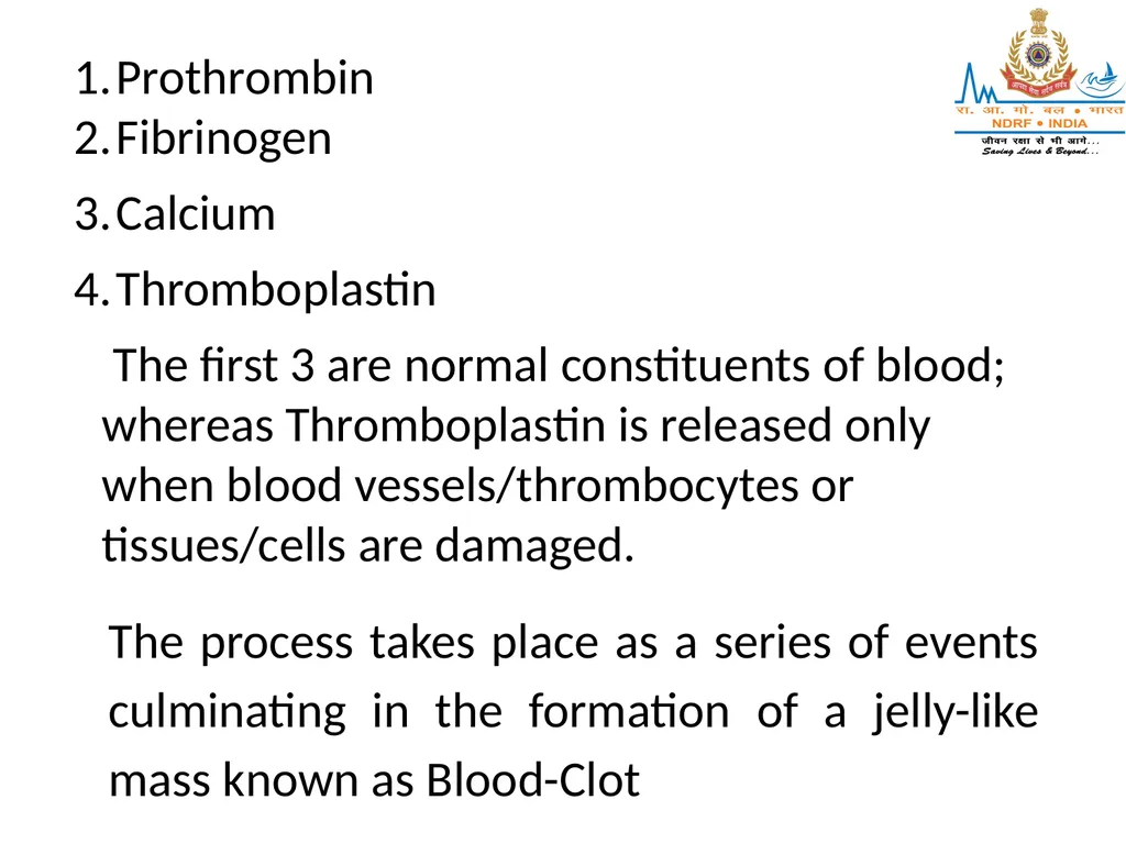
Prothrombin
Fibrinogen
Calcium
Thromboplastin
The first 3 are normal constituents of blood; whereas Thromboplastin is released only when blood vessels/thrombocytes or tissues/cells are damaged. The process takes place as a series of events culminating in the formation of a jelly-like mass known as Blood-Clot<br>
Fibrinogen
Calcium
Thromboplastin
The first 3 are normal constituents of blood; whereas Thromboplastin is released only when blood vessels/thrombocytes or tissues/cells are damaged. The process takes place as a series of events culminating in the formation of a jelly-like mass known as Blood-Clot<br>
21
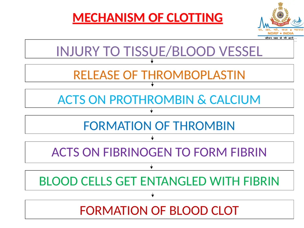
MECHANISM OF CLOTTING RELEASE OF THROMBOPLASTIN ACTS ON PROTHROMBIN & CALCIUM INJURY TO TISSUE/BLOOD VESSEL FORMATION OF THROMBIN ACTS ON FIBRINOGEN TO FORM FIBRIN BLOOD CELLS GET ENTANGLED WITH FIBRIN FORMATION OF BLOOD CLOT<br>
22
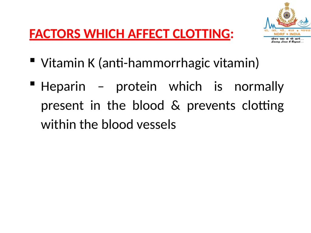
FACTORS WHICH AFFECT CLOTTING:
Vitamin K (anti-hammorrhagic vitamin)
Heparin – protein which is normally present in the blood & prevents clotting within the blood vessels<br>
Vitamin K (anti-hammorrhagic vitamin)
Heparin – protein which is normally present in the blood & prevents clotting within the blood vessels<br>
23
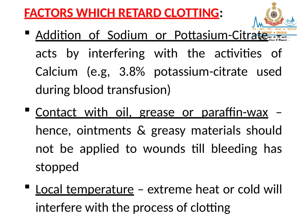
FACTORS WHICH RETARD CLOTTING:
Addition of Sodium or Pottasium-Citrate – acts by interfering with the activities of Calcium (e.g, 3.8% potassium-citrate used during blood transfusion)
Contact with oil, grease or paraffin-wax – hence, ointments & greasy materials should not be applied to wounds till bleeding has stopped
Local temperature – extreme heat or cold will interfere with the process of clotting<br>
Addition of Sodium or Pottasium-Citrate – acts by interfering with the activities of Calcium (e.g, 3.8% potassium-citrate used during blood transfusion)
Contact with oil, grease or paraffin-wax – hence, ointments & greasy materials should not be applied to wounds till bleeding has stopped
Local temperature – extreme heat or cold will interfere with the process of clotting<br>
24
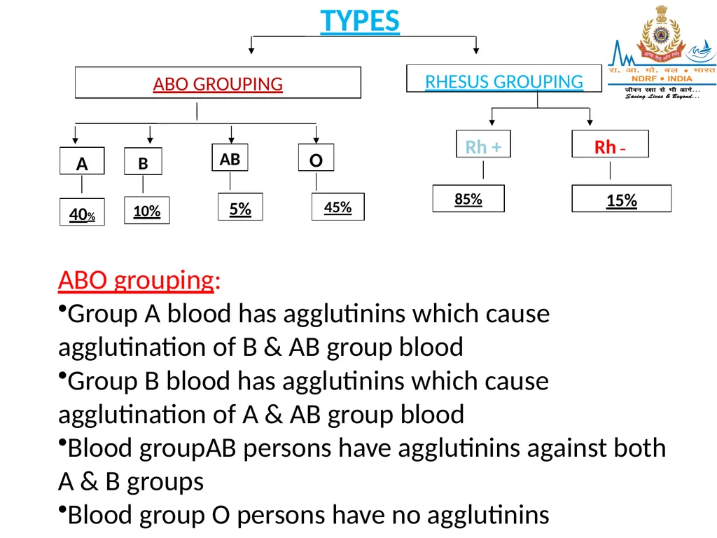
ABO GROUPING RHESUS GROUPING A B O Rh + Rh – 40% 10% 45% 85% 15% AB 5% TYPES ABO grouping:
Group A blood has agglutinins which cause agglutination of B & AB group blood
Group B blood has agglutinins which cause agglutination of A & AB group blood
Blood groupAB persons have agglutinins against both A & B groups
Blood group O persons have no agglutinins<br>
Group A blood has agglutinins which cause agglutination of B & AB group blood
Group B blood has agglutinins which cause agglutination of A & AB group blood
Blood groupAB persons have agglutinins against both A & B groups
Blood group O persons have no agglutinins<br>
25
Rh grouping:
The Rh antigen was first discovered in Rhesus monkeys
Rh antigen is present in RBCs of Rh+ persons & absent in Rh- persons
Plasma normally does not contain any anti-Rh antibodies
If Rh- person receives Rh+ blood for the first time, antibodies are produced by the body’s immune system. If such a person receives Rh+ blood again, these antibodies will cause haemolysis of the donated blood<br>
The Rh antigen was first discovered in Rhesus monkeys
Rh antigen is present in RBCs of Rh+ persons & absent in Rh- persons
Plasma normally does not contain any anti-Rh antibodies
If Rh- person receives Rh+ blood for the first time, antibodies are produced by the body’s immune system. If such a person receives Rh+ blood again, these antibodies will cause haemolysis of the donated blood<br>
26
BLOOD DONATION & TRANSFUSION:
Due to existence of these 2 main types of blood-grouping factors, various combinations like A+, A–, AB+, AB –, O+, etc. are possible
Blood transfusions can be given only with compatible blood of the same group
Even if donor is of the same group, direct cross-matching with blood of recipient has to be done due to presence of numerous other minor blood-group factors<br>
Due to existence of these 2 main types of blood-grouping factors, various combinations like A+, A–, AB+, AB –, O+, etc. are possible
Blood transfusions can be given only with compatible blood of the same group
Even if donor is of the same group, direct cross-matching with blood of recipient has to be done due to presence of numerous other minor blood-group factors<br>
27
Approx 250 – 300 ml of blood is taken during blood-donation Every healthy, young person can donate blood once in 3 – 4 months
People with infectious diseases like Hepatitis (A & B); HIV; etc. should not donate blood due to risk of transmitting disease<br>
People with infectious diseases like Hepatitis (A & B); HIV; etc. should not donate blood due to risk of transmitting disease<br>
28
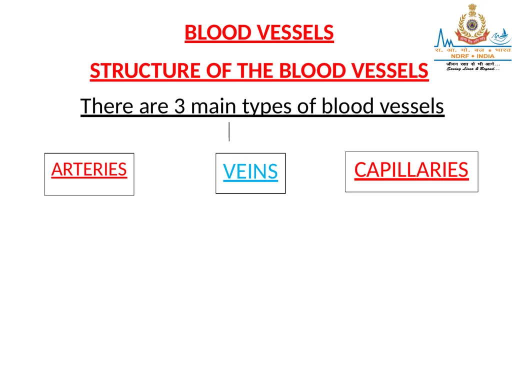
BLOOD VESSELS
STRUCTURE OF THE BLOOD VESSELS There are 3 main types of blood vessels ARTERIES VEINS CAPILLARIES<br>
STRUCTURE OF THE BLOOD VESSELS There are 3 main types of blood vessels ARTERIES VEINS CAPILLARIES<br>
29
I] ARTERIES:
Transport oxygenated blood (except pulmonary artery)
Transport blood away from the heart
Arteries vary considerably in size, but have much the same structure<br>
Transport oxygenated blood (except pulmonary artery)
Transport blood away from the heart
Arteries vary considerably in size, but have much the same structure<br>
30
STRUCTURE:
3 layers of tissue:
Tunica Adventitia – outer layer, formed by fibrous tissue
Tunica Media – intermediate layer, formed by elastic & smooth muscular tissue
Tunica Intima – innermost layer, epithelial tissue (endothelium)<br>
3 layers of tissue:
Tunica Adventitia – outer layer, formed by fibrous tissue
Tunica Media – intermediate layer, formed by elastic & smooth muscular tissue
Tunica Intima – innermost layer, epithelial tissue (endothelium)<br>
31
Amount of muscular & elastic tissue varies according to size. In larger arteries, tunica media contains more elastic & less muscular tissue. The proportion gradually changes & as arteries become smaller, there is very little elastic tissue & high proportion of muscular tissue<br>
32
IIVEINS:
Transport deoxygenated blood (except pulmonary vein)
Transport blood towards the heart
STRUCTURE:
Same 3 layers as in arteries; however, the main difference being that the tunica media is thinner & there is relatively less muscular & elastic tissue in the walls of veins<br>
Transport deoxygenated blood (except pulmonary vein)
Transport blood towards the heart
STRUCTURE:
Same 3 layers as in arteries; however, the main difference being that the tunica media is thinner & there is relatively less muscular & elastic tissue in the walls of veins<br>
33
The walls of the veins being thinner, collapse when cut while the arteries, being thicker, retain their cylindrical shape even when cut Some veins possess valves –
They prevent back-flow of blood
Abundant in veins of the extremities, but absent from the veins of the Thorax & Abdomen
Formed by a fold of tunica intima strengthened by connective tissue<br>
They prevent back-flow of blood
Abundant in veins of the extremities, but absent from the veins of the Thorax & Abdomen
Formed by a fold of tunica intima strengthened by connective tissue<br>
34
Semi-lunar in shape
Defective valves results in veins getting distended due to pooled blood leading to tortuous, enlarged veins called Varicose Veins<br>
Defective valves results in veins getting distended due to pooled blood leading to tortuous, enlarged veins called Varicose Veins<br>
35
III CAPILLARIES:
Connecting link between arteries & veins
Smaller arteries called arterioles further break-up into minute vessels to form capillaries
Capillary walls permit passage of water, substances of small molecular size & gases; but do not permit blood-cells& plasma to escape
Exchange of nutrient material, waste products & O2/CO2 takes place between blood & tissue at the capillary level<br>
Connecting link between arteries & veins
Smaller arteries called arterioles further break-up into minute vessels to form capillaries
Capillary walls permit passage of water, substances of small molecular size & gases; but do not permit blood-cells& plasma to escape
Exchange of nutrient material, waste products & O2/CO2 takes place between blood & tissue at the capillary level<br>
36
STRUCTURE:
The walls are made up of a single layer of endothelium tissue, which is very thin
NERVOUS CONTROL OF BLOOD VESSELS:
Both arteries & veins are controlled by the Autonomic Nervous System
The nerves arise from the Vasomotor Centre of the Medulla-Oblongata<br>
The walls are made up of a single layer of endothelium tissue, which is very thin
NERVOUS CONTROL OF BLOOD VESSELS:
Both arteries & veins are controlled by the Autonomic Nervous System
The nerves arise from the Vasomotor Centre of the Medulla-Oblongata<br>
37
These nerves are responsible for changes in the caliber of the vessels They, thus, control the amount of blood circulating to various parts of the body
Nerves which reduce the lumen of the vessels are called vaso-constrictors
Nerves which increase the lumen of the vessels are called vaso-dilators<br>
Nerves which reduce the lumen of the vessels are called vaso-constrictors
Nerves which increase the lumen of the vessels are called vaso-dilators<br>
38
THE HEART
The Heart is a hollow, muscular organ
Situated in the middle mediastinum of thoracic cavity, between lungs
Lies obliquely towards the left
The base of the heart is on the upper side & apex on the lower side
Roughly 10cm x 8cm x 6cm in size (approx. individual’s fist-sized)
Weighs about 270 gms<br>
The Heart is a hollow, muscular organ
Situated in the middle mediastinum of thoracic cavity, between lungs
Lies obliquely towards the left
The base of the heart is on the upper side & apex on the lower side
Roughly 10cm x 8cm x 6cm in size (approx. individual’s fist-sized)
Weighs about 270 gms<br>
39
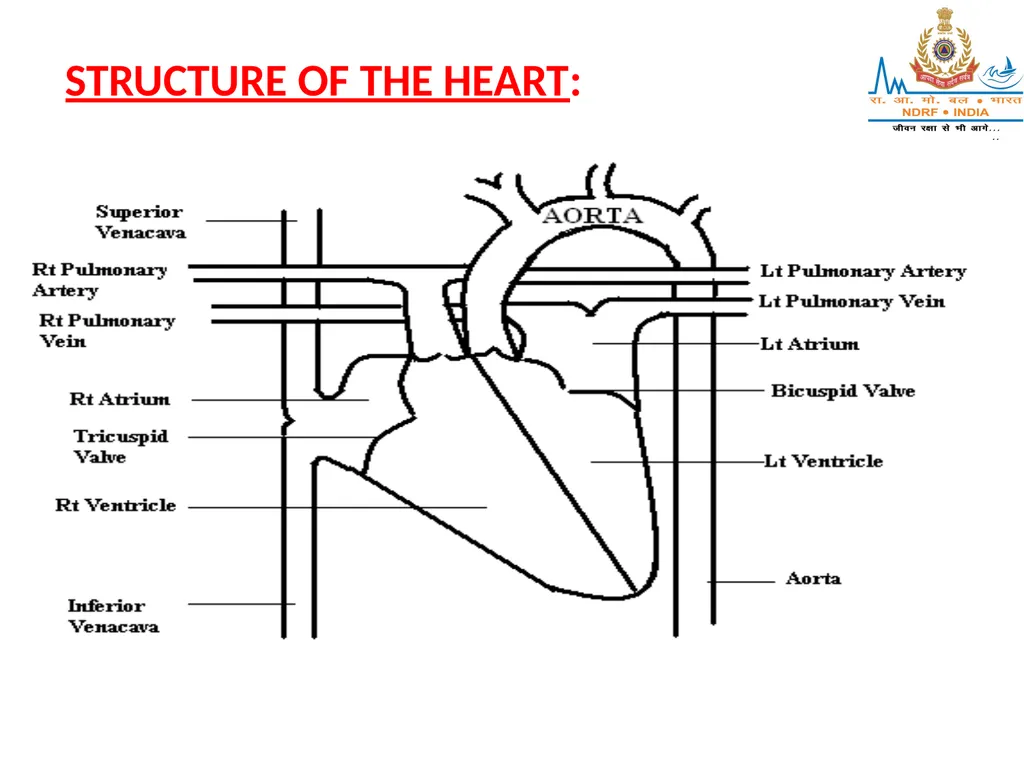
STRUCTURE OF THE HEART:<br>
40
i) From a structural point of view, the heart has 3 layers:
Pericardium – outer layer of fibrous tissue
Myocardium – middle layer of muscular tissue
Endocardium – inner layer of endothelial tissue ii) Divisions:
Divided into left & right sides by a Septum
Both sides divided into upper & lower chambers by valves made of fibrous tissue (prevent back-flow of blood)<br>
Pericardium – outer layer of fibrous tissue
Myocardium – middle layer of muscular tissue
Endocardium – inner layer of endothelial tissue ii) Divisions:
Divided into left & right sides by a Septum
Both sides divided into upper & lower chambers by valves made of fibrous tissue (prevent back-flow of blood)<br>
41
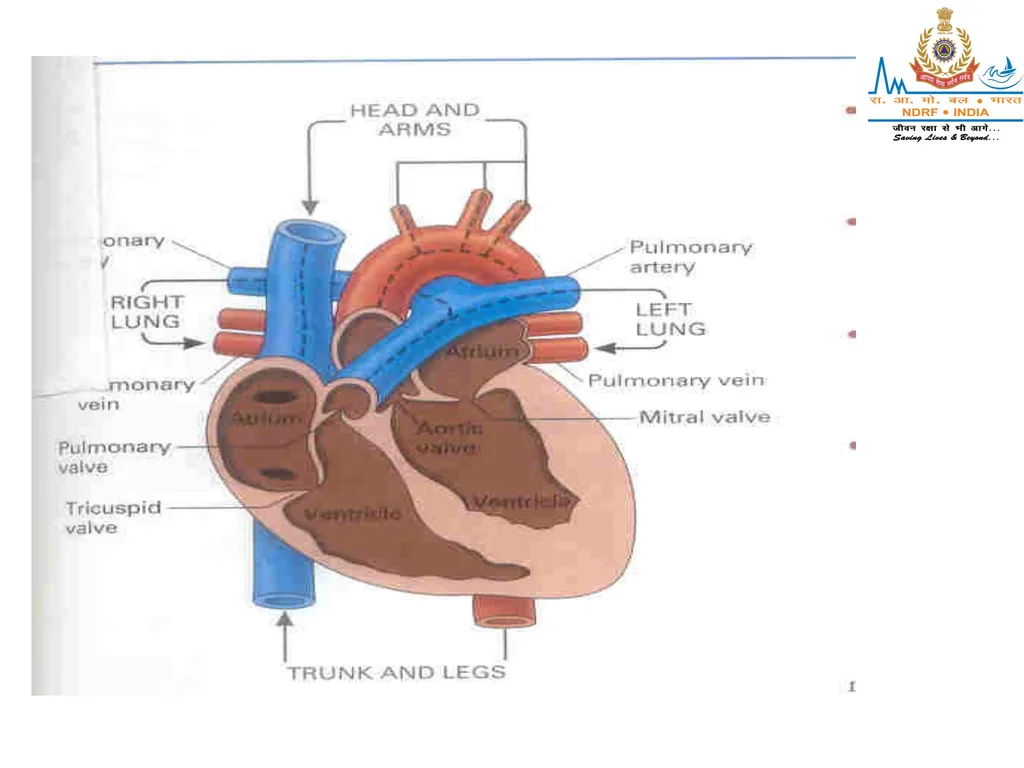
Upper chambers are called Atrium (receiving chambers)
Lower chambers are called Ventricles (pumping chambers)
There is no communication between the right & left sides of the heart
Based on the above, the heart is divided into –
Right Atrium
Right Ventricle
Left Atrium
Left Ventricle<br>
Lower chambers are called Ventricles (pumping chambers)
There is no communication between the right & left sides of the heart
Based on the above, the heart is divided into –
Right Atrium
Right Ventricle
Left Atrium
Left Ventricle<br>
42
iii) Valves:
The heart has 4 valves –
Tricuspid valve : between the Rt Atrium&
Rt Ventricle
Bicuspid Valve : between the Lt Atrium &
Lt Ventricle
Aortic Valve : between the Lt Ventricle
& Aorta
Pulmonary Valve : between the Rt Ventricle
& Pulmonary Trunk<br>
The heart has 4 valves –
Tricuspid valve : between the Rt Atrium&
Rt Ventricle
Bicuspid Valve : between the Lt Atrium &
Lt Ventricle
Aortic Valve : between the Lt Ventricle
& Aorta
Pulmonary Valve : between the Rt Ventricle
& Pulmonary Trunk<br>
43
BLOOD SUPPLY TO THE HEART:
The heart is supplied with arterial blood by the Right & Left Coronary Arteries, which are the first divisions of the Aorta
Venous return is directly into the Right Atrium through the Coronary Sinus<br>
The heart is supplied with arterial blood by the Right & Left Coronary Arteries, which are the first divisions of the Aorta
Venous return is directly into the Right Atrium through the Coronary Sinus<br>
44
NERVOUS CONTROL OF THE HEART:
Heart is controlled by the Autonomic Nervous System
The Nerves arise in the Cardiac Centre of the Medulla Oblongata
Vagus nerve (parasymphatheticfibres) is responsible for slowing down the heart-rate (by slowing down the Sino-atrial node impulses)
Symphathetic Nerves, on the other hand, speed up the heart-rate<br>
Heart is controlled by the Autonomic Nervous System
The Nerves arise in the Cardiac Centre of the Medulla Oblongata
Vagus nerve (parasymphatheticfibres) is responsible for slowing down the heart-rate (by slowing down the Sino-atrial node impulses)
Symphathetic Nerves, on the other hand, speed up the heart-rate<br>
45
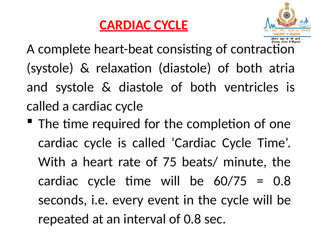
CARDIAC CYCLE A complete heart-beat consisting of contraction (systole) & relaxation (diastole) of both atria and systole & diastole of both ventricles is called a cardiac cycle The time required for the completion of one cardiac cycle is called ‘Cardiac Cycle Time’. With a heart rate of 75 beats/ minute, the cardiac cycle time will be 60/75 = 0.8 seconds, i.e. every event in the cycle will be repeated at an interval of 0.8 sec.<br>
46
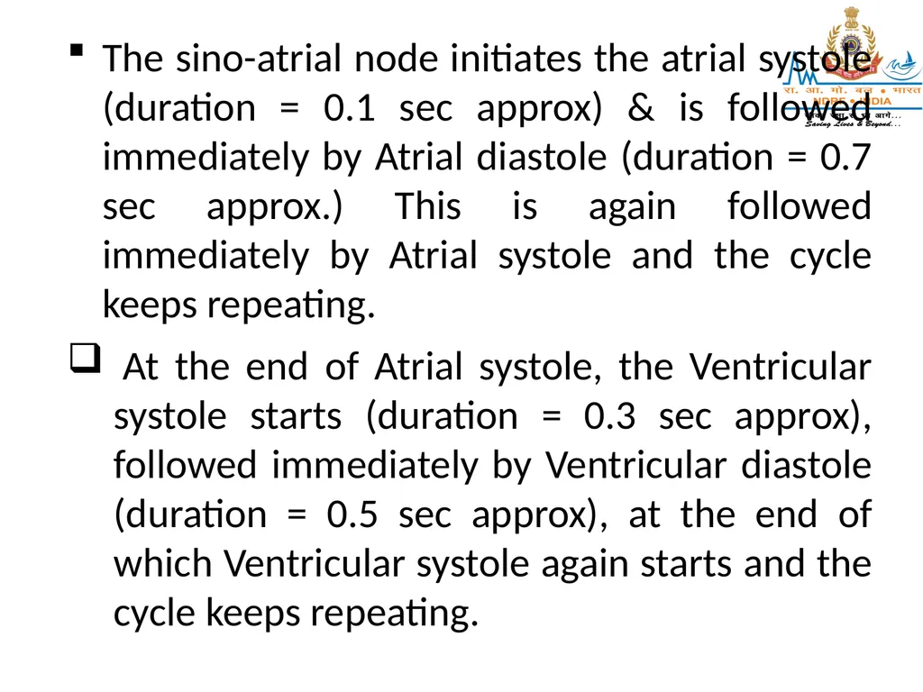
The sino-atrial node initiates the atrial systole (duration = 0.1 sec approx) & is followed immediately by Atrial diastole (duration = 0.7 sec approx.) This is again followed immediately by Atrial systole and the cycle keeps repeating.
At the end of Atrial systole, the Ventricular systole starts (duration = 0.3 sec approx), followed immediately by Ventricular diastole (duration = 0.5 sec approx), at the end of which Ventricular systole again starts and the cycle keeps repeating.<br>
At the end of Atrial systole, the Ventricular systole starts (duration = 0.3 sec approx), followed immediately by Ventricular diastole (duration = 0.5 sec approx), at the end of which Ventricular systole again starts and the cycle keeps repeating.<br>
47
BLOOD PRESSURE
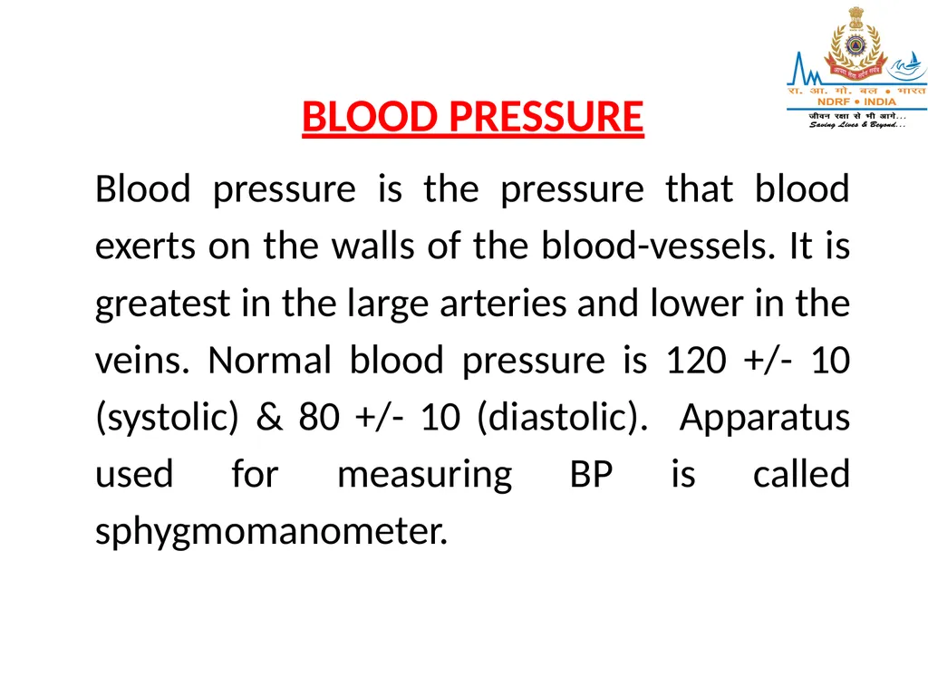
Blood pressure is the pressure that blood exerts on the walls of the blood-vessels. It is greatest in the large arteries and lower in the veins. Normal blood pressure is 120 +/- 10 (systolic) & 80 +/- 10 (diastolic). Apparatus used for measuring BP is called sphygmomanometer.<br>
Blood pressure is the pressure that blood exerts on the walls of the blood-vessels. It is greatest in the large arteries and lower in the veins. Normal blood pressure is 120 +/- 10 (systolic) & 80 +/- 10 (diastolic). Apparatus used for measuring BP is called sphygmomanometer.<br>
48
PULSE
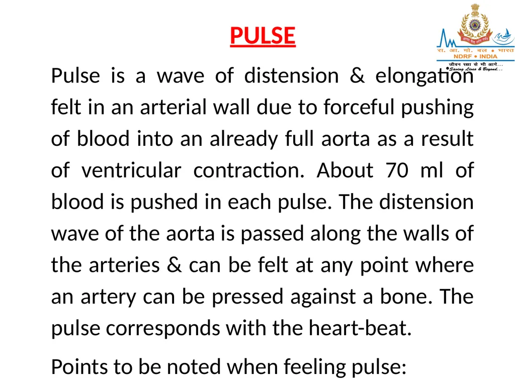
Pulse is a wave of distension & elongation felt in an arterial wall due to forceful pushing of blood into an already full aorta as a result of ventricular contraction. About 70 ml of blood is pushed in each pulse. The distension wave of the aorta is passed along the walls of the arteries & can be felt at any point where an artery can be pressed against a bone. The pulse corresponds with the heart-beat.
Points to be noted when feeling pulse:<br>
Pulse is a wave of distension & elongation felt in an arterial wall due to forceful pushing of blood into an already full aorta as a result of ventricular contraction. About 70 ml of blood is pushed in each pulse. The distension wave of the aorta is passed along the walls of the arteries & can be felt at any point where an artery can be pressed against a bone. The pulse corresponds with the heart-beat.
Points to be noted when feeling pulse:<br>
49
Rate– Number of times the heart beats in 1 minute (Normal: 72/min)
Rhythm – The length of time between each beat indicates regularity of heart-beat
Volume – The strength of the beat<br>
Rhythm – The length of time between each beat indicates regularity of heart-beat
Volume – The strength of the beat<br>
50
FACTORS AFFECTING PULSE-RATE:
(INCREASE IN RATE: TACHYCARDIA; DECRESE IN RATE: BRADYCARDIA)
Exercise speeds up the pulse-rate, while rest slows it down.
Pulse-rate decreases with age (in infants, it is 120 – 140).
Febrile conditions, shock, haemorrhage increase the rate.<br>
(INCREASE IN RATE: TACHYCARDIA; DECRESE IN RATE: BRADYCARDIA)
Exercise speeds up the pulse-rate, while rest slows it down.
Pulse-rate decreases with age (in infants, it is 120 – 140).
Febrile conditions, shock, haemorrhage increase the rate.<br>
51
Bradycardia can occur in head-injuries.
Rate is slightly higher in females than in males.
Some drugs can cause increase or decrease in rate.<br>
Rate is slightly higher in females than in males.
Some drugs can cause increase or decrease in rate.<br>
52
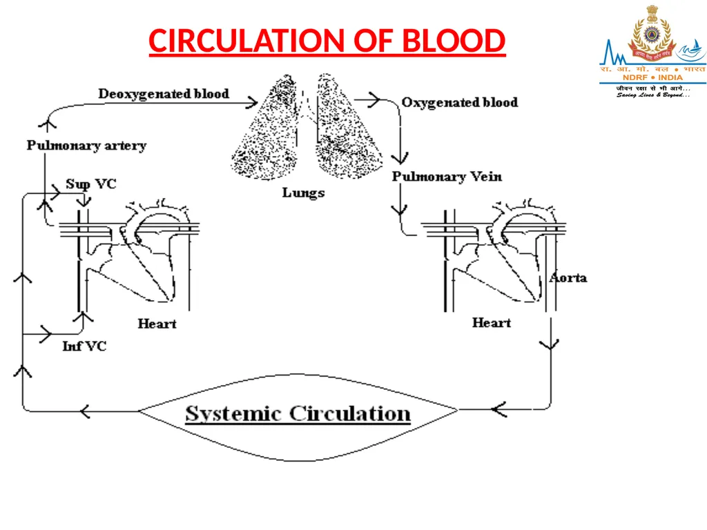
CIRCULATION OF BLOOD<br>
53
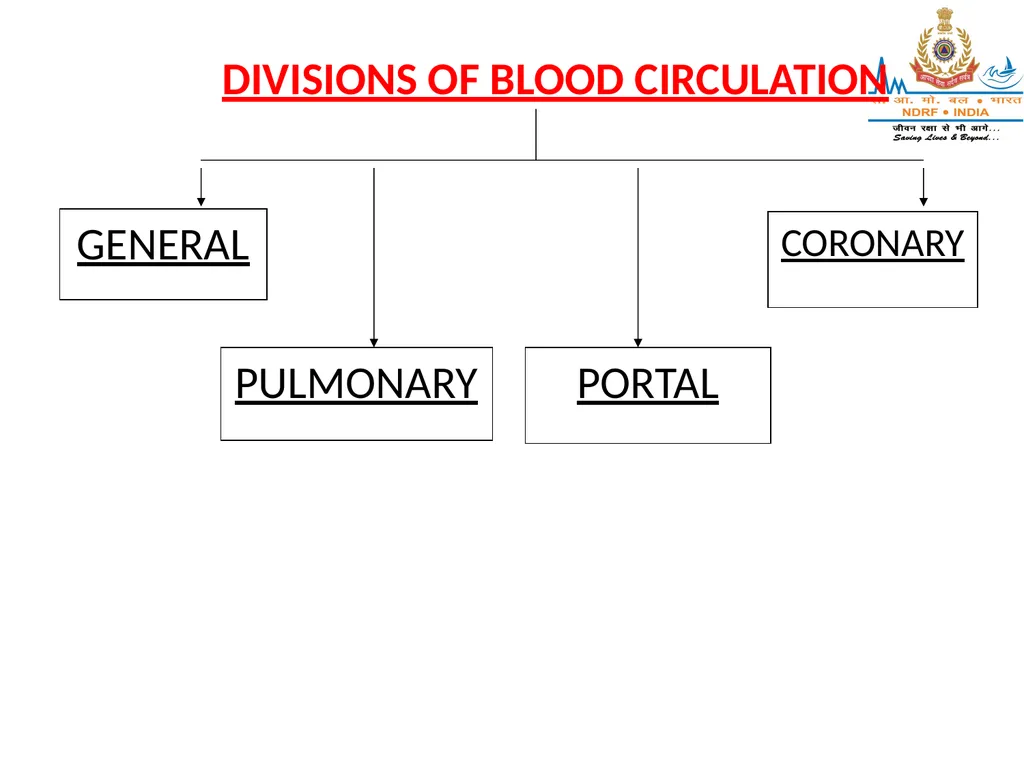
GENERAL PULMONARY PORTAL CORONARY DIVISIONS OF BLOOD CIRCULATION<br>
54
I] GENERAL CIRCULATION:
Venous blood from all parts of the body is brought to the Rt Atrium through the Superior (Head, Neck & Upper Extremity areas) and Inferior (Trunk, Lower Extremity areas) Vena-cavas
Contraction of the Rt Atrium pushes this blood into the Rt Ventricle through the Tricuspid-Valve
Contraction of the Rt Ventricle pushes this blood into the Pulmonary-trunk through the Pulmonary-Valve & further to the lungs<br>
Venous blood from all parts of the body is brought to the Rt Atrium through the Superior (Head, Neck & Upper Extremity areas) and Inferior (Trunk, Lower Extremity areas) Vena-cavas
Contraction of the Rt Atrium pushes this blood into the Rt Ventricle through the Tricuspid-Valve
Contraction of the Rt Ventricle pushes this blood into the Pulmonary-trunk through the Pulmonary-Valve & further to the lungs<br>
55
After oxygenation in the lungs, the blood passes into the Lt Atrium through the Pulmonary Vein
Contraction of the Lt Atrium pushes this blood into the Lt Ventricle through the Bicuspid-Valve
Contraction of the Lt Ventricle pushes this blood into the Aorta through the Aortic-Valve
The Aorta & its branches take the blood to various parts of the body<br>
Contraction of the Lt Atrium pushes this blood into the Lt Ventricle through the Bicuspid-Valve
Contraction of the Lt Ventricle pushes this blood into the Aorta through the Aortic-Valve
The Aorta & its branches take the blood to various parts of the body<br>
56
The arteries break up into smaller arterioles & further into capillaries
Exchange of gases (O2 out & CO2 in) and nutrient material takes place in the capillary beds CO2 & waste products are taken by the venules which join up to form veins and ultimately drain into the superior vena –cava and inferior vena cava This cycle is then repeated<br>
Exchange of gases (O2 out & CO2 in) and nutrient material takes place in the capillary beds CO2 & waste products are taken by the venules which join up to form veins and ultimately drain into the superior vena –cava and inferior vena cava This cycle is then repeated<br>
57
II] PULMONARY CIRCULATION:
Contraction of the Rt Ventricle pushes the de-oxygenated blood into the Pulmonary-trunk through the Pulmonary-Valve
Pulmonary trunk divides into the Rt & Lt Pulmonary Arteries, which pass onto the respective lungs
Gaseous exchange (O2 absorbed & CO2 excreted) takes place in the alvoli of the lungs<br>
Contraction of the Rt Ventricle pushes the de-oxygenated blood into the Pulmonary-trunk through the Pulmonary-Valve
Pulmonary trunk divides into the Rt & Lt Pulmonary Arteries, which pass onto the respective lungs
Gaseous exchange (O2 absorbed & CO2 excreted) takes place in the alvoli of the lungs<br>
58
After oxygenation, the blood passes into the Lt Atrium through the Pulmonary Vein
Pulmonary artery is the only artery carrying de-oxygenated blood
Pulmonary vein is the only vein carrying oxygenated blood<br>
Pulmonary artery is the only artery carrying de-oxygenated blood
Pulmonary vein is the only vein carrying oxygenated blood<br>
59
III] PORTAL CIRCULATION:
Circulation of blood through the liver is called Portal circulation
Blood which has circulated through the stomach, intestines, pancreas & spleen is taken to the liver through the Portal vein Blood is detoxified by enzymes present in the liver
Blood collected from the liver drains through the Hepatic vein into the Inferior Vena-cava<br>
Circulation of blood through the liver is called Portal circulation
Blood which has circulated through the stomach, intestines, pancreas & spleen is taken to the liver through the Portal vein Blood is detoxified by enzymes present in the liver
Blood collected from the liver drains through the Hepatic vein into the Inferior Vena-cava<br>
60
IV] CORONARY CIRCULATION:
Concerned with blood-supply to the Heart- muscle
The Right & Left Coronary arteries branch off from the Ascending Aorta
These further divide into smaller arteries & arterioles which encircle the heart
Impure blood collected from the heart drains directly into the Rt Atrium through the Coronary Sinus<br>
Concerned with blood-supply to the Heart- muscle
The Right & Left Coronary arteries branch off from the Ascending Aorta
These further divide into smaller arteries & arterioles which encircle the heart
Impure blood collected from the heart drains directly into the Rt Atrium through the Coronary Sinus<br>
61
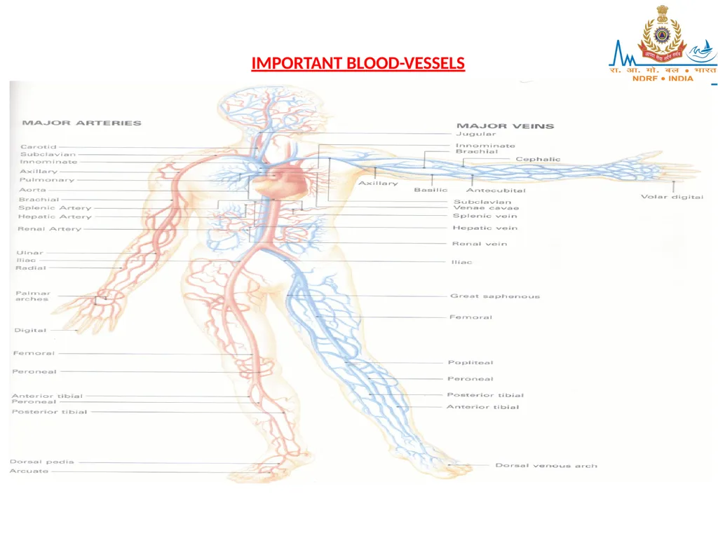
IMPORTANT BLOOD-VESSELS<br>
62
I] AORTA:
Largest artery of the body
Arises from the Lt Ventricle of the heart
Part of Aorta situated in the thorax is known as Thoracic Aorta Part of Aorta situated in the abdomen is known as Abdominal Aorta
Passes through the aortic opening of the diaphragm at the level of the 12th thoracic vertebra<br>
Largest artery of the body
Arises from the Lt Ventricle of the heart
Part of Aorta situated in the thorax is known as Thoracic Aorta Part of Aorta situated in the abdomen is known as Abdominal Aorta
Passes through the aortic opening of the diaphragm at the level of the 12th thoracic vertebra<br>
63
At the level of the 4th lumbar vertebra, it divides into the two main Iliac arteries
Throughout its length, it gives off a number of branches, some paired & others single II] BLOOD SUPPLY OF HEAD & NECK: ARTERIAL SUPPLY:
The Common Carotid Arteries (Rt & Lt) supply the head & neck.
It divides into the External & Internal Carotid Arteries on both sides<br>
Throughout its length, it gives off a number of branches, some paired & others single II] BLOOD SUPPLY OF HEAD & NECK: ARTERIAL SUPPLY:
The Common Carotid Arteries (Rt & Lt) supply the head & neck.
It divides into the External & Internal Carotid Arteries on both sides<br>
64
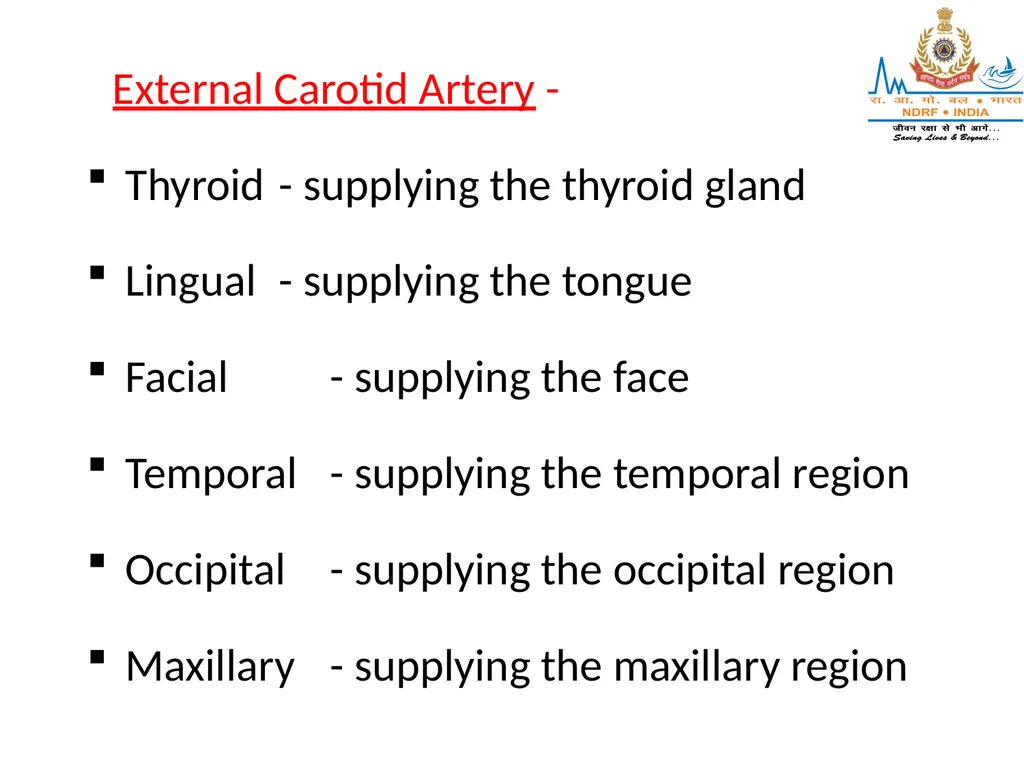
External Carotid Artery -
Thyroid - supplying the thyroid gland
Lingual - supplying the tongue
Facial - supplying the face
Temporal - supplying the temporal region
Occipital - supplying the occipital region
Maxillary - supplying the maxillary region<br>
Thyroid - supplying the thyroid gland
Lingual - supplying the tongue
Facial - supplying the face
Temporal - supplying the temporal region
Occipital - supplying the occipital region
Maxillary - supplying the maxillary region<br>
65
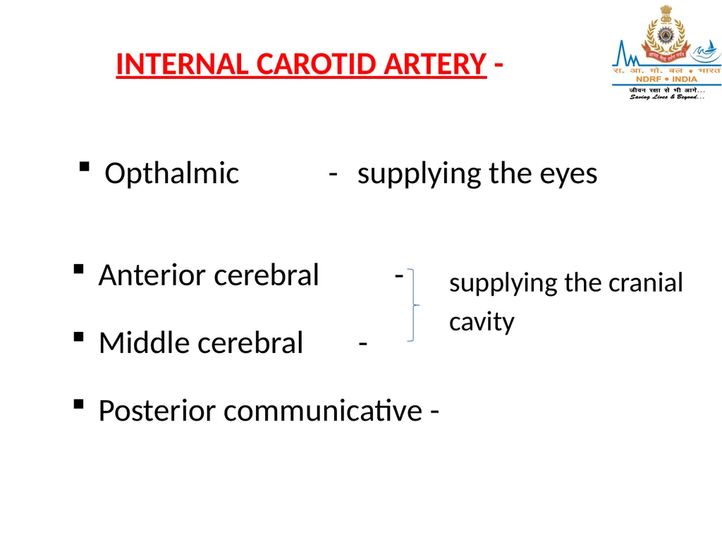
INTERNAL CAROTID ARTERY - Opthalmic - supplying the eyes Anterior cerebral -
Middle cerebral -
Posterior communicative - supplying the cranial cavity<br>
Middle cerebral -
Posterior communicative - supplying the cranial cavity<br>
66
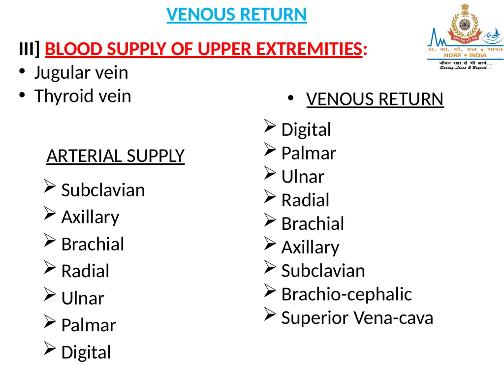
VENOUS RETURN III] BLOOD SUPPLY OF UPPER EXTREMITIES:
Jugular vein
Thyroid vein ARTERIAL SUPPLY
Subclavian
Axillary
Brachial
Radial
Ulnar
Palmar
Digital VENOUS RETURN
Digital
Palmar
Ulnar
Radial
Brachial
Axillary
Subclavian
Brachio-cephalic
Superior Vena-cava<br>
Jugular vein
Thyroid vein ARTERIAL SUPPLY
Subclavian
Axillary
Brachial
Radial
Ulnar
Palmar
Digital VENOUS RETURN
Digital
Palmar
Ulnar
Radial
Brachial
Axillary
Subclavian
Brachio-cephalic
Superior Vena-cava<br>
67
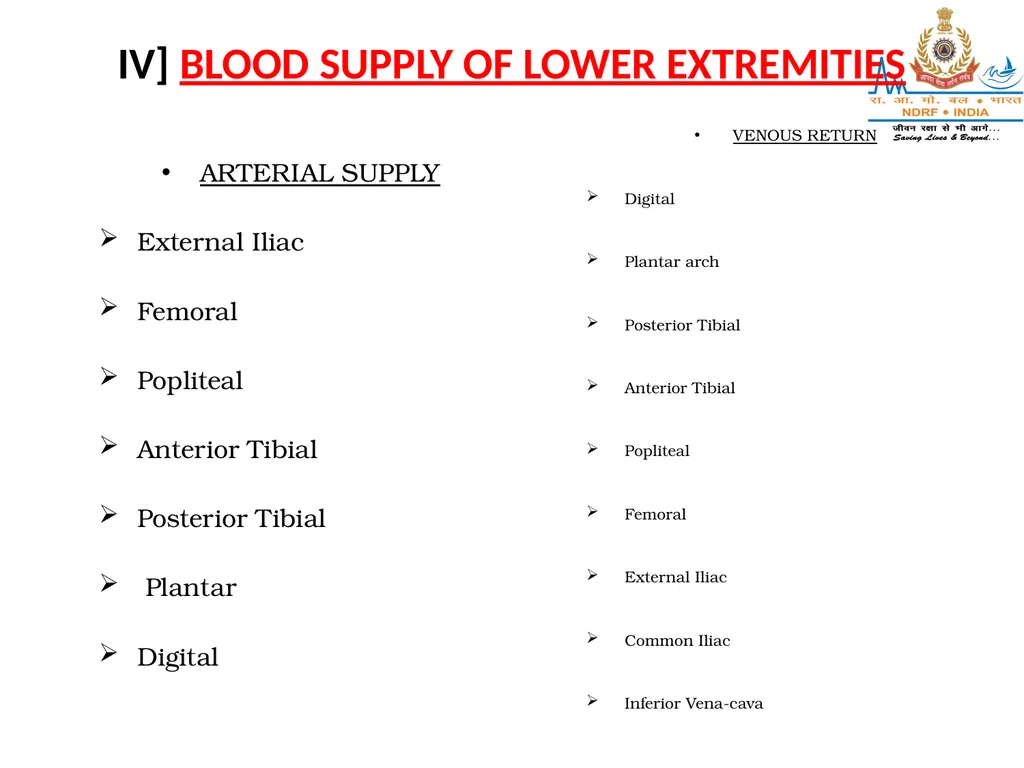
IV] BLOOD SUPPLY OF LOWER EXTREMITIES ARTERIAL SUPPLY
External Iliac
Femoral
Popliteal
Anterior Tibial
Posterior Tibial
Plantar
Digital VENOUS RETURN
Digital
Plantar arch
Posterior Tibial
Anterior Tibial
Popliteal
Femoral
External Iliac
Common Iliac
Inferior Vena-cava<br>
External Iliac
Femoral
Popliteal
Anterior Tibial
Posterior Tibial
Plantar
Digital VENOUS RETURN
Digital
Plantar arch
Posterior Tibial
Anterior Tibial
Popliteal
Femoral
External Iliac
Common Iliac
Inferior Vena-cava<br>
68
ANY QUESTION
?<br>
?<br>
69
THANKS<br>