
Commonly Seen Infectious and Non-Infectious Skin
YM
Published · 86 slides · 0 views

1 / 1
Description
Commonly Seen Infectious and Non-Infectious Skin Disorders in the School-Aged Child Melinda Rodriguez DNP, APRN, FNP-BC Nursing Education Doctors Hospital at Renaissance Health Systems Disclosures Nothing to disclose Objectives Discuss a
Related Topics
Share
Embed code
Download this presentation From Below
"Commonly Seen Infectious and Non-Infectious Skin" is the property of its rightful owner. Permission is granted to download and print the materials on this website for personal, non-commercial use only, and to display it on your personal computer provided you do not modify the materials and that you retain all copyright notices contained in the materials. By downloading content from our website, you accept the terms of this agreement.
Presentation Transcript
01
Commonly Seen Infectious and Non-Infectious Skin Disorders in the School-Aged Child Melinda Rodriguez DNP, APRN, FNP-BC
Nursing Education Doctor’s Hospital at Renaissance Health Systems<br>
Nursing Education Doctor’s Hospital at Renaissance Health Systems<br>
02
Disclosures Nothing to disclose<br>
03
Objectives Discuss a brief overview of the anatomy and physiology of the skin
Discuss the importance of history collection
Discuss infectious skin disorders affecting the school-aged child
Discuss the non-infectious skin disorders affecting the school-aged child
Discuss assessment and management of Acanthosis Nigricans (AN)<br>
Discuss the importance of history collection
Discuss infectious skin disorders affecting the school-aged child
Discuss the non-infectious skin disorders affecting the school-aged child
Discuss assessment and management of Acanthosis Nigricans (AN)<br>
04
Brief Overview of Integumentary System Provides an elastic, rugged, self-regenerating cover for the body
Largest organ of the body
Includes: hair and nails
Maintains and keeps body structures in place<br>
Largest organ of the body
Includes: hair and nails
Maintains and keeps body structures in place<br>
05
Anatomy and Physiology Comprised of several layers
Protects against microbial and foreign substance invasion
Regulates body temperature
Provides sensory perception via nerve endings
Produces vitamin D from precursors in skin
Contributes to blood pressure regulation<br>
Protects against microbial and foreign substance invasion
Regulates body temperature
Provides sensory perception via nerve endings
Produces vitamin D from precursors in skin
Contributes to blood pressure regulation<br>
06
Functions of the Skin Complex organs made up of may cell types
Largest organ of the body
Provides barrier between external and internal environments
Provides protection against organisms
Skin receptors relay: touch, pressure, temperature and pain to CNS
Also provide ability for localization and discrimination McCance & Huether, (2014).<br>
Largest organ of the body
Provides barrier between external and internal environments
Provides protection against organisms
Skin receptors relay: touch, pressure, temperature and pain to CNS
Also provide ability for localization and discrimination McCance & Huether, (2014).<br>
07
Overview of the Skin Assessment Problems may arise from many mechanisms and inflammatory processes
Some causes may be environmental, traumatic and secondary to exposures
Evaluation of skin disorders require a in-depth focus history and PE
Assess for infectious symptoms: fever, itching
Look at the presentation of lesion, configuration and distribution Seidel, Ball, Dains et al., (2015).<br>
Some causes may be environmental, traumatic and secondary to exposures
Evaluation of skin disorders require a in-depth focus history and PE
Assess for infectious symptoms: fever, itching
Look at the presentation of lesion, configuration and distribution Seidel, Ball, Dains et al., (2015).<br>
08
External Clues to Internal Problems Persistent pruritus may indicate chronic renal failure, liver disease, diabetes
Supernumerary nipples located along mammary ridge, may be associated with renal problems
Facial port wine stain may be associated with ocular defects, malformation of meninges<br>
Supernumerary nipples located along mammary ridge, may be associated with renal problems
Facial port wine stain may be associated with ocular defects, malformation of meninges<br>
09
Age-Appropriate History Gather data specific to current skin problems
Family, PMH of similar problems
Skin care routines
Recent changes in skin, hair or nail care
Sun-exposure habits; use of sunscreen
Medication history
Onset, date of occurrence
History of recent travel
Rx medications; OTC medications, lotions used<br>
Family, PMH of similar problems
Skin care routines
Recent changes in skin, hair or nail care
Sun-exposure habits; use of sunscreen
Medication history
Onset, date of occurrence
History of recent travel
Rx medications; OTC medications, lotions used<br>
10
History of Present Illness Note recent or past changes in the skin: pruritus, dryness, sores, rashes, lumps
Symptoms: pain, exudate, bleeding, color changes
Recent drug exposure; chemicals;
Generalized symptoms: fever, travel hx
Use of topical or oral medications Seidel et al., (2015).<br>
Symptoms: pain, exudate, bleeding, color changes
Recent drug exposure; chemicals;
Generalized symptoms: fever, travel hx
Use of topical or oral medications Seidel et al., (2015).<br>
11
History (cont’d) Eating habits; allergies to foods
Communicable disease exposure
Allergic disorders; asthma
Exposure to pets; animals
Skin injury; outdoor exposures
Nail biting
Thinning of hair Seidel et al., (2015).<br>
Communicable disease exposure
Allergic disorders; asthma
Exposure to pets; animals
Skin injury; outdoor exposures
Nail biting
Thinning of hair Seidel et al., (2015).<br>
12
Mechanisms of Self-Defense Bacteria-Derived Chemicals: skin, mucous membranes and GI tract, urethra and vagina have protective microorganisms
Common bacteria on the skin: staph and strept
C-difficile in the GI tract
Lactobacillus protection of the vaginal tract<br>
Common bacteria on the skin: staph and strept
C-difficile in the GI tract
Lactobacillus protection of the vaginal tract<br>
13
Inspection of the Skin Performed by inspection and palpation
Inspection: lighting is essential
Observe for symmetry
Adequate exposure of the skin
Inspect skin thickness
Assess for color variances
Assess for nevi; abnormally shaped; variegated colors Seidel et al., (2015).<br>
Inspection: lighting is essential
Observe for symmetry
Adequate exposure of the skin
Inspect skin thickness
Assess for color variances
Assess for nevi; abnormally shaped; variegated colors Seidel et al., (2015).<br>
14
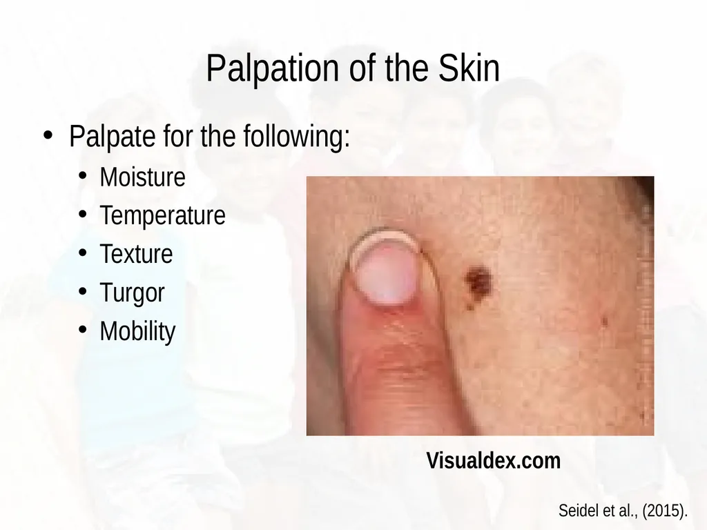
Palpation of the Skin Palpate for the following:
Moisture
Temperature
Texture
Turgor
Mobility Visualdex.com Seidel et al., (2015).<br>
Moisture
Temperature
Texture
Turgor
Mobility Visualdex.com Seidel et al., (2015).<br>
15
Blood supply/nerve innervation Blood supply to skin limited
Include papillary capillaries
Dermis facilitates the regulation of body temperature
Evaporation of sweat cools body
Regulates vasoconstriction McCance & Huether, (2010).<br>
Include papillary capillaries
Dermis facilitates the regulation of body temperature
Evaporation of sweat cools body
Regulates vasoconstriction McCance & Huether, (2010).<br>
16
Morphological Criteria Includes:
Location of lesion
Distribution
Determine whether primary or secondary
Shape of lesion
Margins/borders/irregularities
Pigmentation/color/variations
Palpate texture/consistency
Wear gloves if open lesions present Seidel et al., (2015).<br>
Location of lesion
Distribution
Determine whether primary or secondary
Shape of lesion
Margins/borders/irregularities
Pigmentation/color/variations
Palpate texture/consistency
Wear gloves if open lesions present Seidel et al., (2015).<br>
17
Morphological Characteristics of Lesions Linear (in a line)
Stellate (star shaped)
Reticulate (netlike; lacy)
Mobilliform (maculopapular; confluent)
Irregular borders
Border raised above
Advancing; spreading beyond borders (cellulitis) Seidel et al., (2015).<br>
Stellate (star shaped)
Reticulate (netlike; lacy)
Mobilliform (maculopapular; confluent)
Irregular borders
Border raised above
Advancing; spreading beyond borders (cellulitis) Seidel et al., (2015).<br>
18
Pigmentation Flesh colored
Erythematous/pink
Salmon colored (psoriasis)
Black
Purple
Yellow/waxy
Pearly<br>
Erythematous/pink
Salmon colored (psoriasis)
Black
Purple
Yellow/waxy
Pearly<br>
19
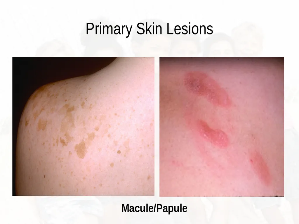
Primary Skin Lesions Macule: flat, circumscribed area; changes to color of skin; less than 1cm in diameter (freckle)
Papule: elevated firm circumscribed area less than 1cm (wart)
Patch: a flat non-palpable irregular shaped macule; more than 1cm (vitiligo)
Plaque: elevated, firm, rough with flat top surface; greater than 1cm in diameter ( psoriasis)
Vesicle: elevated, circumscribed superficial; does not extend to dermis, filled with serous fluid less than 1cm McCance & Huether, (2014).<br>
Papule: elevated firm circumscribed area less than 1cm (wart)
Patch: a flat non-palpable irregular shaped macule; more than 1cm (vitiligo)
Plaque: elevated, firm, rough with flat top surface; greater than 1cm in diameter ( psoriasis)
Vesicle: elevated, circumscribed superficial; does not extend to dermis, filled with serous fluid less than 1cm McCance & Huether, (2014).<br>
20
Primary Skin Lesions Macule/Papule<br>
21
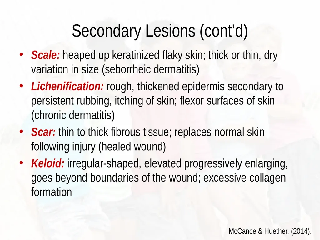
Secondary Lesions (cont’d) Scale: heaped up keratinized flaky skin; thick or thin, dry variation in size (seborrheic dermatitis)
Lichenification: rough, thickened epidermis secondary to persistent rubbing, itching of skin; flexor surfaces of skin (chronic dermatitis)
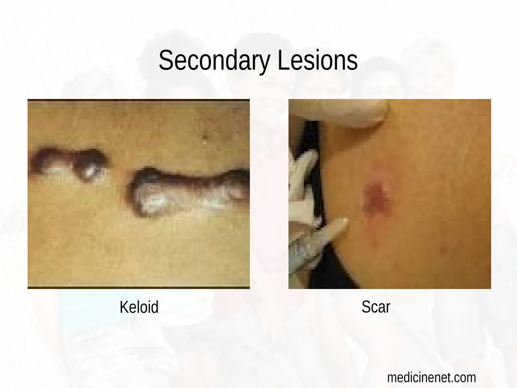
Scar: thin to thick fibrous tissue; replaces normal skin following injury (healed wound)
Keloid: irregular-shaped, elevated progressively enlarging, goes beyond boundaries of the wound; excessive collagen formation McCance & Huether, (2014).<br>
Lichenification: rough, thickened epidermis secondary to persistent rubbing, itching of skin; flexor surfaces of skin (chronic dermatitis)
Scar: thin to thick fibrous tissue; replaces normal skin following injury (healed wound)
Keloid: irregular-shaped, elevated progressively enlarging, goes beyond boundaries of the wound; excessive collagen formation McCance & Huether, (2014).<br>
22
Secondary Lesions Keloid Scar medicinenet.com<br>
23
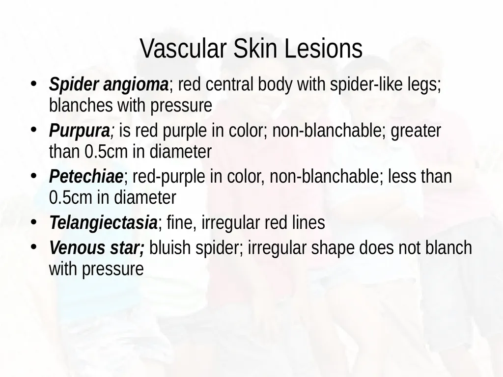
Vascular Skin Lesions Spider angioma; red central body with spider-like legs; blanches with pressure
Purpura; is red purple in color; non-blanchable; greater than 0.5cm in diameter
Petechiae; red-purple in color, non-blanchable; less than 0.5cm in diameter
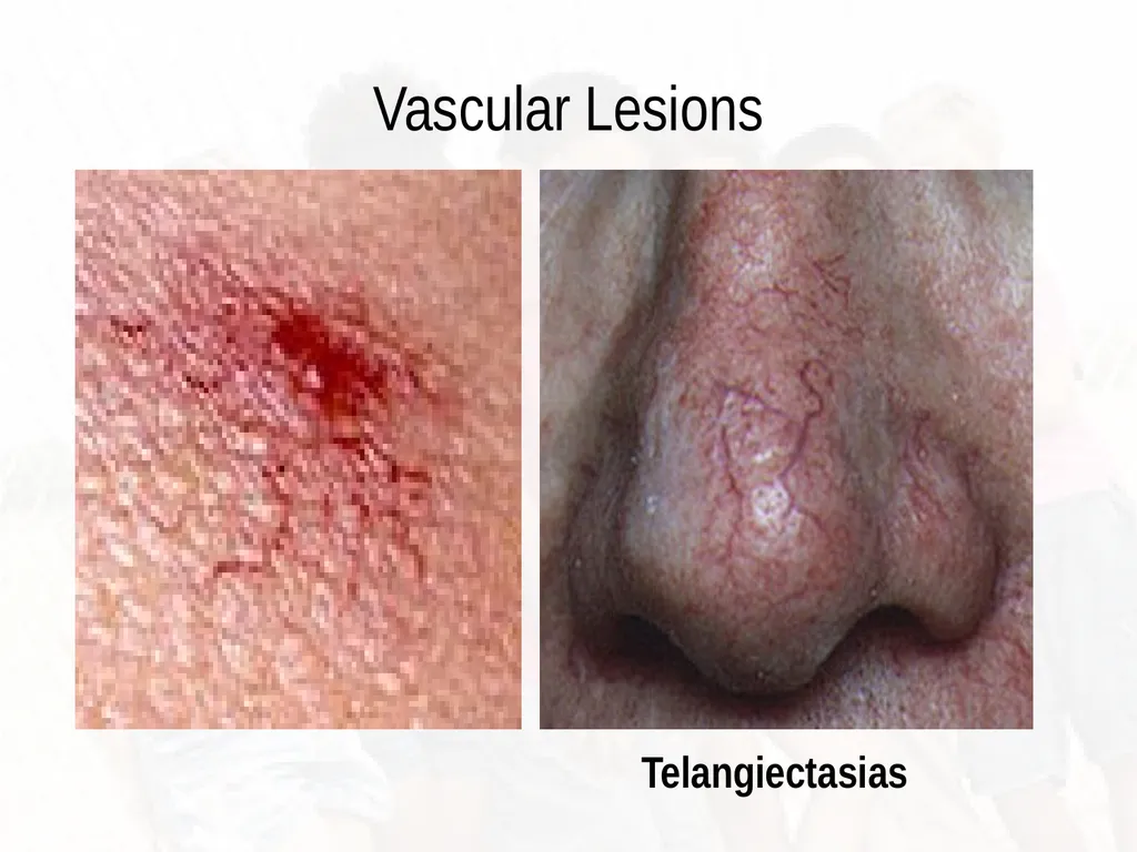
Telangiectasia; fine, irregular red lines
Venous star; bluish spider; irregular shape does not blanch with pressure<br>
Purpura; is red purple in color; non-blanchable; greater than 0.5cm in diameter
Petechiae; red-purple in color, non-blanchable; less than 0.5cm in diameter
Telangiectasia; fine, irregular red lines
Venous star; bluish spider; irregular shape does not blanch with pressure<br>
24
Vascular Lesions Telangiectasias<br>
25
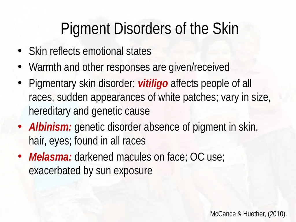
Pigment Disorders of the Skin Skin reflects emotional states
Warmth and other responses are given/received
Pigmentary skin disorder: vitiligo affects people of all races, sudden appearances of white patches; vary in size, hereditary and genetic cause
Albinism: genetic disorder absence of pigment in skin, hair, eyes; found in all races
Melasma: darkened macules on face; OC use; exacerbated by sun exposure McCance & Huether, (2010).<br>
Warmth and other responses are given/received
Pigmentary skin disorder: vitiligo affects people of all races, sudden appearances of white patches; vary in size, hereditary and genetic cause
Albinism: genetic disorder absence of pigment in skin, hair, eyes; found in all races
Melasma: darkened macules on face; OC use; exacerbated by sun exposure McCance & Huether, (2010).<br>
26
Assessment of the Adolescent Increased oiliness or perspiration may be evident
Increased axillary perspiration related to maturity of the apocrine glands
Hair on extremities becomes coarser and darker
Pubic hair develops; secondary sex characteristics<br>
Increased axillary perspiration related to maturity of the apocrine glands
Hair on extremities becomes coarser and darker
Pubic hair develops; secondary sex characteristics<br>
27
Infectious and Non-Infectious Conditions of the Skin Management and Treatment<br>
28
Common Skin Disorders Seen in the Schools Impetigo
Varicella
Scabies/Pediculosis
Herpes simplex
Contact dermatitis/eczema
Molluscum Contagiosum
Hand, Foot and Mouth Disease
Fifth’s Disease (erythema infectiosum)
Rubeola /Measles
Stept Infection (Scarletina)<br>
Varicella
Scabies/Pediculosis
Herpes simplex
Contact dermatitis/eczema
Molluscum Contagiosum
Hand, Foot and Mouth Disease
Fifth’s Disease (erythema infectiosum)
Rubeola /Measles
Stept Infection (Scarletina)<br>
29
Infectious vs. Non-Infectious History of present illness is very important
Events that preceded the skin condition
Need to rule out trauma
Medication history
Previous outbreak
Fever and any other systemic symptoms
Allergies<br>
Events that preceded the skin condition
Need to rule out trauma
Medication history
Previous outbreak
Fever and any other systemic symptoms
Allergies<br>
30
Eczema Characterized by : acute inflammation, erythema, edema and vesiculation
Itching is often severe
Multiple causes; allergic contact
Common culprits: personal care products, fragrances, detergents
Often sudden in onset Habif, (2011).<br>
Itching is often severe
Multiple causes; allergic contact
Common culprits: personal care products, fragrances, detergents
Often sudden in onset Habif, (2011).<br>
31
Prognosis/Management Avoid provoking factors; eruption improves in 7-10 days
Excoriation secondary to itching/scratching could develop bacterial infection
Topical steroids (used sparingly and as directed)
Oral antihistamines (Benadryl)
Treatment often based on elimination of causing factor Habif, (2011).<br>
Excoriation secondary to itching/scratching could develop bacterial infection
Topical steroids (used sparingly and as directed)
Oral antihistamines (Benadryl)
Treatment often based on elimination of causing factor Habif, (2011).<br>
32
Allergic Contact Dermatitis Common T-cell mediated or delayed hypersensitivity
Allergens: chemicals, foreign proteins, poison ivy
Erythema, swelling with itching
Vesicular lesions are where contact is made
Removal is necessary to help with tissue repair
Systemic steroids are one form of treatment
Atopic dermatitis: more common in infancy and childhood, usually associated with asthma, allergic rhinitis McCance & Huether, (2014).<br>
Allergens: chemicals, foreign proteins, poison ivy
Erythema, swelling with itching
Vesicular lesions are where contact is made
Removal is necessary to help with tissue repair
Systemic steroids are one form of treatment
Atopic dermatitis: more common in infancy and childhood, usually associated with asthma, allergic rhinitis McCance & Huether, (2014).<br>
33
Allergic Contact Dermatitis Delayed type hypersensitivity reaction
Caused by skin contact with an allergen
Results in eczematous dermatitis
Common causes include:
Metals (nickel)
Rubber
Shoes
Preservatives in lotions, creams, cosmetics Habif, (2011).<br>
Caused by skin contact with an allergen
Results in eczematous dermatitis
Common causes include:
Metals (nickel)
Rubber
Shoes
Preservatives in lotions, creams, cosmetics Habif, (2011).<br>
34
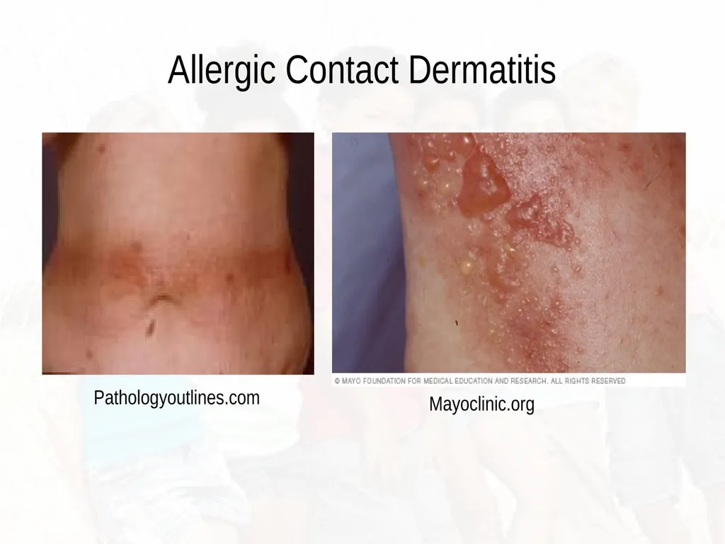
Allergic Contact Dermatitis Pathologyoutlines.com Mayoclinic.org<br>
35
Management/ Treatment Avoidance of the allergenic substance
Identification of allergen (patch testing)
Topical treatment (topical corticosteroids)
Choice of topical corticosteroids depends on body site affected (use sparingly on pediatric population)
3-week tapering course of oral corticosteroids
Education of patient/caregiver Habif, (2011).<br>
Identification of allergen (patch testing)
Topical treatment (topical corticosteroids)
Choice of topical corticosteroids depends on body site affected (use sparingly on pediatric population)
3-week tapering course of oral corticosteroids
Education of patient/caregiver Habif, (2011).<br>
36
Pediatric Considerations Allergies can develop after years of exposure to products/medications
Consider “patch” testing
Re-assessment of recent exposures
Assess the integrity of the skin
Be alert for S/S of infection Habif, (2011).<br>
Consider “patch” testing
Re-assessment of recent exposures
Assess the integrity of the skin
Be alert for S/S of infection Habif, (2011).<br>
37
Bacterial Infections of the Skin Can result from primary skin lesions
Any break in the integrity of the skin
May result in erythema, edema, pain, pus
May result in systemic symptoms such as:
Fever
Malaise
Myalgias
Nausea and vomiting<br>
Any break in the integrity of the skin
May result in erythema, edema, pain, pus
May result in systemic symptoms such as:
Fever
Malaise
Myalgias
Nausea and vomiting<br>
38
Impetigo Highly contagious superficial skin infection
Caused by strept or staph
80% of cases caused by staph aureus
Occurs after minor skin injury, insect bite
Bacteria may colonize in the nasal passages
Warm climates and poor hygiene contribute to it
Lesions may be localized or wide spread; common on face Habif, (2011).<br>
Caused by strept or staph
80% of cases caused by staph aureus
Occurs after minor skin injury, insect bite
Bacteria may colonize in the nasal passages
Warm climates and poor hygiene contribute to it
Lesions may be localized or wide spread; common on face Habif, (2011).<br>
39
Skin Findings Vesicles/pustules present
Red a moist base
Erythematous
Lesions often coalesce
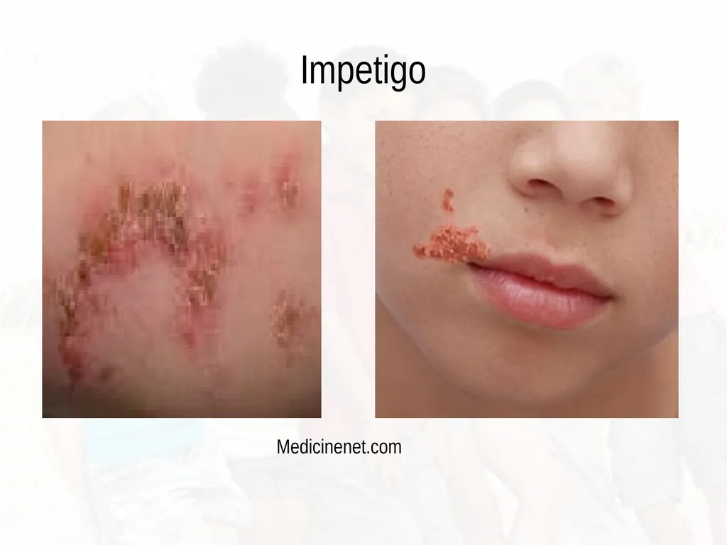
Develop an adherent crust honey-yellow to white-brown in color
Thin-roofed bullae may develop Habif, (2011).<br>
Red a moist base
Erythematous
Lesions often coalesce
Develop an adherent crust honey-yellow to white-brown in color
Thin-roofed bullae may develop Habif, (2011).<br>
40
Impetigo Medicinenet.com<br>
41
Pediatric Considerations Most common bacterial infection in children
Rarely post-streptococcal glomerulonephritis may follow infection
Antibacterial soaps are recommended to be used twice daily for chronic cases
Bacterial culture may be indicated for chronic cases Habif, (2011).<br>
Rarely post-streptococcal glomerulonephritis may follow infection
Antibacterial soaps are recommended to be used twice daily for chronic cases
Bacterial culture may be indicated for chronic cases Habif, (2011).<br>
42
Treatment/Management Disease is self-limiting; could spread
Localized infections: Mupirocin 2% topical
Oral antibiotics: doxycycline, clarithromycin, cephalexin (Keflex) x 10-14 days of treatment
Recurrent impetigo may require topical Mupirocin in the nares
Good handwashing Habif, (2011).<br>
Localized infections: Mupirocin 2% topical
Oral antibiotics: doxycycline, clarithromycin, cephalexin (Keflex) x 10-14 days of treatment
Recurrent impetigo may require topical Mupirocin in the nares
Good handwashing Habif, (2011).<br>
43
Viral Infections Verucca: warts, common benign papillomas; caused by HPV; transmitted by direct contact
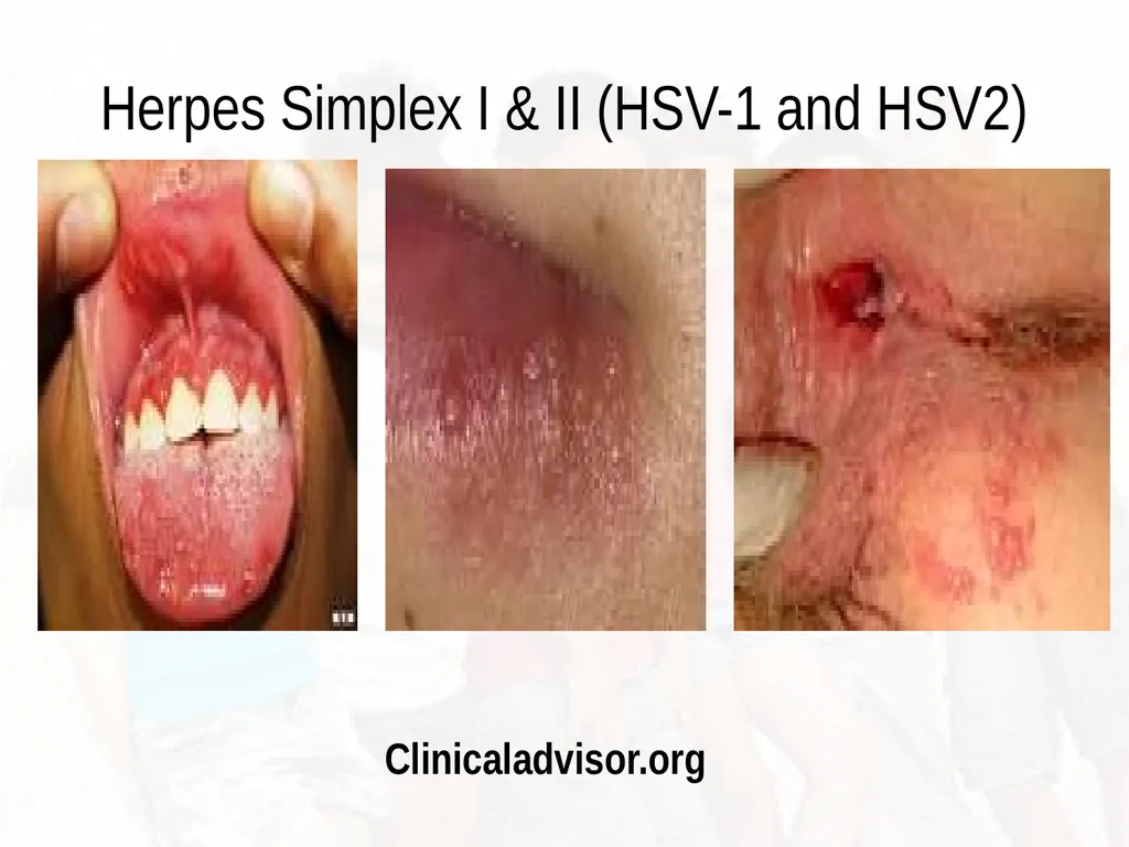
Herpes simplex: (HSV) infection of skin and mucous membranes; two types HSV 1 and HSV 2; symptoms begin with burning or tingling; umbilicated vesicles and erythema
Herpes Zoster: shingles; acute localized vesicular eruption distributed along dermatomal segment; prevention via Zostavax vaccine McCance & Huether, (2014).<br>
Herpes simplex: (HSV) infection of skin and mucous membranes; two types HSV 1 and HSV 2; symptoms begin with burning or tingling; umbilicated vesicles and erythema
Herpes Zoster: shingles; acute localized vesicular eruption distributed along dermatomal segment; prevention via Zostavax vaccine McCance & Huether, (2014).<br>
44
Verruca Vulgaris Also known as “warts”
Benign epidermal proliferations
Caused by human papilloma virus (HPV)
Over 150 different types of HPV
Transmission is by simple contact; often on non-intact skin
Local spread is caused by autoinoculation
Peak incidence ages 12-16 yrs Habif, (2011).<br>
Benign epidermal proliferations
Caused by human papilloma virus (HPV)
Over 150 different types of HPV
Transmission is by simple contact; often on non-intact skin
Local spread is caused by autoinoculation
Peak incidence ages 12-16 yrs Habif, (2011).<br>
45
Skin Findings Flesh-colored papules evolve into dome shaped, gray to brown, hyperkeratotic , rough papules
Common sites:
Hands
Skin
Periungual
Knees, plantar surfaces Habif, (2011).<br>
Common sites:
Hands
Skin
Periungual
Knees, plantar surfaces Habif, (2011).<br>
46
Management/Treatment Course is highly variable
Spontaneous resolution with time
2/3 of warts in children regress within 2 years
Multiple treatments are available
OTC topical salicylic acid preparations
Duration of treatment is usually 8-12 weeks
Cryotherapy
Imiquimod 5% cream (Aldara) Habif, (2011).<br>
Spontaneous resolution with time
2/3 of warts in children regress within 2 years
Multiple treatments are available
OTC topical salicylic acid preparations
Duration of treatment is usually 8-12 weeks
Cryotherapy
Imiquimod 5% cream (Aldara) Habif, (2011).<br>
47
Herpes Simplex Double-stranded DNA virus; two virus types (types 1 & 2)
Type I associated with vesicular, ulcerative oral infections
Type II associated with genital infections
Primary infection can be asymptomatic
Spread by respiratory droplets, direct contact with active lesion
Contact with virus containing fluid: saliva, cervical secretions in people with no active disease
Symptoms occur 3-7 days after contact Habif, (2011).<br>
Type I associated with vesicular, ulcerative oral infections
Type II associated with genital infections
Primary infection can be asymptomatic
Spread by respiratory droplets, direct contact with active lesion
Contact with virus containing fluid: saliva, cervical secretions in people with no active disease
Symptoms occur 3-7 days after contact Habif, (2011).<br>
48
Herpes Simplex I & II (HSV-1 and HSV2) Clinicaladvisor.org<br>
49
Primary Infection Tenderness, pain, mild paresthesias or burning before onset of lesion
Grouped vesicles on erythematous base appear; subsequently erode
Lesions on the mucus membrane accumulate exudate; on skin may form a crust
Lesions last 2-6 weeks and heal without scarring Habif, (2011).<br>
Grouped vesicles on erythematous base appear; subsequently erode
Lesions on the mucus membrane accumulate exudate; on skin may form a crust
Lesions last 2-6 weeks and heal without scarring Habif, (2011).<br>
50
Recurrent Infection Recurrence rate is same as primary infection
Local skin trauma, systemic changes (fatigue, fever) reactivate the virus
Travels down the peripheral nerve to site of initial infection
Prodromal symptoms may last 2-24 hours
Many can experience a decrease in outbreaks with time Habif, (2011).<br>
Local skin trauma, systemic changes (fatigue, fever) reactivate the virus
Travels down the peripheral nerve to site of initial infection
Prodromal symptoms may last 2-24 hours
Many can experience a decrease in outbreaks with time Habif, (2011).<br>
51
Management and Treatment Education on how to prevent transmission
Avoid contact with open lesions
Infections can resolve without treatment
Children should be advised to avoid sharing drinks, eating utensils; kissing
Topical agents may be over-the-counter (OTC) or prescribed
Antiviral medications Habif, (2011).<br>
Avoid contact with open lesions
Infections can resolve without treatment
Children should be advised to avoid sharing drinks, eating utensils; kissing
Topical agents may be over-the-counter (OTC) or prescribed
Antiviral medications Habif, (2011).<br>
52
Molluscum Contagiosum Localized, self-limiting viral infection
Transmitted by self inoculation; skin to skin contact
Cause is DNA virus of the poxvirus family
May occur at any age: peaks between 3-9yrs and 16-24
Tenderness and itching of lesions may occur
Transmitted by close contact Habif, (2011).<br>
Transmitted by self inoculation; skin to skin contact
Cause is DNA virus of the poxvirus family
May occur at any age: peaks between 3-9yrs and 16-24
Tenderness and itching of lesions may occur
Transmitted by close contact Habif, (2011).<br>
53
Skin Findings Begins as 1-2 shiny, white to flesh-colored dome shaped firm papule
Small central whitish umbilication (depression)
Untreated lesions persist for 6-9 months
Inflammation surrounding the lesion implies host immune response and nearing resolution
Children have lesions in the upper trunk, extremities and on face Habif, (2011).<br>
Small central whitish umbilication (depression)
Untreated lesions persist for 6-9 months
Inflammation surrounding the lesion implies host immune response and nearing resolution
Children have lesions in the upper trunk, extremities and on face Habif, (2011).<br>
54
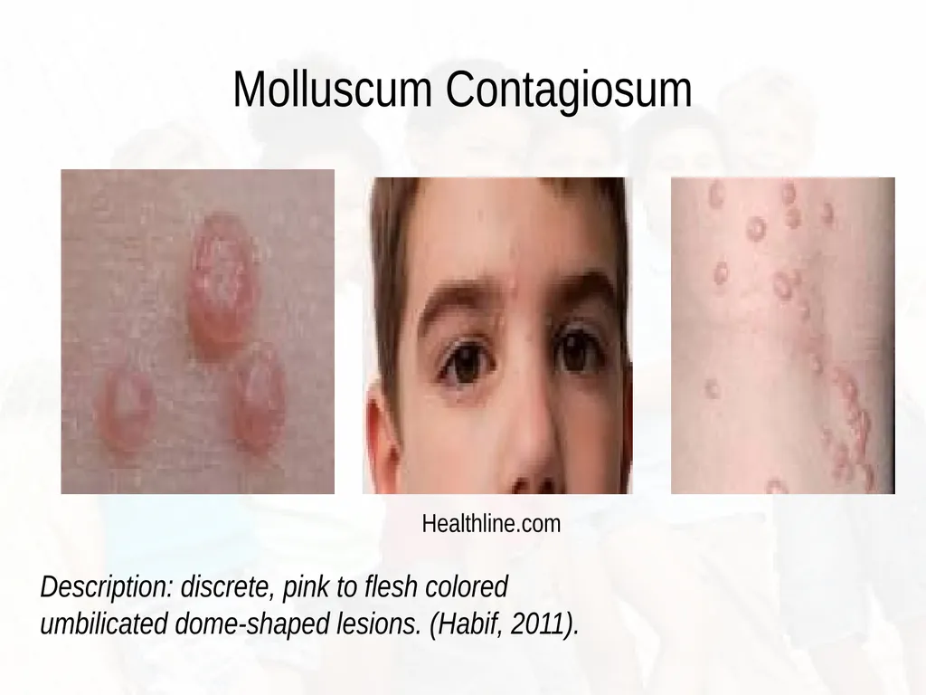
Molluscum Contagiosum Healthline.com Description: discrete, pink to flesh colored umbilicated dome-shaped lesions. (Habif, 2011).<br>
55
Management and Treatment Should be kept covered by clothing
Minimize transmission of the virus
Curettage to remove fairly painless and decreases recurrence
Imiquimod 5% cream (Aldara)
***** This lesion in young adults could indicate a sexual transmission. If seen in pediatric population in genitalia suspect for sexual abuse. Habif, (2011).<br>
Minimize transmission of the virus
Curettage to remove fairly painless and decreases recurrence
Imiquimod 5% cream (Aldara)
***** This lesion in young adults could indicate a sexual transmission. If seen in pediatric population in genitalia suspect for sexual abuse. Habif, (2011).<br>
56
Pediatric Considerations Autoinoculation around eye is common
Lesions will resolve spontaneously with cell-mediated immunity
Primarily a sexually transmitted disease in young adults
Lesions will occur in the lower abdomen, genitalia and thighs Habif, (2011).<br>
Lesions will resolve spontaneously with cell-mediated immunity
Primarily a sexually transmitted disease in young adults
Lesions will occur in the lower abdomen, genitalia and thighs Habif, (2011).<br>
57
Varicella Highly contagious infection
Caused by varicella virus
Caused by human herpes virus type 3
Transmission is via airborne droplets or vesicular fluid
Patients are contagious 2-days prior to outbreak of lesions
Prodromal symptoms include:
Low-grade fever
Headache
Generalized vesicular rash Habif, (2011).<br>
Caused by varicella virus
Caused by human herpes virus type 3
Transmission is via airborne droplets or vesicular fluid
Patients are contagious 2-days prior to outbreak of lesions
Prodromal symptoms include:
Low-grade fever
Headache
Generalized vesicular rash Habif, (2011).<br>
58
Skin Findings Simultaneous presentation of lesions in various stages of development (vesicles, pustules, crusts)
Begin as 2-4mm red papule, then evolve to a thin-walled clear vesicle
Vesicle becomes umbilicated; fluid can become cloudy
Lesions eruption ceases within 4 days
Crusts fall within 7 days Habif, (2011).<br>
Begin as 2-4mm red papule, then evolve to a thin-walled clear vesicle
Vesicle becomes umbilicated; fluid can become cloudy
Lesions eruption ceases within 4 days
Crusts fall within 7 days Habif, (2011).<br>
59
Management and Treatment Symptomatic treatment includes use of bland, antipruritic lotions and antihistamines
Hydration
Tylenol or ibuprofen for fever
Cut nails short to avoid self-inoculation or skin infection (impetigo)
Varicella vaccine is 96% effective
Seroconversion is 71-91% in healthy children
Children immunized with live attenuated virus may have a mild febrile illness; few vesicles 2 weeks after vaccine Habif, (2011).<br>
Hydration
Tylenol or ibuprofen for fever
Cut nails short to avoid self-inoculation or skin infection (impetigo)
Varicella vaccine is 96% effective
Seroconversion is 71-91% in healthy children
Children immunized with live attenuated virus may have a mild febrile illness; few vesicles 2 weeks after vaccine Habif, (2011).<br>
60
Hand, Foot and Mouth Disease Highly contagious viral infection
Causes aphthae-like oral erosions
Vesicular lesions on hands and feet
Self limiting
Associated with coxsackie virus A-16
Incubation period 4-6 days
Spread is by nose and throat discharge
Mild symptoms of sore throat and malaise; abdominal pain 1-2 days
20% develop cervical lymphadenopathy Habif, (2011).<br>
Causes aphthae-like oral erosions
Vesicular lesions on hands and feet
Self limiting
Associated with coxsackie virus A-16
Incubation period 4-6 days
Spread is by nose and throat discharge
Mild symptoms of sore throat and malaise; abdominal pain 1-2 days
20% develop cervical lymphadenopathy Habif, (2011).<br>
61
Skin Findings Oral aphthae-like erosions vary 10 or more
Cutaneous lesions occur in 2/3 of patients
Begin as 3-7mm red macules, becoming pale, white oval vesicles with red areola
Healing occurs in approximately 7 days Habif, (2011).<br>
Cutaneous lesions occur in 2/3 of patients
Begin as 3-7mm red macules, becoming pale, white oval vesicles with red areola
Healing occurs in approximately 7 days Habif, (2011).<br>
62
Management and Treatment Children may be isolated during most contagious period (3-7 days)
Fever/pain controlled with Tylenol
Cool fluids; acidic food avoided
Need to keep child well hydrated
Antiviral medication Habif, (2011).<br>
Fever/pain controlled with Tylenol
Cool fluids; acidic food avoided
Need to keep child well hydrated
Antiviral medication Habif, (2011).<br>
63
Erythema Infectiosum (Fifth’s Disease) Also known as “slapped cheek” syndrome
Viral exanthem
Occurs mostly in the winter and spring
Caused by parvovirus B19
Transmitted via respiratory secretions, blood or vertically from mother to fetus
Peak age is between 5-14 years
Prodromal symptoms: low grade fever, pruritus, malaise, sore throat Habif, (2011).<br>
Viral exanthem
Occurs mostly in the winter and spring
Caused by parvovirus B19
Transmitted via respiratory secretions, blood or vertically from mother to fetus
Peak age is between 5-14 years
Prodromal symptoms: low grade fever, pruritus, malaise, sore throat Habif, (2011).<br>
64
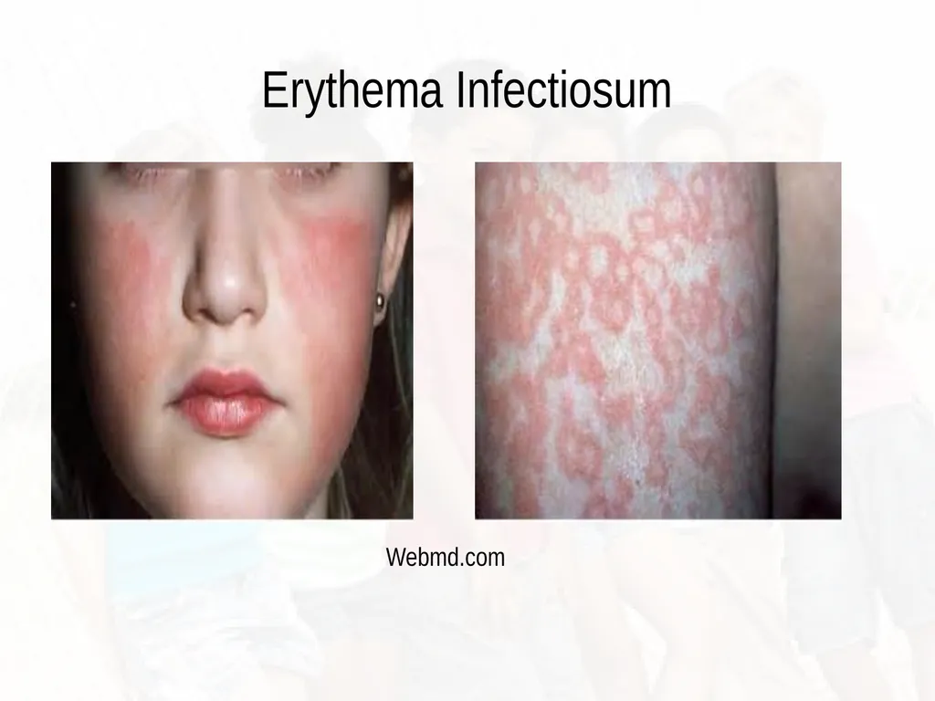
Skin Findings Facial erythema (slapped cheek)
Red papules on cheeks that coalesce
2-days after onset of facial rash, lacy, erythema in a “fish-net” pattern on trunk and proximal extremities, buttocks
Fades within 6-14 days
May appear 2-3 weeks; factors such as sunlight, hot water and emotional/physical activity
Adults may experience myalgias Habif, (2011).<br>
Red papules on cheeks that coalesce
2-days after onset of facial rash, lacy, erythema in a “fish-net” pattern on trunk and proximal extremities, buttocks
Fades within 6-14 days
May appear 2-3 weeks; factors such as sunlight, hot water and emotional/physical activity
Adults may experience myalgias Habif, (2011).<br>
65
Prognosis/Treatment Exposed pregnant women should seek serological testing and follow up with PCP
Child is not considered infectious once rash develops; may return to school
Most infections are self-limiting without consequence
NSAIDS can control myalgias
Control fever, hydration
Pregnant woman exposed should seek OB/GYN care Habif, (2011).<br>
Child is not considered infectious once rash develops; may return to school
Most infections are self-limiting without consequence
NSAIDS can control myalgias
Control fever, hydration
Pregnant woman exposed should seek OB/GYN care Habif, (2011).<br>
66
Erythema Infectiosum Webmd.com<br>
67
Kawasaki Disease Also known as mucocutaneous lymph node syndrome
Morbidity and mortality associated with cardiovascular complications
Ages range from 7 weeks to 12 years; adult cases rare
Recurrence is rare
Diagnosis based on having the following:
Fever of unknown origin
Bilateral conjuntiva injection
Cervical lymphadenopathy
Exanthem with vesicles and or crusts
Coronary artery aneurysms<br>
Morbidity and mortality associated with cardiovascular complications
Ages range from 7 weeks to 12 years; adult cases rare
Recurrence is rare
Diagnosis based on having the following:
Fever of unknown origin
Bilateral conjuntiva injection
Cervical lymphadenopathy
Exanthem with vesicles and or crusts
Coronary artery aneurysms<br>
68
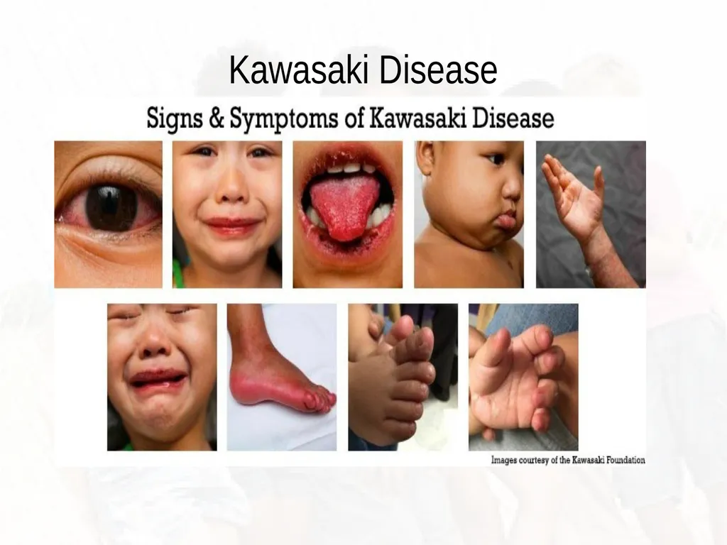
Skin Findings Conjunctival injection
Uveitis
Lips and oral pharynx erythematous, dry fissured, cracked and crusted
Hypertrophic tongue papillae (strawberry tongue)
Extremities (2-5 days) feet become edematous and tender
Desquamation of the hands and feet; peeling of skin
Rash is polymorphous, macular, papular, urticarial-like lesions; diaper dermatitis<br>
Uveitis
Lips and oral pharynx erythematous, dry fissured, cracked and crusted
Hypertrophic tongue papillae (strawberry tongue)
Extremities (2-5 days) feet become edematous and tender
Desquamation of the hands and feet; peeling of skin
Rash is polymorphous, macular, papular, urticarial-like lesions; diaper dermatitis<br>
69
Non-Skin Findings Fever without chills or seats can last 15-30 days
Fever begins abruptly and spikes dos not respond to antibiotics or antipyretics
Cervical lymphadenopathy, often limited to one
Cardiac involvement; myocarditis, tachycardia and arrhythmias
Coronary artery aneurysms
Acute phase leukocytosis<br>
Fever begins abruptly and spikes dos not respond to antibiotics or antipyretics
Cervical lymphadenopathy, often limited to one
Cardiac involvement; myocarditis, tachycardia and arrhythmias
Coronary artery aneurysms
Acute phase leukocytosis<br>
70
Treatment I.V. immune globulin (IVIG)
Methylprednisolone an alternative to IVIG
Close monitoring of patient
Hydration
Oral care
Rest; control of fever<br>
Methylprednisolone an alternative to IVIG
Close monitoring of patient
Hydration
Oral care
Rest; control of fever<br>
71
Kawasaki Disease<br>
72
Streptococcal Infection/Scarletina Posterior pharynx is erythematous
Enlarged palatine tonsils
Cervical lymphadenopathy
Fever, malaise
Post infection:
Skin develops dry sandpaper appearance<br>
Enlarged palatine tonsils
Cervical lymphadenopathy
Fever, malaise
Post infection:
Skin develops dry sandpaper appearance<br>
73
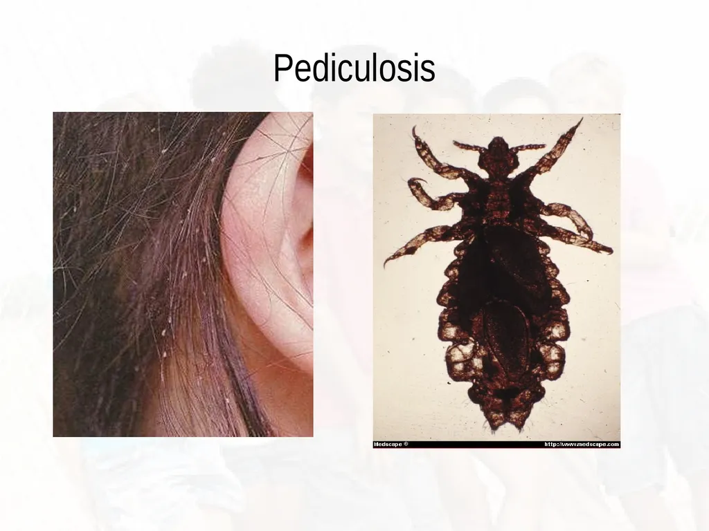
Pediculosis (head lice) Flattened, wingless, insects; infest hair of scalp, body and pubic region
Attach to the skin and feed on human blood
Lay eggs (nits) on shaft of hair
Highly contagious
Direct contact primary source of transmission
Lice live about 30 days
Females lay 7-10 nits daily
Lay nits 1cm from scalp Habif, (2011).<br>
Attach to the skin and feed on human blood
Lay eggs (nits) on shaft of hair
Highly contagious
Direct contact primary source of transmission
Lice live about 30 days
Females lay 7-10 nits daily
Lay nits 1cm from scalp Habif, (2011).<br>
74
Pediculosis<br>
75
Management and Treatment Standard is topical with Permethrin rinse 1% OTC
Permethrin 5% is administered for treatment failures
Home remedies include:
Application of Vaseline
Mayonnaise or pomades
Apply shower cap and keep overnight
Hair clean 1-2-3 kills lice on contact
Oral prescribed treatments; Bactrim
Oral antibiotics for secondary infection
Nit removal (may use vinegar with 50% water) Habif, (2011).<br>
Permethrin 5% is administered for treatment failures
Home remedies include:
Application of Vaseline
Mayonnaise or pomades
Apply shower cap and keep overnight
Hair clean 1-2-3 kills lice on contact
Oral prescribed treatments; Bactrim
Oral antibiotics for secondary infection
Nit removal (may use vinegar with 50% water) Habif, (2011).<br>
76
Scabies Parasitic infection caused by mite Sarcoptes scabiei
Complaints are of intense itching, unremitting
Common presentation in one member of the family
Can be seen in families
Skin findings: curved burrow, can be linear and S-shaped; slightly elevated vesicle or papule 1-2mm in size
Can be found in intertriginous areas, webs of fingers, wrists, sides of hands, feet, lateral fingers and toes. genitalia Habif, (2011).<br>
Complaints are of intense itching, unremitting
Common presentation in one member of the family
Can be seen in families
Skin findings: curved burrow, can be linear and S-shaped; slightly elevated vesicle or papule 1-2mm in size
Can be found in intertriginous areas, webs of fingers, wrists, sides of hands, feet, lateral fingers and toes. genitalia Habif, (2011).<br>
77
Management and Treatment Permethrin or lindane applied to entire skin surface from the neck down
Patient should bathe after 12 hours of application
Avoid eyes and mouth
Benzyl benzoate bath and lotion
All clothes must be washed; bed linen
Post treatment pruritus can occur
Assessment for areas of topical infection from scratching Habif, (2011).<br>
Patient should bathe after 12 hours of application
Avoid eyes and mouth
Benzyl benzoate bath and lotion
All clothes must be washed; bed linen
Post treatment pruritus can occur
Assessment for areas of topical infection from scratching Habif, (2011).<br>
78
Lyme Disease Tick-borne disease; Borrelia burgdorferi
Evolves through 3 stages; affects almost all organ systems
Cutaneous eruption of Lyme disease is called erythema migrans
Onset of disease is 3-28 days after tick bite
3 Stages:
Stage I: expanding target like patch; flu like symptoms
Stage II: cardiac and neurological problems
Stage III: arthritis and continuous neuro problems persist Habif, (2011).<br>
Evolves through 3 stages; affects almost all organ systems
Cutaneous eruption of Lyme disease is called erythema migrans
Onset of disease is 3-28 days after tick bite
3 Stages:
Stage I: expanding target like patch; flu like symptoms
Stage II: cardiac and neurological problems
Stage III: arthritis and continuous neuro problems persist Habif, (2011).<br>
79
Skin Findings Initial tick bite, inflamed bite reaction
Tick must stay attached for at least 24 hours
Skin changes (erythema migrans)
Begins with a small papule with slowly enlarging ring of erythema
20-50% of people have multiple rings Habif, (2011).<br>
Tick must stay attached for at least 24 hours
Skin changes (erythema migrans)
Begins with a small papule with slowly enlarging ring of erythema
20-50% of people have multiple rings Habif, (2011).<br>
80
Management and Treatment Prevention of tick bites/exposure
Wearing protective garments
Frequent assessment of skin
N-diethyl-meta-tolumide on skin/permethrin on clothes (need to check with PCP for safety)
Early symptoms of disease treated with 21 days of treatment with Doxycycline, or Ceftin, or Amoxicillin
Seek PCP care ASAP Habif, (2011).<br>
Wearing protective garments
Frequent assessment of skin
N-diethyl-meta-tolumide on skin/permethrin on clothes (need to check with PCP for safety)
Early symptoms of disease treated with 21 days of treatment with Doxycycline, or Ceftin, or Amoxicillin
Seek PCP care ASAP Habif, (2011).<br>
81
Acanthosis Nigricans Elevated, velvety hyperpigmentation of the flexural skin, neck, axillae and groin
Commonly associated with obesity, diabetes, endocrinopathies
Patients complain about an asymptomatic dirty appearance to skin folds; not removed by vigorous washing
May be a family hx of eruption Habif, (2011).<br>
Commonly associated with obesity, diabetes, endocrinopathies
Patients complain about an asymptomatic dirty appearance to skin folds; not removed by vigorous washing
May be a family hx of eruption Habif, (2011).<br>
82
Skin/Non-Skin Findings AN is a cutaneous marker of tissue insulin resistance
Patients without DM have increased levels of circulating insulin
Glucose levels may be elevated
Impaired response to exogenous insulin
Can be caused by estrogens and nicotinic acid
Less common: tumors of the lung, prostate, breast and ovary Habif, (2011).<br>
Patients without DM have increased levels of circulating insulin
Glucose levels may be elevated
Impaired response to exogenous insulin
Can be caused by estrogens and nicotinic acid
Less common: tumors of the lung, prostate, breast and ovary Habif, (2011).<br>
83
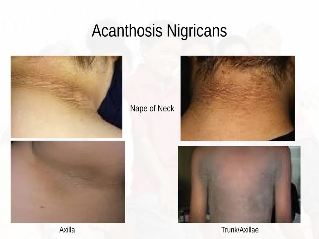
Acanthosis Nigricans Nape of Neck Axilla Trunk/Axillae<br>
84
Management and Treatment Skin eruption does not cause require treatment
Treatment is necessary for obesity
Evaluation for the presence of diabetes
Evaluation of blood pressure, measurement of body mass index (BMI)
Goal of therapy is to correct underlying disease process
Correction of hyperinsulemia (metabolic syndrome)
Weight reduction Habif, (2011).<br>
Treatment is necessary for obesity
Evaluation for the presence of diabetes
Evaluation of blood pressure, measurement of body mass index (BMI)
Goal of therapy is to correct underlying disease process
Correction of hyperinsulemia (metabolic syndrome)
Weight reduction Habif, (2011).<br>
85
Summary Proper assessment of skin disorder
Assess for system involvement; fever, malaise
Educate children and families on the importance of:
Proper hygiene
Early evaluation by PCP and follow-up
Referral and reporting of communicable diseases
Refer to communicable disease reference chart<br>
Assess for system involvement; fever, malaise
Educate children and families on the importance of:
Proper hygiene
Early evaluation by PCP and follow-up
Referral and reporting of communicable diseases
Refer to communicable disease reference chart<br>
86
References American Diabetes Association (2017). Standards of medical care in diabetes-2017.
Diabetes Care: The Journal of Clinical and Applied Research and Education. 40(1),
pgs. 1-142.
Habif, T.P., Campbell, J.L., Chapman, M.S., Dinulos, J.G.H, & Zug, K.A. (2011). Skin
disease: Diagnosis & Treatment, 3rd Ed. Saunders Elsevier, New York, NY
McCance, K.L., Huether, S.E., Brashers, V.L, & Rote,N.S. (2014). Pathophysiology:
The biological basis for disease in adults and children. 7th Ed. St. Louis, Missouri.
National Institute of Diabetes and Digestive and Kidney Diseases [NIDDK]. (2017).
Retrieved from: http://www.niddk.nih.gov.health-information/diabetes/overview/
Seidel, H.M., Ball, J.W., Dains, J.E., Flynn, J.A., Solomon, B.S., & Stewart, R.W.
(2015). Mosby’s Guide to Physical Examination, 8th Ed., Elsevier, St. Louis, Missouri.<br>
Diabetes Care: The Journal of Clinical and Applied Research and Education. 40(1),
pgs. 1-142.
Habif, T.P., Campbell, J.L., Chapman, M.S., Dinulos, J.G.H, & Zug, K.A. (2011). Skin
disease: Diagnosis & Treatment, 3rd Ed. Saunders Elsevier, New York, NY
McCance, K.L., Huether, S.E., Brashers, V.L, & Rote,N.S. (2014). Pathophysiology:
The biological basis for disease in adults and children. 7th Ed. St. Louis, Missouri.
National Institute of Diabetes and Digestive and Kidney Diseases [NIDDK]. (2017).
Retrieved from: http://www.niddk.nih.gov.health-information/diabetes/overview/
Seidel, H.M., Ball, J.W., Dains, J.E., Flynn, J.A., Solomon, B.S., & Stewart, R.W.
(2015). Mosby’s Guide to Physical Examination, 8th Ed., Elsevier, St. Louis, Missouri.<br>