
Dr. Irfan Hussain ( Assistant Professor)
SB
Published · 85 slides · 0 views

1 / 1






Description
Dr. Irfan Hussain ( Assistant Professor) Department of Orthopaedics Acetabular Fracture Introduction Elderly pt.- Due to fall As isolated injury Younger pt.- MVA High incidence of major associated injuries Anatomy Can be conceptualized as
Related Topics
Share
Embed code
Download this presentation From Below
"Dr. Irfan Hussain ( Assistant Professor)" is the property of its rightful owner. Permission is granted to download and print the materials on this website for personal, non-commercial use only, and to display it on your personal computer provided you do not modify the materials and that you retain all copyright notices contained in the materials. By downloading content from our website, you accept the terms of this agreement.
Presentation Transcript
01
Dr. Irfan Hussain
( Assistant Professor)
Department of Orthopaedics Acetabular Fracture<br>
( Assistant Professor)
Department of Orthopaedics Acetabular Fracture<br>
02
Introduction Elderly pt.-
Due to fall
As isolated injury
Younger pt.-
MVA
High incidence of major associated injuries<br>
Due to fall
As isolated injury
Younger pt.-
MVA
High incidence of major associated injuries<br>
03
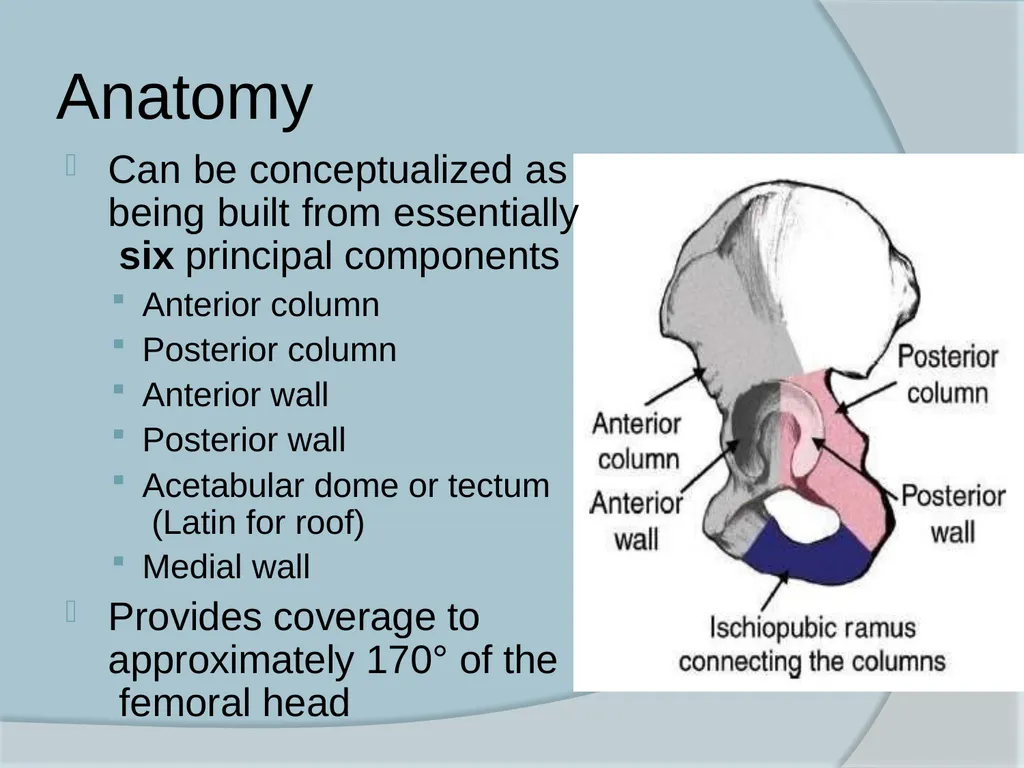
Anatomy Can be conceptualized as being built from essentially six principal components
Anterior column
Posterior column
Anterior wall
Posterior wall
Acetabular dome or tectum (Latin for roof)
Medial wall
Provides coverage to approximately 170° of the femoral head<br>
Anterior column
Posterior column
Anterior wall
Posterior wall
Acetabular dome or tectum (Latin for roof)
Medial wall
Provides coverage to approximately 170° of the femoral head<br>
04
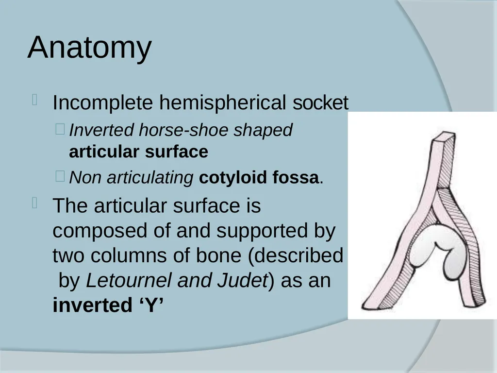
Anatomy Incomplete hemispherical socket
Inverted horse-shoe shaped
articular surface
Non articulating cotyloid fossa.
The articular surface is composed of and supported by two columns of bone (described by Letournel and Judet) as an
inverted ‘Y’<br>
Inverted horse-shoe shaped
articular surface
Non articulating cotyloid fossa.
The articular surface is composed of and supported by two columns of bone (described by Letournel and Judet) as an
inverted ‘Y’<br>
05
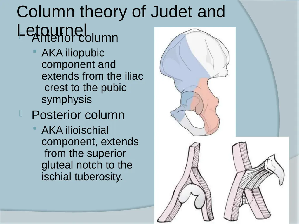
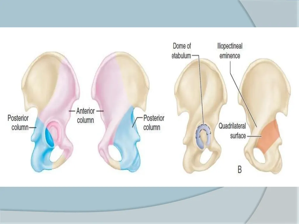
Column theory of Judet and Letournel Anterior column
AKA iliopubic component and extends from the iliac crest to the pubic symphysis
Posterior column
AKA ilioischial component, extends from the superior gluteal notch to the ischial tuberosity.<br>
AKA iliopubic component and extends from the iliac crest to the pubic symphysis
Posterior column
AKA ilioischial component, extends from the superior gluteal notch to the ischial tuberosity.<br>
06
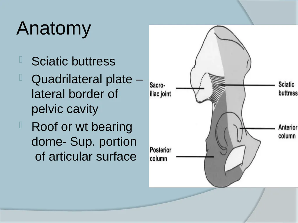
Anatomy Sciatic buttress
Quadrilateral plate – lateral border of pelvic cavity
Roof or wt bearing dome- Sup. portion of articular surface<br>
Quadrilateral plate – lateral border of pelvic cavity
Roof or wt bearing dome- Sup. portion of articular surface<br>
07
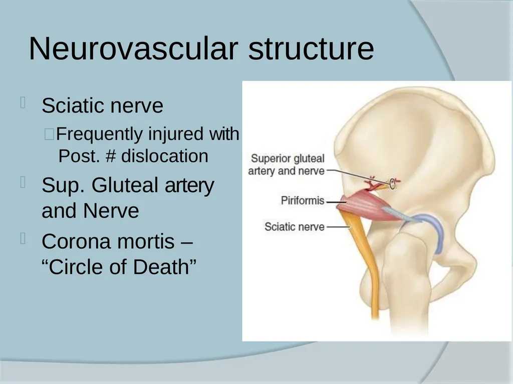
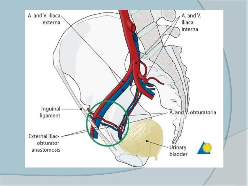
Neurovascular structure Sciatic nerve
Frequently injured with Post. # dislocation
Sup. Gluteal artery and Nerve
Corona mortis – “Circle of Death”<br>
Frequently injured with Post. # dislocation
Sup. Gluteal artery and Nerve
Corona mortis – “Circle of Death”<br>
08
The classification of acetabular fracture Letournel and Judet - commonly used
5 elementary and 5 asscociated types
Simple types –
Posterior wall #
Posterior column #
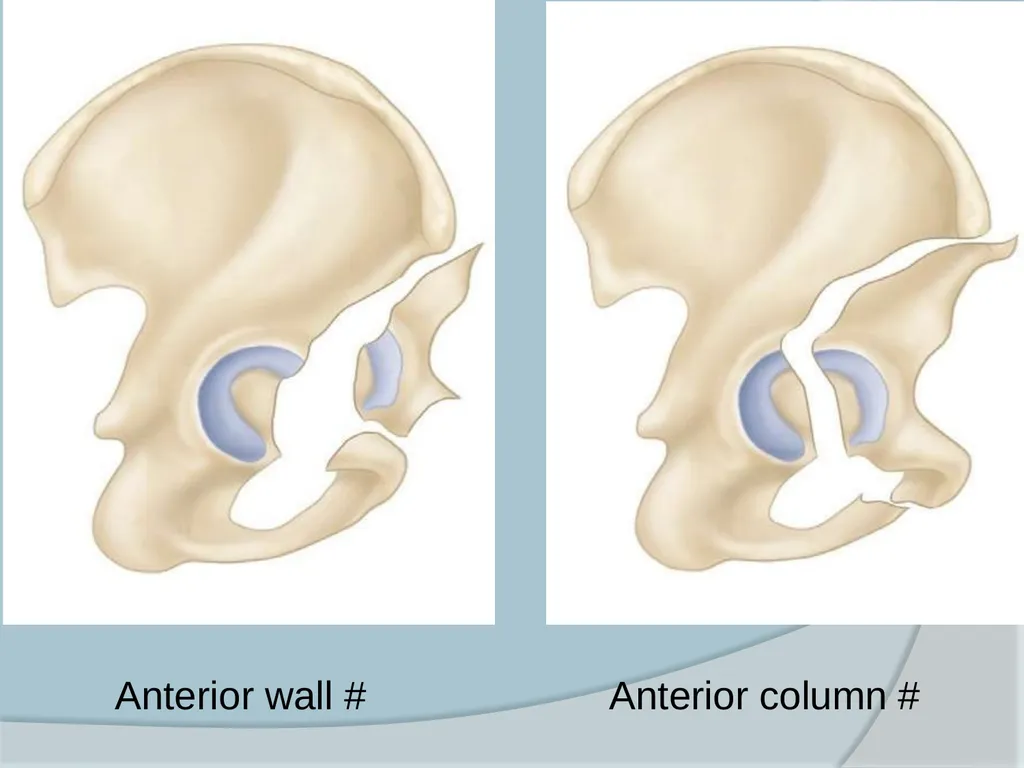
Anterior wall #
Anterior column #
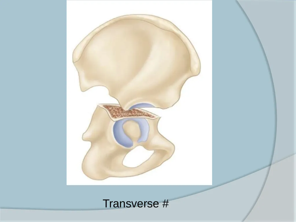
Trasverse #<br>
5 elementary and 5 asscociated types
Simple types –
Posterior wall #
Posterior column #
Anterior wall #
Anterior column #
Trasverse #<br>
09
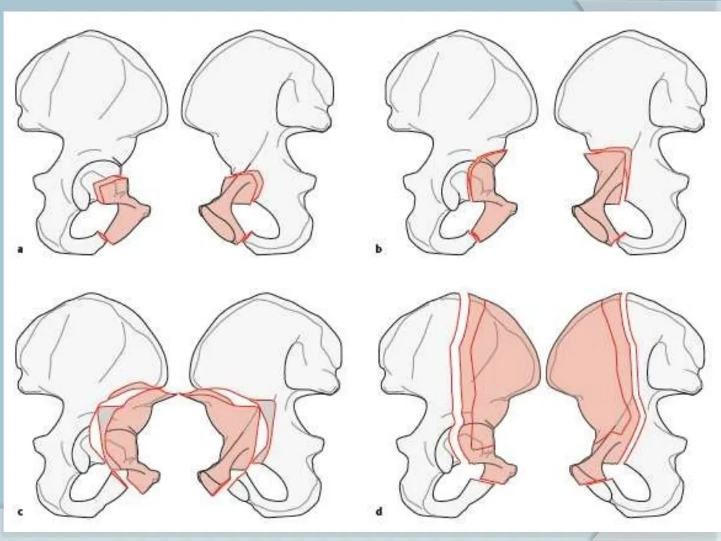
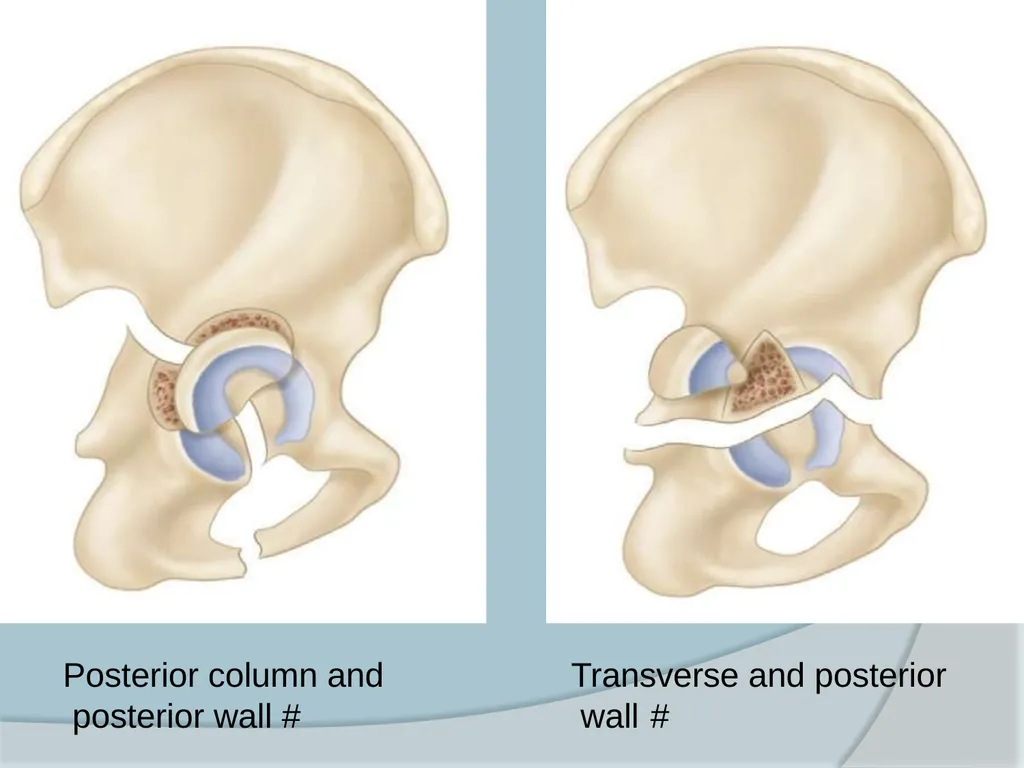
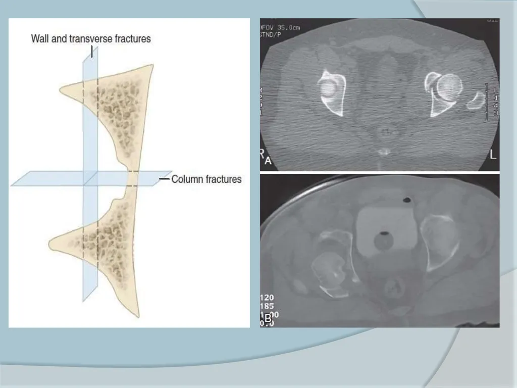
Associated # types Posterior column and posterior wall #
Transverse and posterior wall #
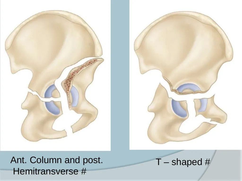
Anterior column (or wall) and posterior hemitransverse #
T-shaped #
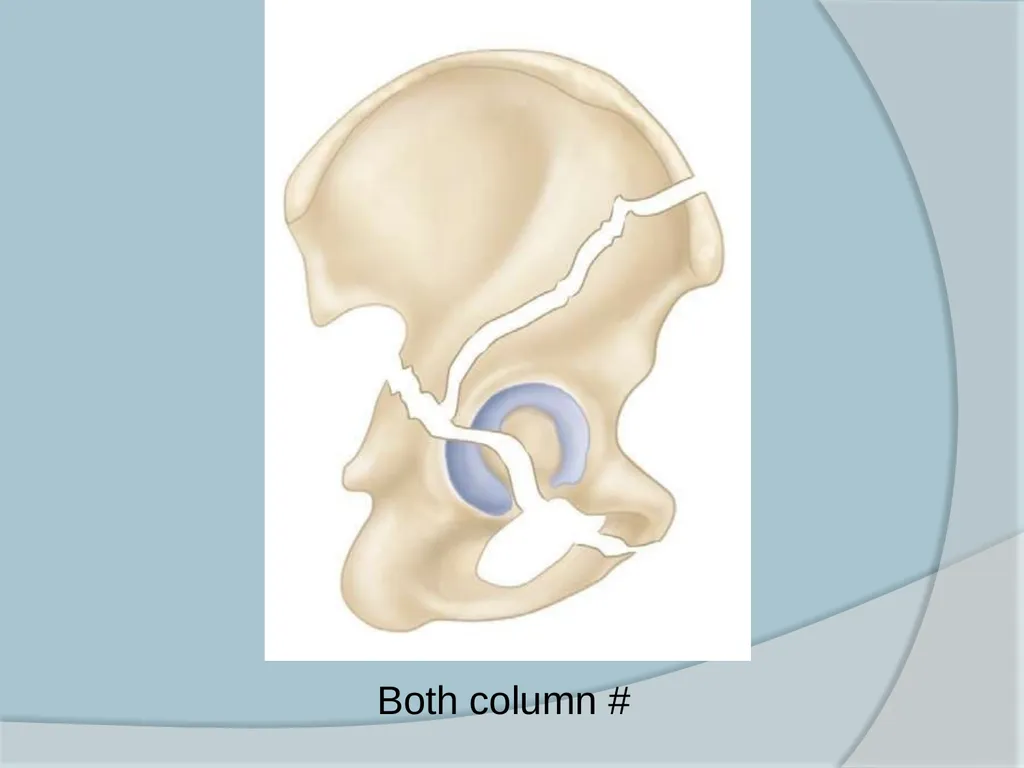
Both-column #<br>
Transverse and posterior wall #
Anterior column (or wall) and posterior hemitransverse #
T-shaped #
Both-column #<br>
10
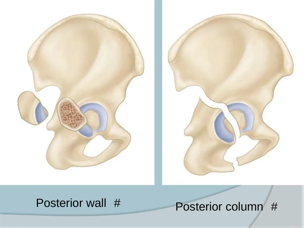
Posterior wall # Posterior column #<br>
11
Anterior wall # Anterior column #<br>
12
Transverse #<br>
13
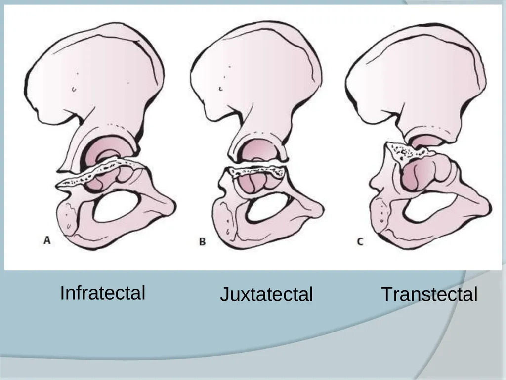
Infratectal Juxtatectal Transtectal<br>
14
Posterior column and posterior wall # Transverse and posterior wall #<br>
15
T – shaped # Ant. Column and post. Hemitransverse #<br>
16
Both column #<br>
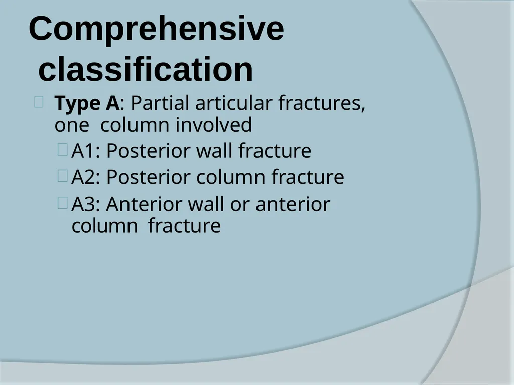
17
Comprehensive classification Type A: Partial articular fractures, one column involved
A1: Posterior wall fracture
A2: Posterior column fracture
A3: Anterior wall or anterior column fracture<br>
A1: Posterior wall fracture
A2: Posterior column fracture
A3: Anterior wall or anterior column fracture<br>
18
Comprehensive classification Type B: Partial articular fractures (transverse or T-type fracture, both columns involved)
B1: Transverse fracture
B2: T-shaped fracture
B3: Anterior column plus posterior hemitransverse fracture<br>
B1: Transverse fracture
B2: T-shaped fracture
B3: Anterior column plus posterior hemitransverse fracture<br>
19
Comprehensive classification Type C: Complete articular fracture (both column fracture; floating acetabulum)
C1: Both column #, high variety (anterior column fracture extends to the iliac crest)
C2: Both column #, low variety (anterior column fracture extends to the anterior border of the ilium)
C3: Both column # involving the sacroiliac joint<br>
C1: Both column #, high variety (anterior column fracture extends to the iliac crest)
C2: Both column #, low variety (anterior column fracture extends to the anterior border of the ilium)
C3: Both column # involving the sacroiliac joint<br>
20
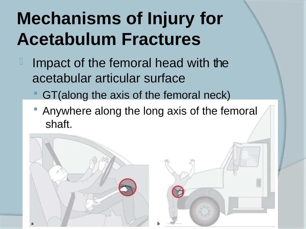
Mechanisms of Injury for Acetabulum Fractures Impact of the femoral head with the acetabular articular surface
GT(along the axis of the femoral neck)
Anywhere along the long axis of the femoral shaft.<br>
GT(along the axis of the femoral neck)
Anywhere along the long axis of the femoral shaft.<br>
21
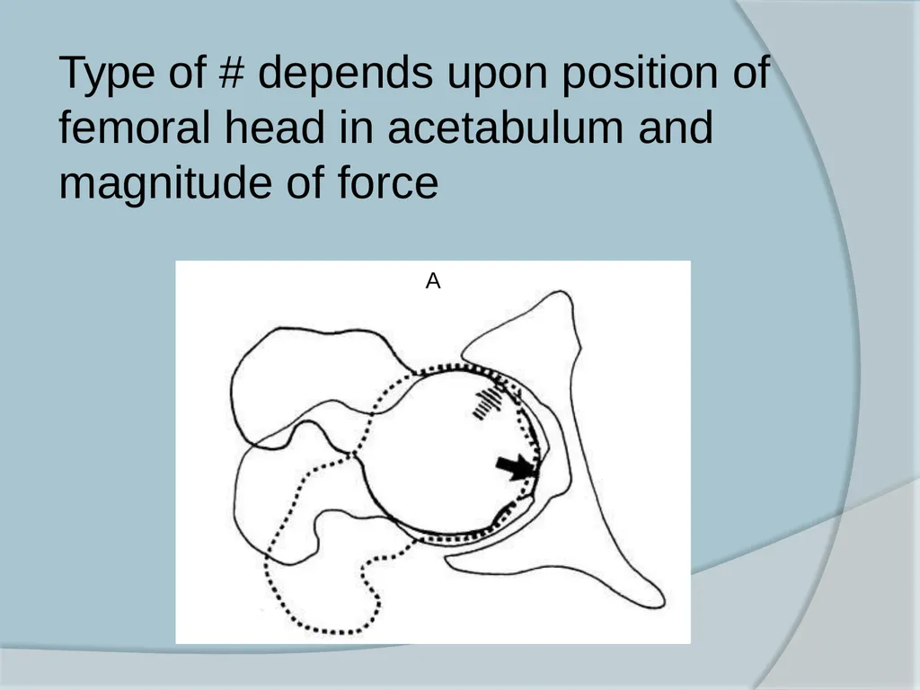
Type of # depends upon position of femoral head in acetabulum and magnitude of force A<br>
22
Signs and symptoms History – cause of injury
ATLS protocol
Visceral injuries common
Associated fractures common
Elderly pt. (underlying cardiac or neurologic condition)
Assess and document NV status<br>
ATLS protocol
Visceral injuries common
Associated fractures common
Elderly pt. (underlying cardiac or neurologic condition)
Assess and document NV status<br>
23
Signs and symptoms Shortening present if hip is dislocated
Flexion, adduction, and internal rotation of the hip may not be present<br>
Flexion, adduction, and internal rotation of the hip may not be present<br>
24
Radiographic Evaluation X-Rays- 3 views
Standard AP view
450 Obturator oblique view
450 Iliac oblique view
CT scan<br>
Standard AP view
450 Obturator oblique view
450 Iliac oblique view
CT scan<br>
25
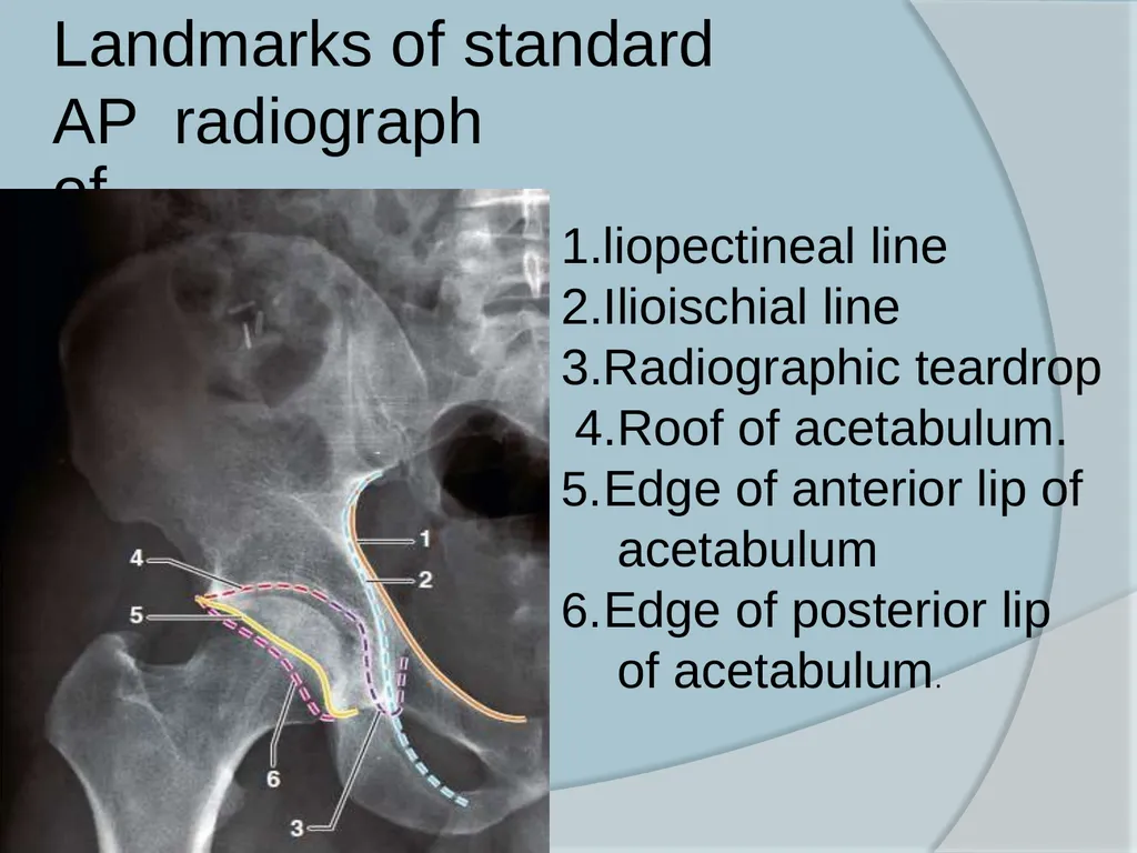
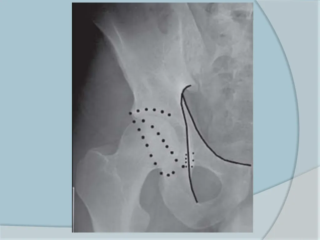
Landmarks of standard AP radiograph of hip 1.liopectineal line 2.Ilioischial line 3.Radiographic teardrop 4.Roof of acetabulum.
Edge of anterior lip of acetabulum
Edge of posterior lip of acetabulum.<br>
Edge of anterior lip of acetabulum
Edge of posterior lip of acetabulum.<br>
26
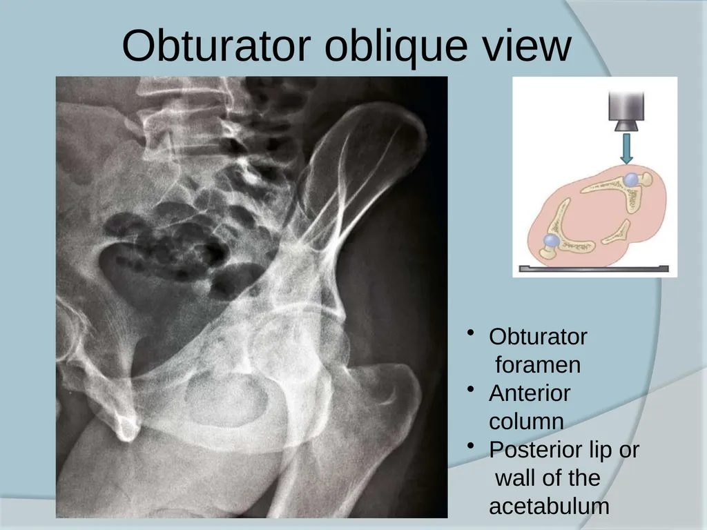
Obturator oblique view Obturator foramen
Anterior column
Posterior lip or wall of the acetabulum<br>
Anterior column
Posterior lip or wall of the acetabulum<br>
27
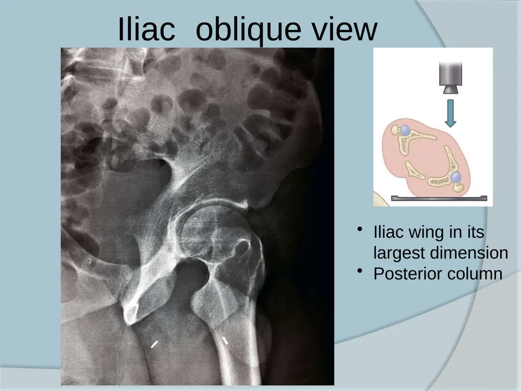
Iliac oblique view Iliac wing in its largest dimension
Posterior column<br>
Posterior column<br>
28
CT scan Axial cuts should be taken with thin slices
Extent and location of acetabular wall fractures
Presence of intra-articular free fragments
Orientation of fracture lines and Marginal impaction
Femoral head defect
Rotation of fracture fragments
Pelvic hematoma
Sacroiliac joint integrity<br>
Extent and location of acetabular wall fractures
Presence of intra-articular free fragments
Orientation of fracture lines and Marginal impaction
Femoral head defect
Rotation of fracture fragments
Pelvic hematoma
Sacroiliac joint integrity<br>
29
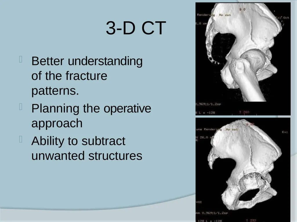
3-D CT Better understanding of the fracture patterns.
Planning the operative approach
Ability to subtract unwanted structures<br>
Planning the operative approach
Ability to subtract unwanted structures<br>
30
Management Initial treatment – follow ATLS protocols
Operative treatment are usually not performed as an emergency
Closed reduction of hip dislocations should be performed Skeletal traction
Allow soft tissue healing initially
Maintain limb length
Maintain femoral head reduction<br>
Operative treatment are usually not performed as an emergency
Closed reduction of hip dislocations should be performed Skeletal traction
Allow soft tissue healing initially
Maintain limb length
Maintain femoral head reduction<br>
31
Management The goal of the treatment is restoration of articular surface, prevent post traumatic arthritis and to mobilise the patient as early as possible<br>
32
Management Factors to be considered are-
Patient factors
Fracture factors
Expertise available<br>
Patient factors
Fracture factors
Expertise available<br>
33
Patients factors Age
Pre injury activity level
Medical comorbidities
Associated injuries
Functional demands<br>
Pre injury activity level
Medical comorbidities
Associated injuries
Functional demands<br>
34
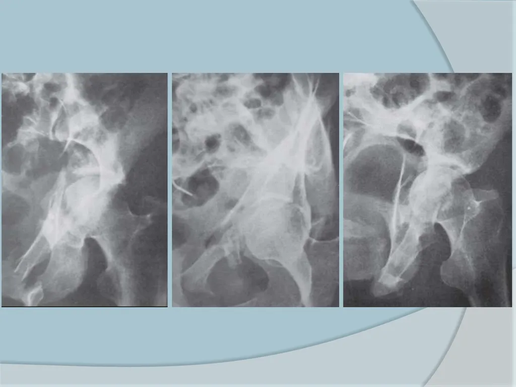
Non-operative Indications Hip stable and congruous.
Patient factors
Medical contraindications
Severe osteoporosis in elderly
Undisplaced/Minimally displaced fractures
Fractures with secondary congruence (both-column)
Preexistent arthritis of hip
Local soft tissue problems
Morel Lavelle’ lesion
Open wound
Suprapubic catheter (C/I to Ilioinguinal and Stoppa)<br>
Patient factors
Medical contraindications
Severe osteoporosis in elderly
Undisplaced/Minimally displaced fractures
Fractures with secondary congruence (both-column)
Preexistent arthritis of hip
Local soft tissue problems
Morel Lavelle’ lesion
Open wound
Suprapubic catheter (C/I to Ilioinguinal and Stoppa)<br>
35
Morel Lavalle lesion Localized area of subcutaneous fat necrosis over the lateral aspect of the hip
Operating through it has been associated with a higher rate of postoperative infection<br>
Operating through it has been associated with a higher rate of postoperative infection<br>
36
Non operative Treatment Bed rest is necessary in the acute injury phase only
Mobilization as soon as symptoms allow
Patients should begin with touch-down partial weight-bearing
Radiographs at frequent intervals
Gradually progress to full weight bearing when there is adequate fracture healing, usually by 6 to 12 weeks
Joint mobilization should be continued throughout the rehabilitation period<br>
Mobilization as soon as symptoms allow
Patients should begin with touch-down partial weight-bearing
Radiographs at frequent intervals
Gradually progress to full weight bearing when there is adequate fracture healing, usually by 6 to 12 weeks
Joint mobilization should be continued throughout the rehabilitation period<br>
37
Operative Treatment Instability
Incongruity.<br>
Incongruity.<br>
38
Instability Usually associated with posterior fracture types
Less commonly anterior
May be central if large fragment of quadrilateral plate is fractured<br>
Less commonly anterior
May be central if large fragment of quadrilateral plate is fractured<br>
39
Incongruity The curve of femoral head should fit exactly in to dome of acetabulum in all three radiographic views
Matta has recommended use of roof arc measurements to measure incongruity<br>
Matta has recommended use of roof arc measurements to measure incongruity<br>
40
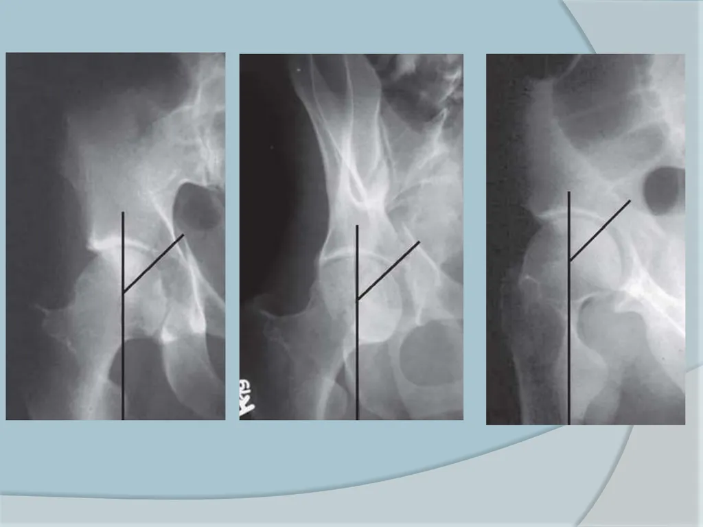
Roof arc angle Is an estimation of amount of WBD involved
Measured on all three radiograph views with the leg out of traction
Medial roof arc angle is measured on AP view
Anterior roof arc angle is measured on obturator oblique view
Posterior roof arc angle is measured on Iliac oblique view
WBD is considered to be intact and hip joint congruous if measurement > 45o (Matta)<br>
Measured on all three radiograph views with the leg out of traction
Medial roof arc angle is measured on AP view
Anterior roof arc angle is measured on obturator oblique view
Posterior roof arc angle is measured on Iliac oblique view
WBD is considered to be intact and hip joint congruous if measurement > 45o (Matta)<br>
41
Criteria for non operative treatment
CT subchondral arc is intact in the superior
10 mm of the acetabulum
Femoral head remains congruent with the superior acetabulum in AP and 45 degree oblique views (out of traction)
No evidence of posterior hip instability<br>
CT subchondral arc is intact in the superior
10 mm of the acetabulum
Femoral head remains congruent with the superior acetabulum in AP and 45 degree oblique views (out of traction)
No evidence of posterior hip instability<br>
42
Operative Indications Hip joint instability and Incongruity
Significant displacement (≥ 2 mm) of the weight-bearing dome on any of the three standard x-rays
Fractures judged to be unstable on image intensification stress examination under anesthesia
Posterior wall fractures involving more than 50% of the posterior wall
Incarcerated bone fragments in the joint
Lack of secondary congruence
Displaced associated femoral head fractures<br>
Significant displacement (≥ 2 mm) of the weight-bearing dome on any of the three standard x-rays
Fractures judged to be unstable on image intensification stress examination under anesthesia
Posterior wall fractures involving more than 50% of the posterior wall
Incarcerated bone fragments in the joint
Lack of secondary congruence
Displaced associated femoral head fractures<br>
43
Timing of surgery Delay of 3-5 days advisable to allow
patient’s general condition to stabilize<br>
patient’s general condition to stabilize<br>
44
Indications for Emergency ORIF Recurrent hip dislocation following reduction despite traction
Irreducible hip dislocation
Progressive sciatic nerve deficit
Associated vascular injury
Open fractures
Ipsilateral femoral neck fracture<br>
Irreducible hip dislocation
Progressive sciatic nerve deficit
Associated vascular injury
Open fractures
Ipsilateral femoral neck fracture<br>
45
Surgical Approaches Main determinants
Fracture type
Elapsed time from injury
Magnitude and location of maximal fracture displacement
Single surgical approach is generally selected<br>
Fracture type
Elapsed time from injury
Magnitude and location of maximal fracture displacement
Single surgical approach is generally selected<br>
46
Surgical approaches Posterior (Kocher-Langenbach) approach
Ilioinguinal approach
Stoppa’s approach
Combined approach
Iliofemoral approach
Extended Iliofemoral approach
Triradiate approach<br>
Ilioinguinal approach
Stoppa’s approach
Combined approach
Iliofemoral approach
Extended Iliofemoral approach
Triradiate approach<br>
47
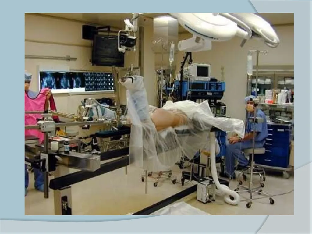
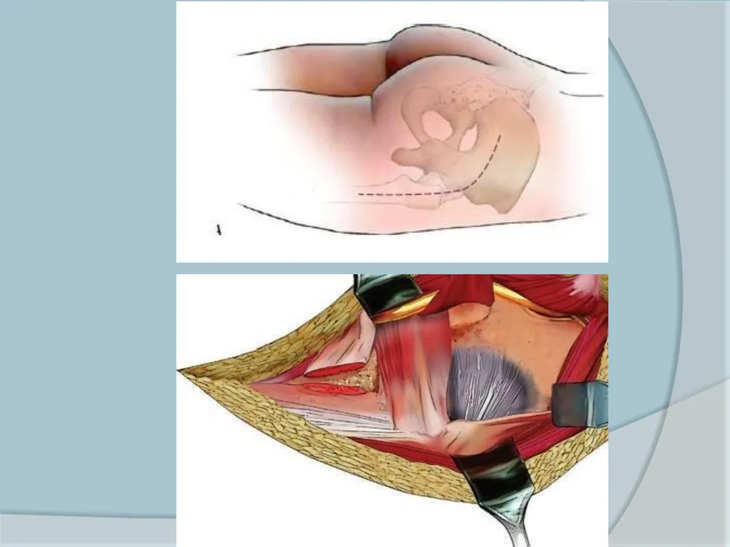
Kocher-Langenbeck approach Position- Prone or lateral with hip in slight extension and knee flexed
Provides access to posterior column and entire posterior wall
Advantages-
Familiarty to most surgeons
Muscle dissection and blood loss minimal
Disadvantages are-
Limited exposure
Possible injury to sciatic nerve and superior gluteal artery& nerve,Pudendal nerve, medial circumflex femoral artery
Increased risk of HO<br>
Provides access to posterior column and entire posterior wall
Advantages-
Familiarty to most surgeons
Muscle dissection and blood loss minimal
Disadvantages are-
Limited exposure
Possible injury to sciatic nerve and superior gluteal artery& nerve,Pudendal nerve, medial circumflex femoral artery
Increased risk of HO<br>
48
Ilioinguinal approach Position- Supine
Approach of choice for all anterior wall and anterior column fractures
Creates three working portals or windows
Lateral window exposes entire iliac fossa, S I joint, sacral ala, and the superior iliopectineal eminence
Middle window exposes the pelvic brim and quadrilateral surface
Medial window provides access to pubic ramus, pubic symphysis and the retropubic space of Retzius<br>
Approach of choice for all anterior wall and anterior column fractures
Creates three working portals or windows
Lateral window exposes entire iliac fossa, S I joint, sacral ala, and the superior iliopectineal eminence
Middle window exposes the pelvic brim and quadrilateral surface
Medial window provides access to pubic ramus, pubic symphysis and the retropubic space of Retzius<br>
49
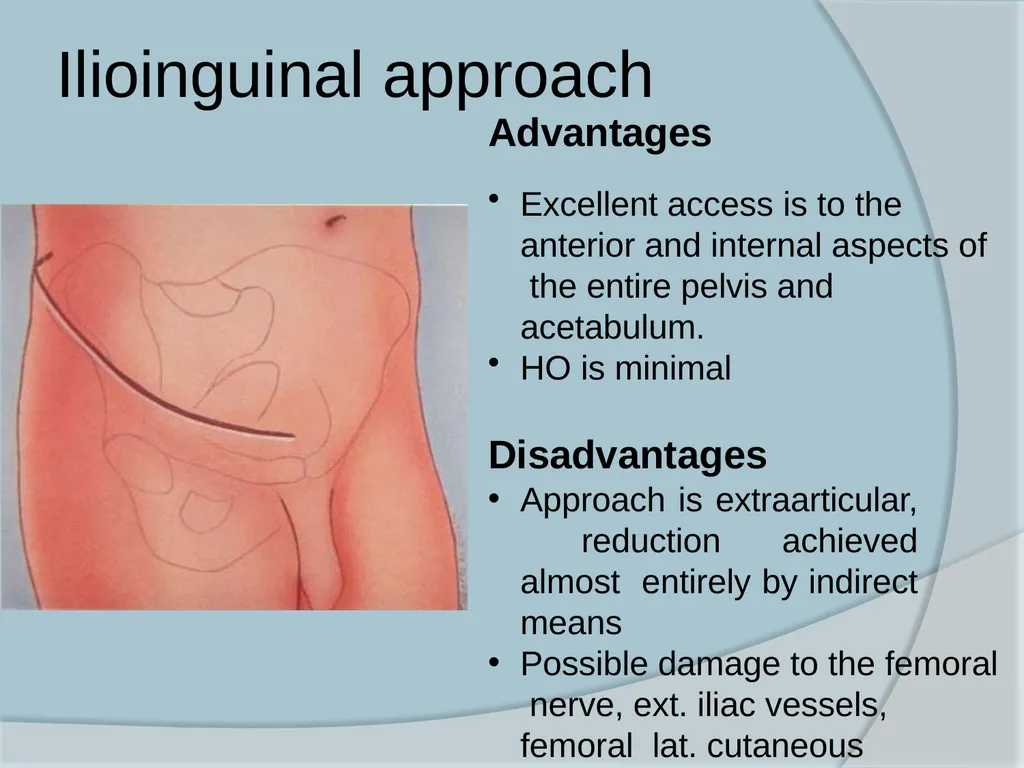
Ilioinguinal approach Advantages
Excellent access is to the anterior and internal aspects of the entire pelvis and acetabulum.
HO is minimal
Disadvantages
Approach is extraarticular, reduction achieved almost entirely by indirect means
Possible damage to the femoral nerve, ext. iliac vessels, femoral lat. cutaneous Nerve,inguinal canal contents<br>
Excellent access is to the anterior and internal aspects of the entire pelvis and acetabulum.
HO is minimal
Disadvantages
Approach is extraarticular, reduction achieved almost entirely by indirect means
Possible damage to the femoral nerve, ext. iliac vessels, femoral lat. cutaneous Nerve,inguinal canal contents<br>
50
Extended Iliofemoral approach Indicated for selected complex acetabular fracture types and for surgery delayed more than 2 weeks following injury
Direct access to the iliac crest and the entire internal iliac fossa
Position- Lateral and the knee in flexed position to relax the sciatic nerve
Provides access to entire posterior column, external ilium, S I joint, and anterior column up to iliopectineal eminence<br>
Direct access to the iliac crest and the entire internal iliac fossa
Position- Lateral and the knee in flexed position to relax the sciatic nerve
Provides access to entire posterior column, external ilium, S I joint, and anterior column up to iliopectineal eminence<br>
51
Extended Iliofemoral approach Advantage
Exellent access to
entire hemipelvis
Disadvantages
High incidence of
HO
Possible damage to superior gluteal and femoral NV bundles<br>
Exellent access to
entire hemipelvis
Disadvantages
High incidence of
HO
Possible damage to superior gluteal and femoral NV bundles<br>
52
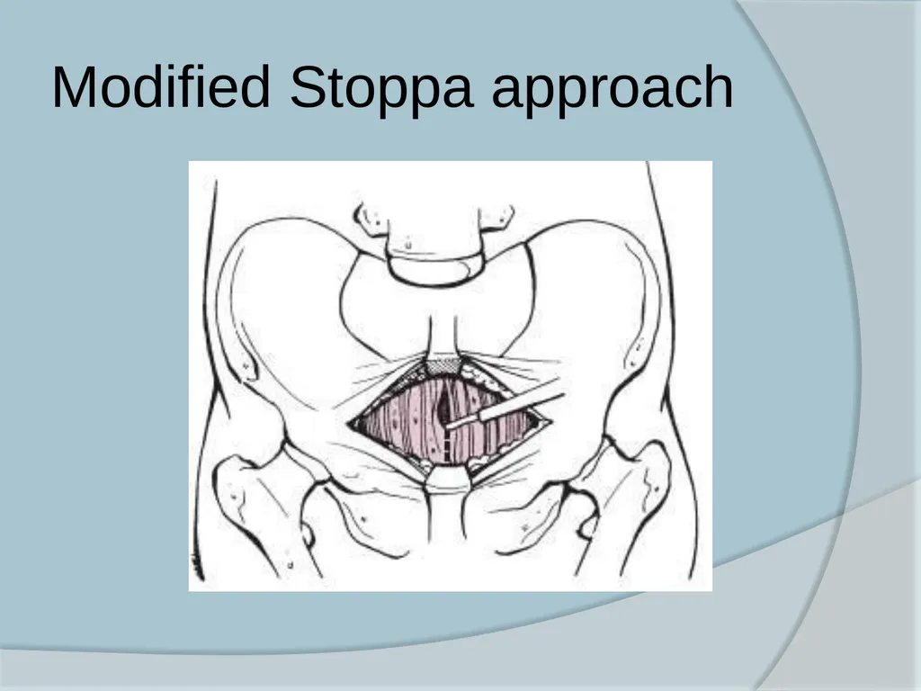
Modified Stoppa Approach Position- Supine
Provides improved exposure of the quadrilateral surface and posterior column
Commonly used in conjunction with the lateral window
Advantage over the ilioinguinal is that dissection of the iliac vessels is not required
Risk of injury to Corona mortis and bladder<br>
Provides improved exposure of the quadrilateral surface and posterior column
Commonly used in conjunction with the lateral window
Advantage over the ilioinguinal is that dissection of the iliac vessels is not required
Risk of injury to Corona mortis and bladder<br>
53
Modified Stoppa approach<br>
54
Treatment of specific fracture pattern Posterior wall fractures
Kocher–Langenbeck approach is ideal for posterior wall fractures
Special circumstances like large wall fragments incarcerated in the joint trochanteric flip osteotomy may be needed<br>
Kocher–Langenbeck approach is ideal for posterior wall fractures
Special circumstances like large wall fragments incarcerated in the joint trochanteric flip osteotomy may be needed<br>
55
Treatment of specific fracture pattern Posterior column fracture
Kocher–Langenbeck
Anterior Column Fractures and Anterior Wall Fractures
Ilioinguinal approach
If the quadrilateral surface is comminuted the modified Stoppa approach may be a better choice<br>
Kocher–Langenbeck
Anterior Column Fractures and Anterior Wall Fractures
Ilioinguinal approach
If the quadrilateral surface is comminuted the modified Stoppa approach may be a better choice<br>
56
Treatment of specific fracture pattern Transverse Fractures
Kocher–Langenbeck approach as posterior column usually is the site of greatest fracture displacement
Transverse and Post. Wall Fractures
Kocher–Langenbeck approach<br>
Kocher–Langenbeck approach as posterior column usually is the site of greatest fracture displacement
Transverse and Post. Wall Fractures
Kocher–Langenbeck approach<br>
57
Treatment of specific fracture pattern Anterior Column (or Wall) and Posterior Hemitransverse Fractures
Posterior column component is only minimally displaced in this fracture type. Therefore ilioinguinal approach is used.
Modified Stoppa approach alternative
Posterior column is widely displaced (more than 5 mm), or it cannot be reduced through the anterior approach, the Kocher– Langenbeck is added<br>
Posterior column component is only minimally displaced in this fracture type. Therefore ilioinguinal approach is used.
Modified Stoppa approach alternative
Posterior column is widely displaced (more than 5 mm), or it cannot be reduced through the anterior approach, the Kocher– Langenbeck is added<br>
58
Treatment of specific fracture pattern T- shaped Fractures
Kocher–Langenbeck approach
If the anterior column fracture cannot be reduced ,subsequent patient repositioning and an anterior approach is required (Ilioinguinal or Modified Stoppa)
Both column Fractures
Commonly operated on using an anterior approach (ilioinguinal or modified Stoppa) with the patient supine<br>
Kocher–Langenbeck approach
If the anterior column fracture cannot be reduced ,subsequent patient repositioning and an anterior approach is required (Ilioinguinal or Modified Stoppa)
Both column Fractures
Commonly operated on using an anterior approach (ilioinguinal or modified Stoppa) with the patient supine<br>
59
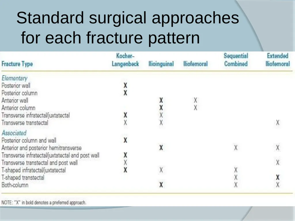
Standard surgical approaches for each fracture pattern<br>
60
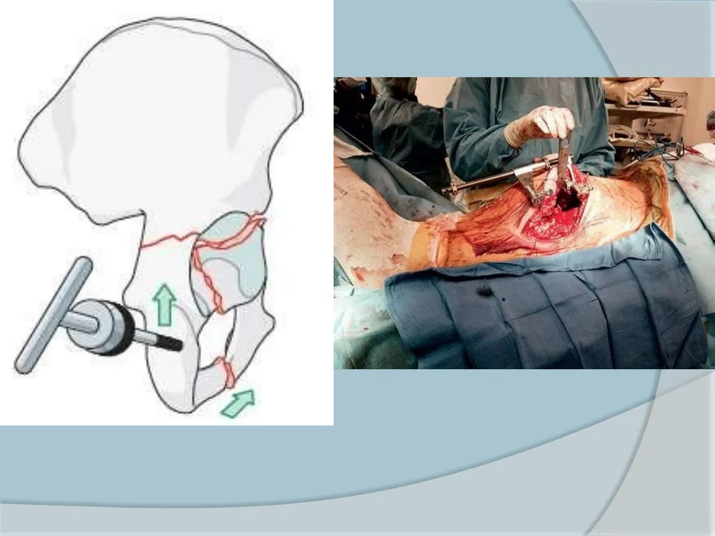
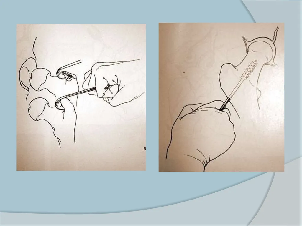
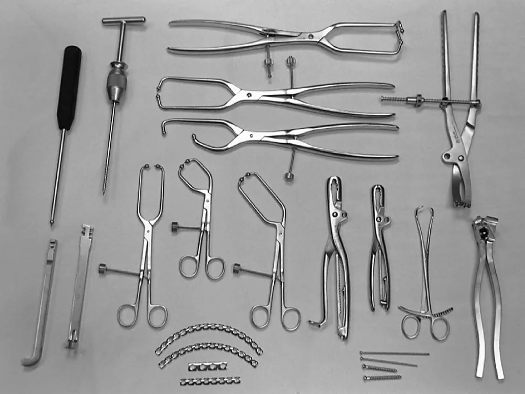
Methods of Reduction Traction-
With Traction table
Manual Traction-
Traction on extremity by manual pull
Corkscrew
Large sharp hook
Schanz pin<br>
With Traction table
Manual Traction-
Traction on extremity by manual pull
Corkscrew
Large sharp hook
Schanz pin<br>
61
Post operative care Suction drain
Antibiotic for 48 – 72 hours
Thromboprophylaxis- Mechanical compressive device with LMWH
Indomethacin 25 mg tds beginning within 24 hours of surgery and continued for 4 to 6 weeks to prevent HO
Passive motion of the hip on the 2nd or 3rd day.
Touch down ambulation & crutches on 2nd to 4th day
Progression to FWB must be tailored to the individual<br>
Antibiotic for 48 – 72 hours
Thromboprophylaxis- Mechanical compressive device with LMWH
Indomethacin 25 mg tds beginning within 24 hours of surgery and continued for 4 to 6 weeks to prevent HO
Passive motion of the hip on the 2nd or 3rd day.
Touch down ambulation & crutches on 2nd to 4th day
Progression to FWB must be tailored to the individual<br>
62
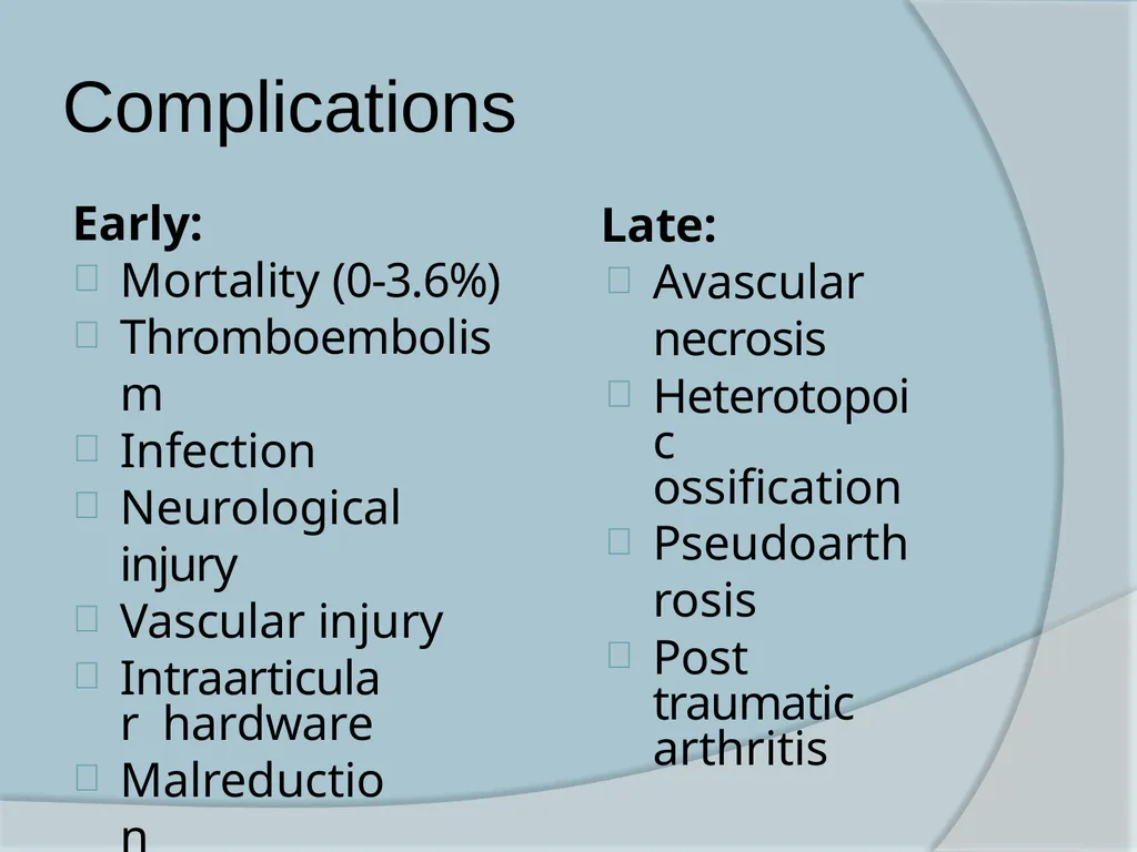
Complications Early:
Mortality (0-3.6%)
Thromboembolism
Infection
Neurological injury
Vascular injury
Intraarticular hardware
Malreduction
Loss of reduction Late:
Avascular necrosis
Heterotopoic ossification
Pseudoarthrosis
Post traumatic arthritis<br>
Mortality (0-3.6%)
Thromboembolism
Infection
Neurological injury
Vascular injury
Intraarticular hardware
Malreduction
Loss of reduction Late:
Avascular necrosis
Heterotopoic ossification
Pseudoarthrosis
Post traumatic arthritis<br>
63
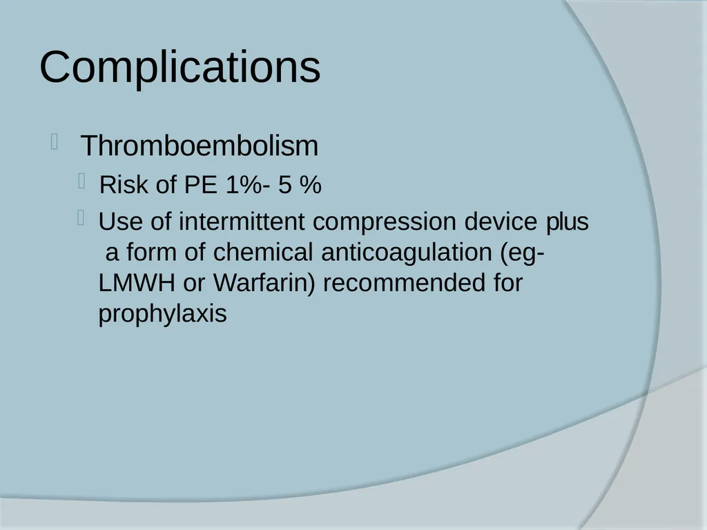
Complications Thromboembolism
Risk of PE 1%- 5 %
Use of intermittent compression device plus a form of chemical anticoagulation (eg- LMWH or Warfarin) recommended for prophylaxis<br>
Risk of PE 1%- 5 %
Use of intermittent compression device plus a form of chemical anticoagulation (eg- LMWH or Warfarin) recommended for prophylaxis<br>
64
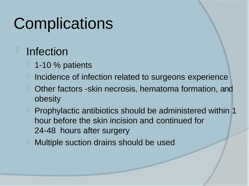
Complications Infection
1-10 % patients
Incidence of infection related to surgeons experience
Other factors -skin necrosis, hematoma formation, and obesity
Prophylactic antibiotics should be administered within 1 hour before the skin incision and continued for 24-48 hours after surgery
Multiple suction drains should be used<br>
1-10 % patients
Incidence of infection related to surgeons experience
Other factors -skin necrosis, hematoma formation, and obesity
Prophylactic antibiotics should be administered within 1 hour before the skin incision and continued for 24-48 hours after surgery
Multiple suction drains should be used<br>
65
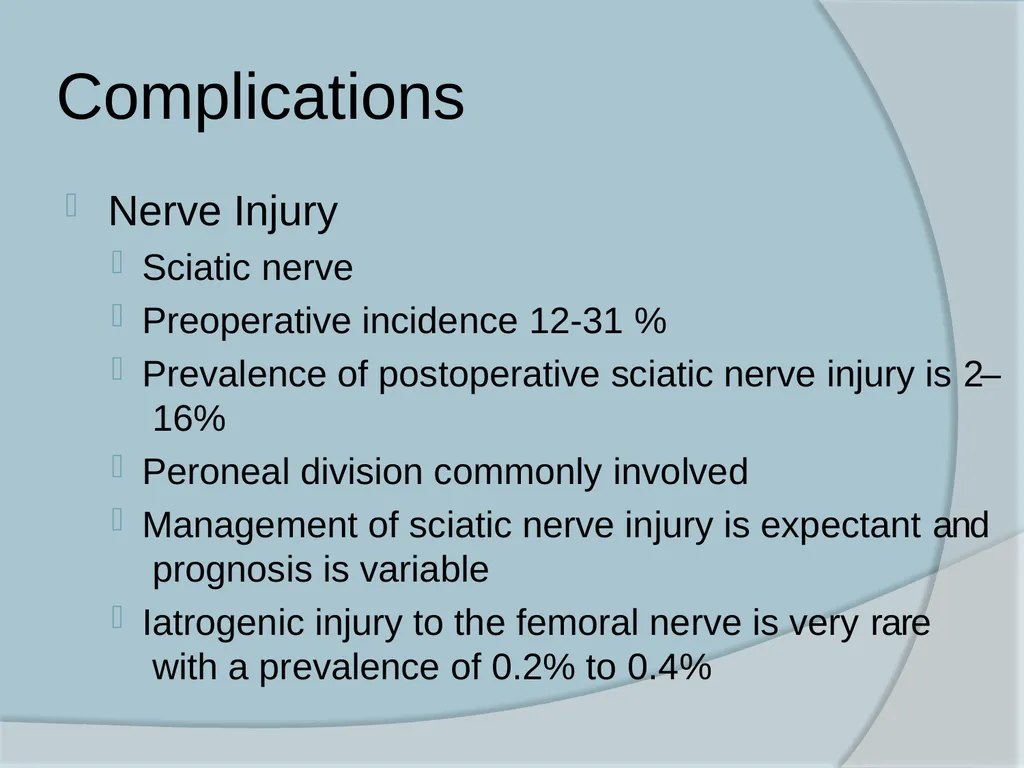
Complications Nerve Injury
Sciatic nerve
Preoperative incidence 12-31 %
Prevalence of postoperative sciatic nerve injury is 2– 16%
Peroneal division commonly involved
Management of sciatic nerve injury is expectant and prognosis is variable
Iatrogenic injury to the femoral nerve is very rare with a prevalence of 0.2% to 0.4%<br>
Sciatic nerve
Preoperative incidence 12-31 %
Prevalence of postoperative sciatic nerve injury is 2– 16%
Peroneal division commonly involved
Management of sciatic nerve injury is expectant and prognosis is variable
Iatrogenic injury to the femoral nerve is very rare with a prevalence of 0.2% to 0.4%<br>
66
Complications Heterotropic ossification
Incidence varies from 3–69%
Related to extensile surgical exposures, male gender, associated head injury, significant delays to surgery, fracture type, the severity of the injury
Rare with ilioinguinal approach
“Significant HO” -loss of active range of motion
>20% of normal.
Indomethacin 25 mg tds for 6 weeks or 75 mg SR capsule OD for 6 weeks
Radiation therapy- 7–8 Gy in a single dose or 10 Gy in five doses<br>
Incidence varies from 3–69%
Related to extensile surgical exposures, male gender, associated head injury, significant delays to surgery, fracture type, the severity of the injury
Rare with ilioinguinal approach
“Significant HO” -loss of active range of motion
>20% of normal.
Indomethacin 25 mg tds for 6 weeks or 75 mg SR capsule OD for 6 weeks
Radiation therapy- 7–8 Gy in a single dose or 10 Gy in five doses<br>
67
Complications Posttraumatic Arthritis
Prevalence of osteoarthritis 4–48%
Quality of the fracture reduction main determinant
Incidence following perfect reduction was 10%
Anatomical reduction and restoration of joint congruency is the best prophylaxis<br>
Prevalence of osteoarthritis 4–48%
Quality of the fracture reduction main determinant
Incidence following perfect reduction was 10%
Anatomical reduction and restoration of joint congruency is the best prophylaxis<br>
68
Conclusion High velocity injuries with significant accompanying injuries
ATLS protocol
Fracture management depends on
Proper case selection for surgical and conservative
treatment
3 dimensional understanding of anatomy,
Proper interpretation of radiographs
Realistic expectations
Specialized instrumentation
Surgical expertise<br>
ATLS protocol
Fracture management depends on
Proper case selection for surgical and conservative
treatment
3 dimensional understanding of anatomy,
Proper interpretation of radiographs
Realistic expectations
Specialized instrumentation
Surgical expertise<br>
69
Conclusion Many perioperative complications are a consequence of the injury
Acetabular fracture fixation surgery is complex and demanding and has the potential for many serious complications
Anatomical reduction of the WBD , congruent reduction of the femoral head and timely surgical intervention are the keys to success<br>
Acetabular fracture fixation surgery is complex and demanding and has the potential for many serious complications
Anatomical reduction of the WBD , congruent reduction of the femoral head and timely surgical intervention are the keys to success<br>
70
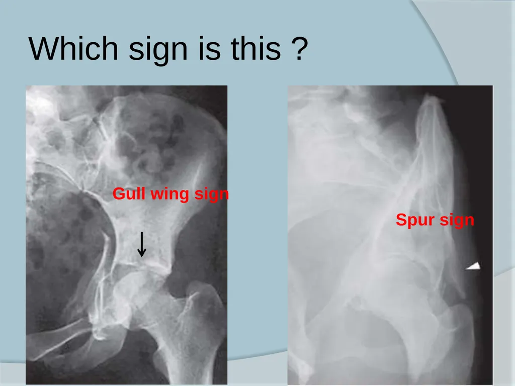
Which sign is this ? Gull wing sign Spur sign<br>
71
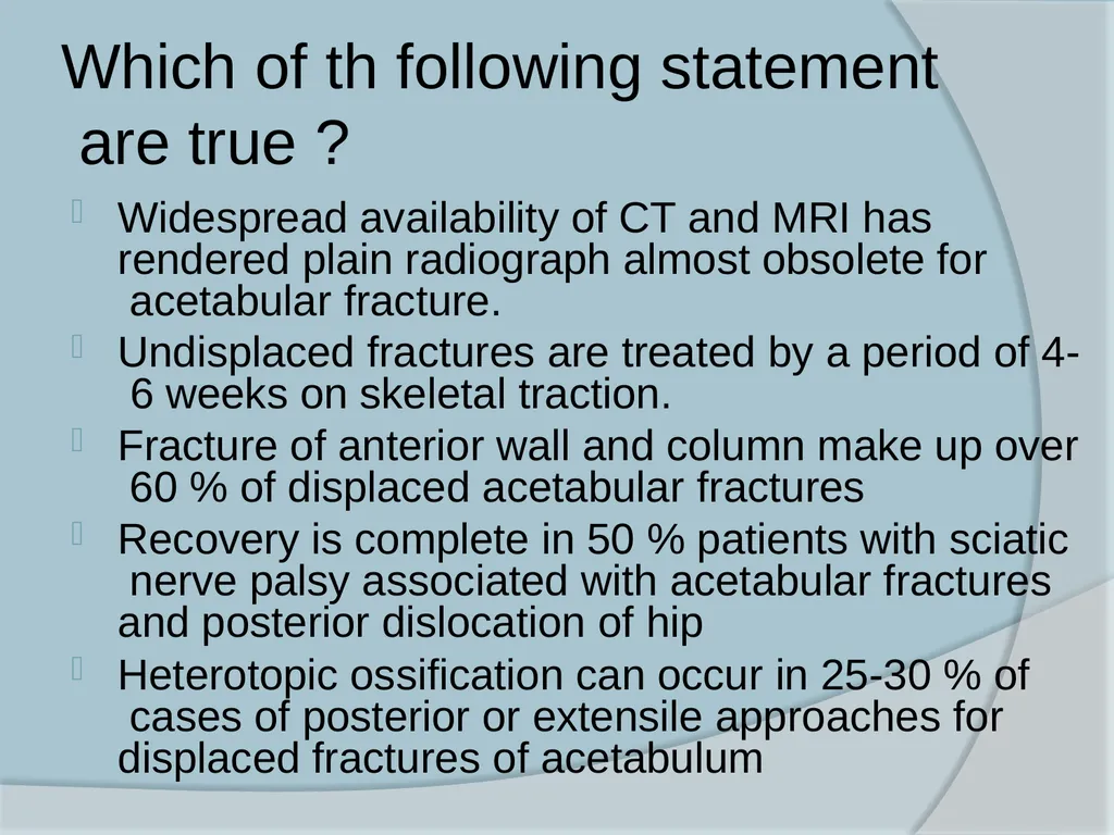
Which of th following statement are true ? Widespread availability of CT and MRI has rendered plain radiograph almost obsolete for acetabular fracture.
Undisplaced fractures are treated by a period of 4- 6 weeks on skeletal traction.
Fracture of anterior wall and column make up over 60 % of displaced acetabular fractures
Recovery is complete in 50 % patients with sciatic nerve palsy associated with acetabular fractures and posterior dislocation of hip
Heterotopic ossification can occur in 25-30 % of cases of posterior or extensile approaches for displaced fractures of acetabulum<br>
Undisplaced fractures are treated by a period of 4- 6 weeks on skeletal traction.
Fracture of anterior wall and column make up over 60 % of displaced acetabular fractures
Recovery is complete in 50 % patients with sciatic nerve palsy associated with acetabular fractures and posterior dislocation of hip
Heterotopic ossification can occur in 25-30 % of cases of posterior or extensile approaches for displaced fractures of acetabulum<br>
72
Criteria for non operative management of acetabular fracture includes all except : Stability demonstrated by dynamic stress radiographs
Femoral head subluxation of 3 mm
Congruence of femoral head with acetabular roof on AP and judet views
Roof arc measurement of greater than or equal to 45
Acetabular articular surface is intact in the superior 10 mm of the joint on CT evaluation<br>
Femoral head subluxation of 3 mm
Congruence of femoral head with acetabular roof on AP and judet views
Roof arc measurement of greater than or equal to 45
Acetabular articular surface is intact in the superior 10 mm of the joint on CT evaluation<br>