
Figure 2 Figure 2. Comparative restriction enzyme
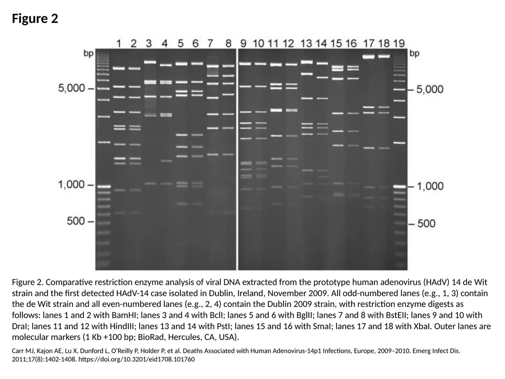
Description: Figure 2 Figure 2. Comparative restriction enzyme analysis of viral DNA extracted from the prototype human adenovirus (HAdV) 14 de Wit strain and the first detected HAdV-14 case isolated in Dublin, Ireland, November 2009. All odd-numbered
Related Topics
Download Presentation
"Figure 2 Figure 2. Comparative restriction enzyme" is the property of its rightful owner. Permission is granted to download and print the materials on this website for personal, non-commercial use only, and to display it on your personal computer provided you do not modify the materials and that you retain all copyright notices contained in the materials. By downloading content from our website, you accept the terms of this agreement.
Presentation Transcript
slide1. Figure 2 Figure 2. Comparative restriction enzyme analysis of viral DNA extracted from the prototype human adenovirus (HAdV) 14 de Wit strain and the first detected HAdV-14 case isolated in Dublin, Ireland, November 2009. All odd-numbered lanes (e.g., 1, 3) contain the de Wit strain and all even-numbered lanes (e.g., 2, 4) contain the Dublin 2009 strain, with restriction enzyme digests as follows: lanes 1 and 2 with BamHI; lanes 3 and 4 with BclI; lanes 5 and 6 with BglII; lanes 7 and 8 with BstEII; lanes 9 and 10 with DraI; lanes 11 and 12 with HindIII; lanes 13 and 14 with PstI; lanes 15 and 16 with SmaI; lanes 17 and 18 with XbaI. Outer lanes are molecular markers (1 Kb +100 bp; BioRad, Hercules, CA, USA). Carr MJ, Kajon AE, Lu X, Dunford L, O’Reilly P, Holder P, et al. Deaths Associated with Human Adenovirus-14p1 Infections, Europe, 2009–2010. Emerg Infect Dis. 2011;17(8):1402-1408. https://doi.org/10.3201/eid1708.101760<br>