GOUT Unit:3 Dr. Anil Kumar (Medicine) Asst. Professor Dept. of VCC GOUT Introduction: The term gout in general term refers to a form of inflammatory arthritis. Gout is defined as abnormal accumulation of uric acid in the bloodstream and
"GOUT Unit:3 Dr. Anil Kumar (Medicine) Asst." is the property of its rightful owner. Permission is granted to
download and print the materials on this website for personal, non-commercial use only, and to display it
on your personal computer provided you do not modify the materials and that you retain all copyright
notices contained in the materials. By downloading content from our website, you accept the terms of this
agreement.
Presentation Transcript
01
GOUT
Unit:3 Dr. Anil Kumar
(Medicine) Asst. Professor
Dept. of VCC<br>
02
GOUT
Introduction:
The term “gout” in general term refers to a form of inflammatory arthritis.
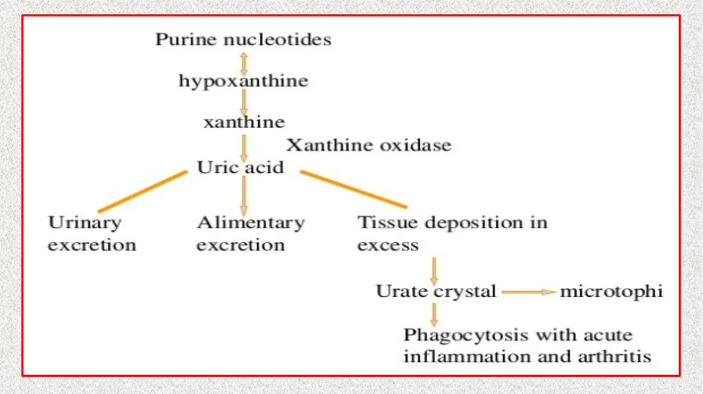
Gout is defined as abnormal accumulation of uric acid in the bloodstream and consequent deposition of uric acid on and within visceral tissues and articular surfaces.
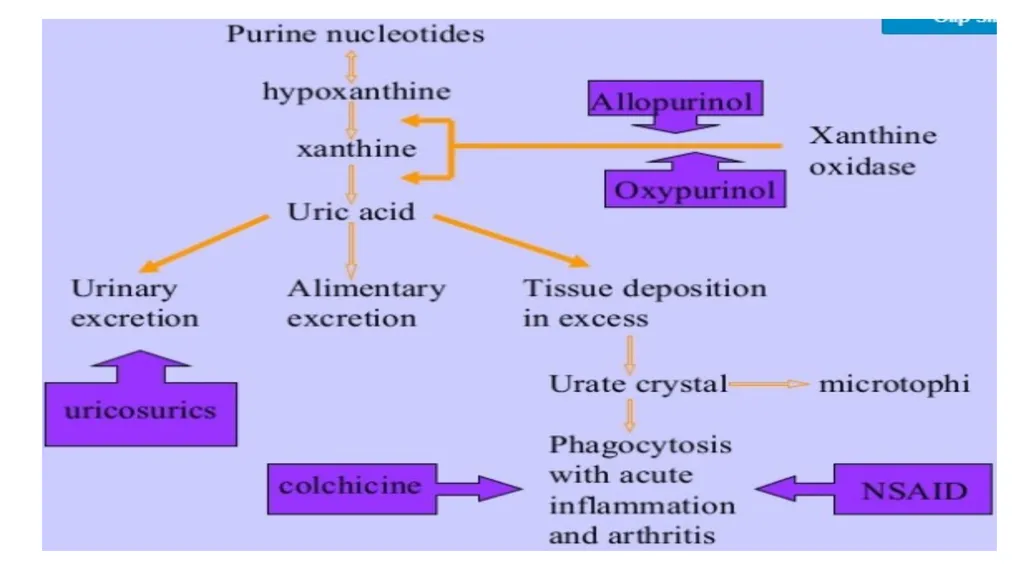
Gout is an inherited disorder of purine metabolism that causes hyperuricemia.
Gout is rare in most other animals due to their ability to produce uricase, which breaks down uric acid
Uric acid is the end product of nitrogen metabolism in birds, and it is produced in the liver.
Host: Birds
Etiology:
Renal disease
Increased protein ingestion
Prolonged dehydration
Reduced renal excretion of urates.
All these will lead to formation Monosodium urate crystal precipitation in tissues<br>
03
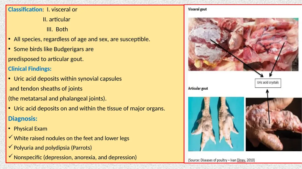
Classification: I. visceral or
II. articular
III. Both
All species, regardless of age and sex, are susceptible.
Some birds like Budgerigars are
predisposed to articular gout.
Clinical Findings:
Uric acid deposits within synovial capsules
and tendon sheaths of joints
(the metatarsal and phalangeal joints).
Uric acid deposits on and within the tissue of major organs.
Diagnosis:
Physical Exam
White raised nodules on the feet and lower legs
Polyuria and polydipsia (Parrots)
Nonspecific (depression, anorexia, and depression)<br>
04
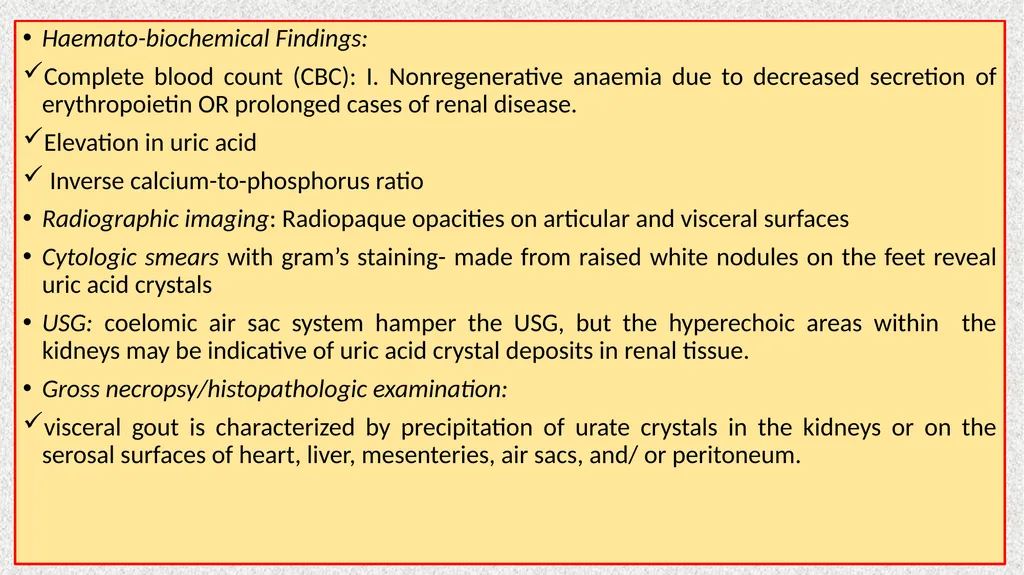
Haemato-biochemical Findings:
Complete blood count (CBC): I. Nonregenerative anaemia due to decreased secretion of erythropoietin OR prolonged cases of renal disease.
Elevation in uric acid
Inverse calcium-to-phosphorus ratio
Radiographic imaging: Radiopaque opacities on articular and visceral surfaces
Cytologic smears with gram’s staining- made from raised white nodules on the feet reveal uric acid crystals
USG: coelomic air sac system hamper the USG, but the hyperechoic areas within the kidneys may be indicative of uric acid crystal deposits in renal tissue.
Gross necropsy/histopathologic examination:
visceral gout is characterized by precipitation of urate crystals in the kidneys or on the serosal surfaces of heart, liver, mesenteries, air sacs, and/ or peritoneum.<br>
05
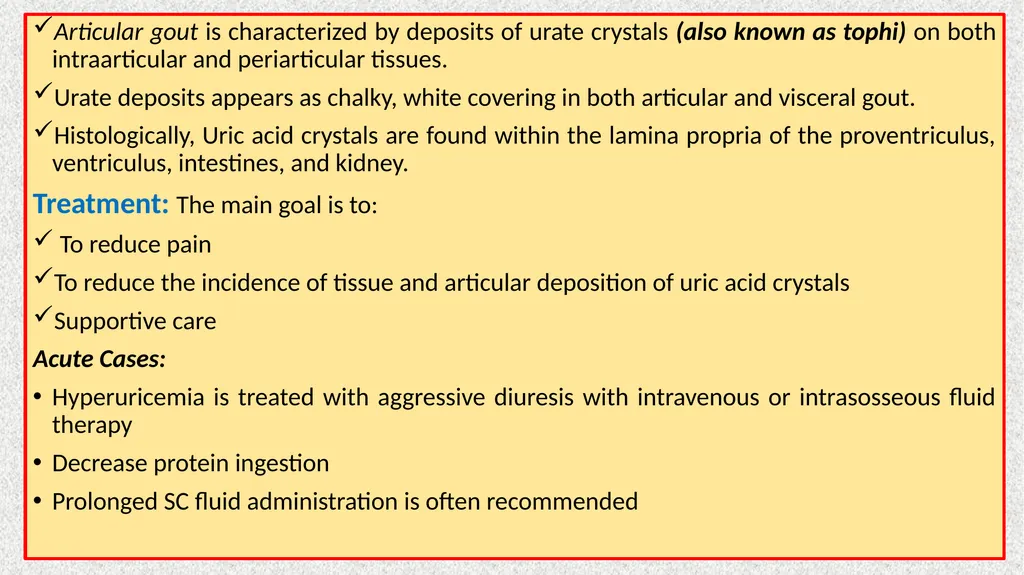
Articular gout is characterized by deposits of urate crystals (also known as tophi) on both intraarticular and periarticular tissues.
Urate deposits appears as chalky, white covering in both articular and visceral gout.
Histologically, Uric acid crystals are found within the lamina propria of the proventriculus, ventriculus, intestines, and kidney.
Treatment: The main goal is to:
To reduce pain
To reduce the incidence of tissue and articular deposition of uric acid crystals
Supportive care
Acute Cases:
Hyperuricemia is treated with aggressive diuresis with intravenous or intrasosseous fluid therapy
Decrease protein ingestion
Prolonged SC fluid administration is often recommended<br>
06
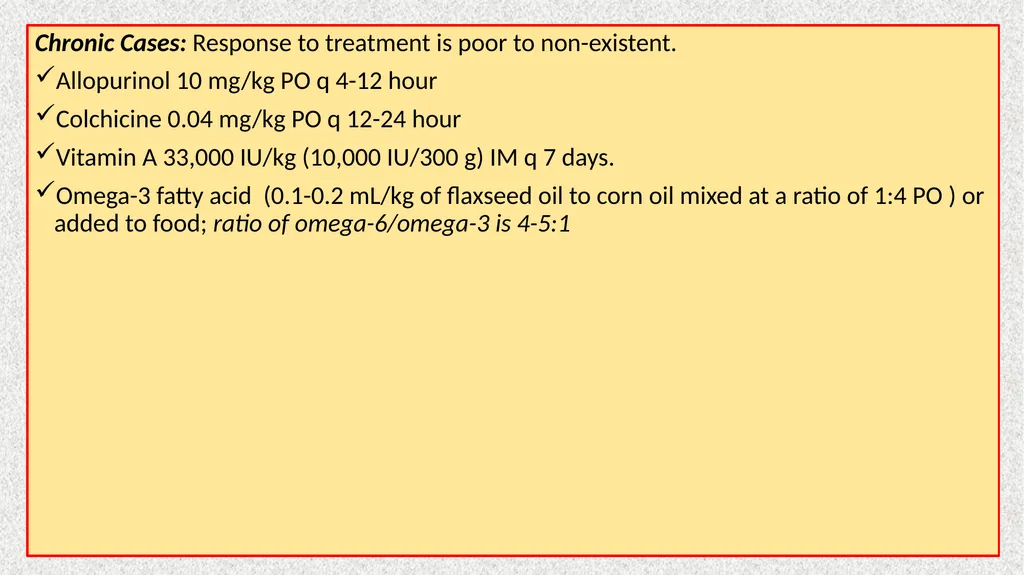
Chronic Cases: Response to treatment is poor to non-existent.
Allopurinol 10 mg/kg PO q 4-12 hour
Colchicine 0.04 mg/kg PO q 12-24 hour
Vitamin A 33,000 IU/kg (10,000 IU/300 g) IM q 7 days.
Omega-3 fatty acid (0.1-0.2 mL/kg of flaxseed oil to corn oil mixed at a ratio of 1:4 PO ) or added to food; ratio of omega-6/omega-3 is 4-5:1<br>