
Lec.3 Liver Function Tests Dr. Shatha AL-Khateeb
KB
Published · 14 slides · 0 views
1 / 1


Description
Lec.3 Liver Function Tests Dr. Shatha AL-Khateeb Algorithm of Approach in Abnormal Liver Function Test Cholestasis It is defined as a decrease in bile flow due to impaired secretion by hepatocytes or to obstruction of bile flow through
Related Topics
Share
Embed code
Download this presentation From Below
"Lec.3 Liver Function Tests Dr. Shatha AL-Khateeb" is the property of its rightful owner. Permission is granted to download and print the materials on this website for personal, non-commercial use only, and to display it on your personal computer provided you do not modify the materials and that you retain all copyright notices contained in the materials. By downloading content from our website, you accept the terms of this agreement.
Presentation Transcript
01
Lec.3 Liver Function Tests
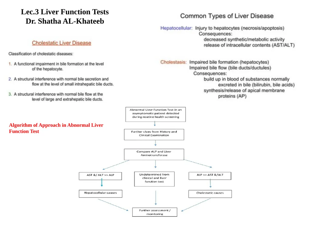
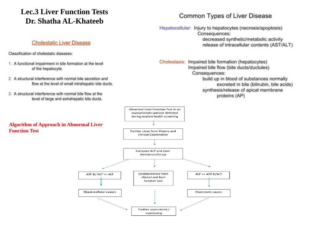
Dr. Shatha AL-Khateeb Algorithm of Approach in Abnormal Liver Function Test<br>
Dr. Shatha AL-Khateeb Algorithm of Approach in Abnormal Liver Function Test<br>
02
Cholestasis
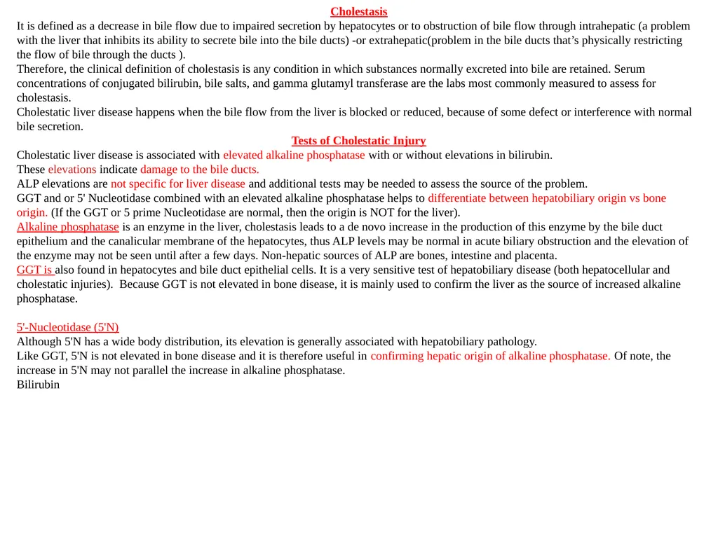
It is defined as a decrease in bile flow due to impaired secretion by hepatocytes or to obstruction of bile flow through intrahepatic (a problem with the liver that inhibits its ability to secrete bile into the bile ducts) -or extrahepatic(problem in the bile ducts that’s physically restricting the flow of bile through the ducts ).
Therefore, the clinical definition of cholestasis is any condition in which substances normally excreted into bile are retained. Serum concentrations of conjugated bilirubin, bile salts, and gamma glutamyl transferase are the labs most commonly measured to assess for cholestasis.
Cholestatic liver disease happens when the bile flow from the liver is blocked or reduced, because of some defect or interference with normal bile secretion.
Tests of Cholestatic Injury
Cholestatic liver disease is associated with elevated alkaline phosphatase with or without elevations in bilirubin.
These elevations indicate damage to the bile ducts.
ALP elevations are not specific for liver disease and additional tests may be needed to assess the source of the problem.
GGT and or 5' Nucleotidase combined with an elevated alkaline phosphatase helps to differentiate between hepatobiliary origin vs bone origin. (If the GGT or 5 prime Nucleotidase are normal, then the origin is NOT for the liver).
Alkaline phosphatase is an enzyme in the liver, cholestasis leads to a de novo increase in the production of this enzyme by the bile duct epithelium and the canalicular membrane of the hepatocytes, thus ALP levels may be normal in acute biliary obstruction and the elevation of the enzyme may not be seen until after a few days. Non-hepatic sources of ALP are bones, intestine and placenta.
GGT is also found in hepatocytes and bile duct epithelial cells. It is a very sensitive test of hepatobiliary disease (both hepatocellular and cholestatic injuries). Because GGT is not elevated in bone disease, it is mainly used to confirm the liver as the source of increased alkaline phosphatase.
5'-Nucleotidase (5'N)
Although 5'N has a wide body distribution, its elevation is generally associated with hepatobiliary pathology.
Like GGT, 5'N is not elevated in bone disease and it is therefore useful in confirming hepatic origin of alkaline phosphatase. Of note, the increase in 5'N may not parallel the increase in alkaline phosphatase.
Bilirubin<br>
It is defined as a decrease in bile flow due to impaired secretion by hepatocytes or to obstruction of bile flow through intrahepatic (a problem with the liver that inhibits its ability to secrete bile into the bile ducts) -or extrahepatic(problem in the bile ducts that’s physically restricting the flow of bile through the ducts ).
Therefore, the clinical definition of cholestasis is any condition in which substances normally excreted into bile are retained. Serum concentrations of conjugated bilirubin, bile salts, and gamma glutamyl transferase are the labs most commonly measured to assess for cholestasis.
Cholestatic liver disease happens when the bile flow from the liver is blocked or reduced, because of some defect or interference with normal bile secretion.
Tests of Cholestatic Injury
Cholestatic liver disease is associated with elevated alkaline phosphatase with or without elevations in bilirubin.
These elevations indicate damage to the bile ducts.
ALP elevations are not specific for liver disease and additional tests may be needed to assess the source of the problem.
GGT and or 5' Nucleotidase combined with an elevated alkaline phosphatase helps to differentiate between hepatobiliary origin vs bone origin. (If the GGT or 5 prime Nucleotidase are normal, then the origin is NOT for the liver).
Alkaline phosphatase is an enzyme in the liver, cholestasis leads to a de novo increase in the production of this enzyme by the bile duct epithelium and the canalicular membrane of the hepatocytes, thus ALP levels may be normal in acute biliary obstruction and the elevation of the enzyme may not be seen until after a few days. Non-hepatic sources of ALP are bones, intestine and placenta.
GGT is also found in hepatocytes and bile duct epithelial cells. It is a very sensitive test of hepatobiliary disease (both hepatocellular and cholestatic injuries). Because GGT is not elevated in bone disease, it is mainly used to confirm the liver as the source of increased alkaline phosphatase.
5'-Nucleotidase (5'N)
Although 5'N has a wide body distribution, its elevation is generally associated with hepatobiliary pathology.
Like GGT, 5'N is not elevated in bone disease and it is therefore useful in confirming hepatic origin of alkaline phosphatase. Of note, the increase in 5'N may not parallel the increase in alkaline phosphatase.
Bilirubin<br>
03
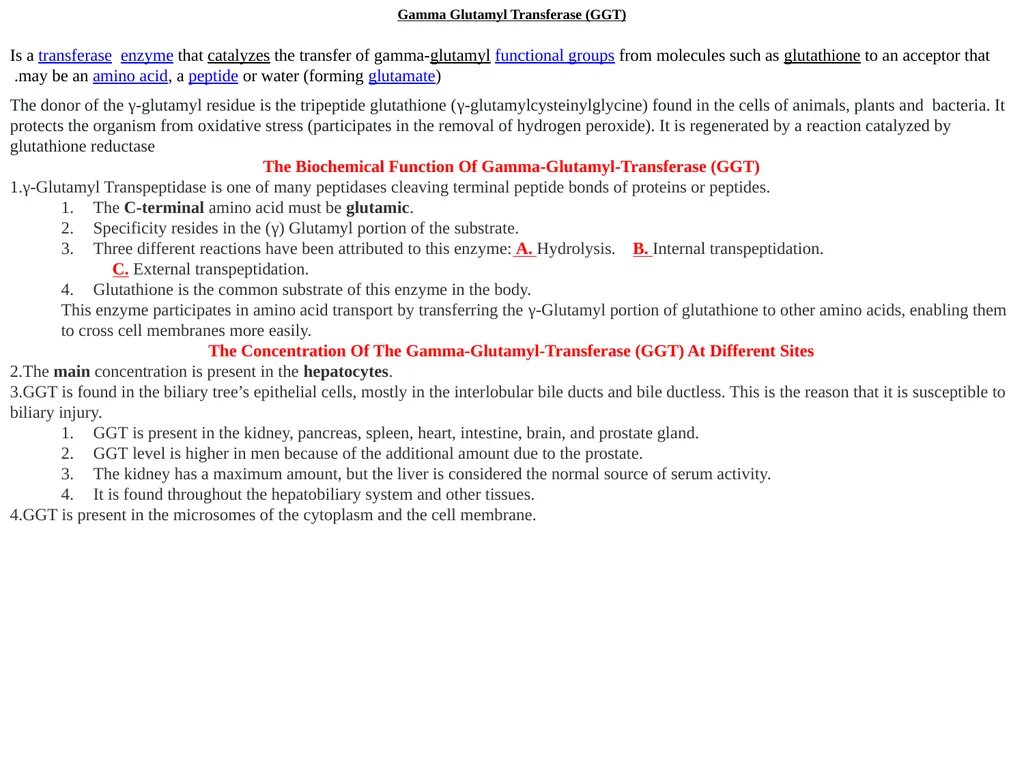
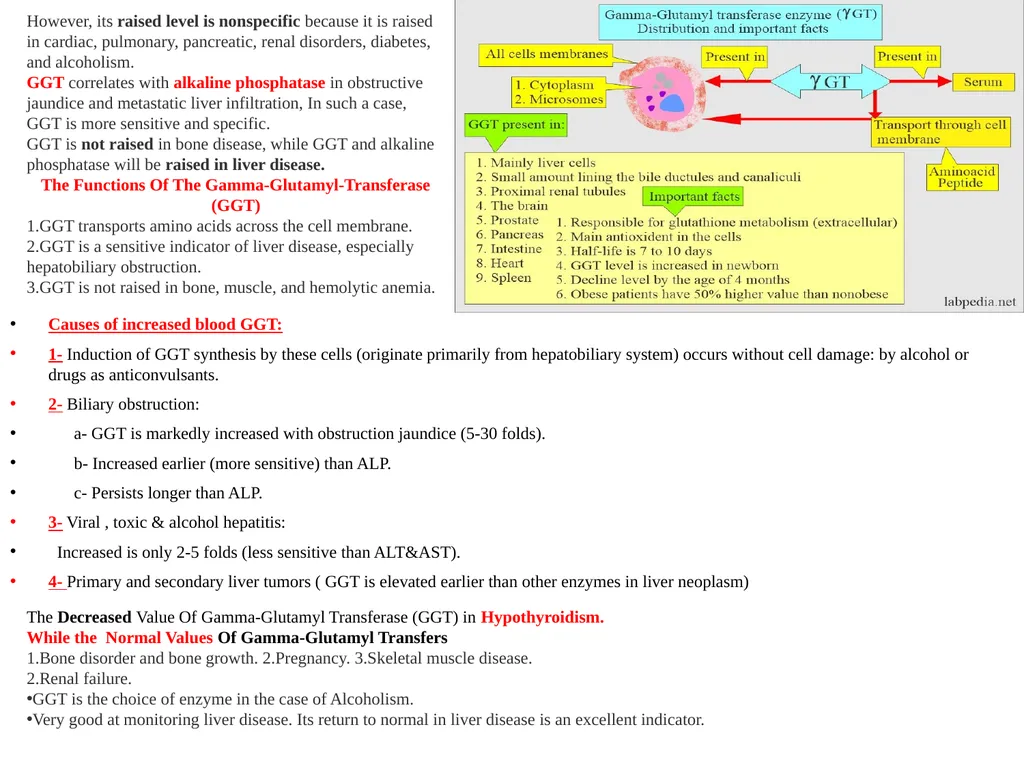
Gamma Glutamyl Transferase (GGT) Is a transferase enzyme that catalyzes the transfer of gamma-glutamyl functional groups from molecules such as glutathione to an acceptor that may be an amino acid, a peptide or water (forming glutamate).
The donor of the γ-glutamyl residue is the tripeptide glutathione (γ-glutamylcysteinylglycine) found in the cells of animals, plants and bacteria. It protects the organism from oxidative stress (participates in the removal of hydrogen peroxide). It is regenerated by a reaction catalyzed by glutathione reductase
The Biochemical Function Of Gamma-Glutamyl-Transferase (GGT)
γ-Glutamyl Transpeptidase is one of many peptidases cleaving terminal peptide bonds of proteins or peptides.
The C-terminal amino acid must be glutamic.
Specificity resides in the (γ) Glutamyl portion of the substrate.
Three different reactions have been attributed to this enzyme: A. Hydrolysis. B. Internal transpeptidation.
C. External transpeptidation.
Glutathione is the common substrate of this enzyme in the body.
This enzyme participates in amino acid transport by transferring the γ-Glutamyl portion of glutathione to other amino acids, enabling them to cross cell membranes more easily.
The Concentration Of The Gamma-Glutamyl-Transferase (GGT) At Different Sites
The main concentration is present in the hepatocytes.
GGT is found in the biliary tree’s epithelial cells, mostly in the interlobular bile ducts and bile ductless. This is the reason that it is susceptible to biliary injury.
GGT is present in the kidney, pancreas, spleen, heart, intestine, brain, and prostate gland.
GGT level is higher in men because of the additional amount due to the prostate.
The kidney has a maximum amount, but the liver is considered the normal source of serum activity.
It is found throughout the hepatobiliary system and other tissues.
GGT is present in the microsomes of the cytoplasm and the cell membrane.<br>
The donor of the γ-glutamyl residue is the tripeptide glutathione (γ-glutamylcysteinylglycine) found in the cells of animals, plants and bacteria. It protects the organism from oxidative stress (participates in the removal of hydrogen peroxide). It is regenerated by a reaction catalyzed by glutathione reductase
The Biochemical Function Of Gamma-Glutamyl-Transferase (GGT)
γ-Glutamyl Transpeptidase is one of many peptidases cleaving terminal peptide bonds of proteins or peptides.
The C-terminal amino acid must be glutamic.
Specificity resides in the (γ) Glutamyl portion of the substrate.
Three different reactions have been attributed to this enzyme: A. Hydrolysis. B. Internal transpeptidation.
C. External transpeptidation.
Glutathione is the common substrate of this enzyme in the body.
This enzyme participates in amino acid transport by transferring the γ-Glutamyl portion of glutathione to other amino acids, enabling them to cross cell membranes more easily.
The Concentration Of The Gamma-Glutamyl-Transferase (GGT) At Different Sites
The main concentration is present in the hepatocytes.
GGT is found in the biliary tree’s epithelial cells, mostly in the interlobular bile ducts and bile ductless. This is the reason that it is susceptible to biliary injury.
GGT is present in the kidney, pancreas, spleen, heart, intestine, brain, and prostate gland.
GGT level is higher in men because of the additional amount due to the prostate.
The kidney has a maximum amount, but the liver is considered the normal source of serum activity.
It is found throughout the hepatobiliary system and other tissues.
GGT is present in the microsomes of the cytoplasm and the cell membrane.<br>
04
However, its raised level is nonspecific because it is raised in cardiac, pulmonary, pancreatic, renal disorders, diabetes, and alcoholism.
GGT correlates with alkaline phosphatase in obstructive jaundice and metastatic liver infiltration, In such a case, GGT is more sensitive and specific.
GGT is not raised in bone disease, while GGT and alkaline phosphatase will be raised in liver disease.
The Functions Of The Gamma-Glutamyl-Transferase (GGT)
GGT transports amino acids across the cell membrane.
GGT is a sensitive indicator of liver disease, especially hepatobiliary obstruction.
GGT is not raised in bone, muscle, and hemolytic anemia. Causes of increased blood GGT:
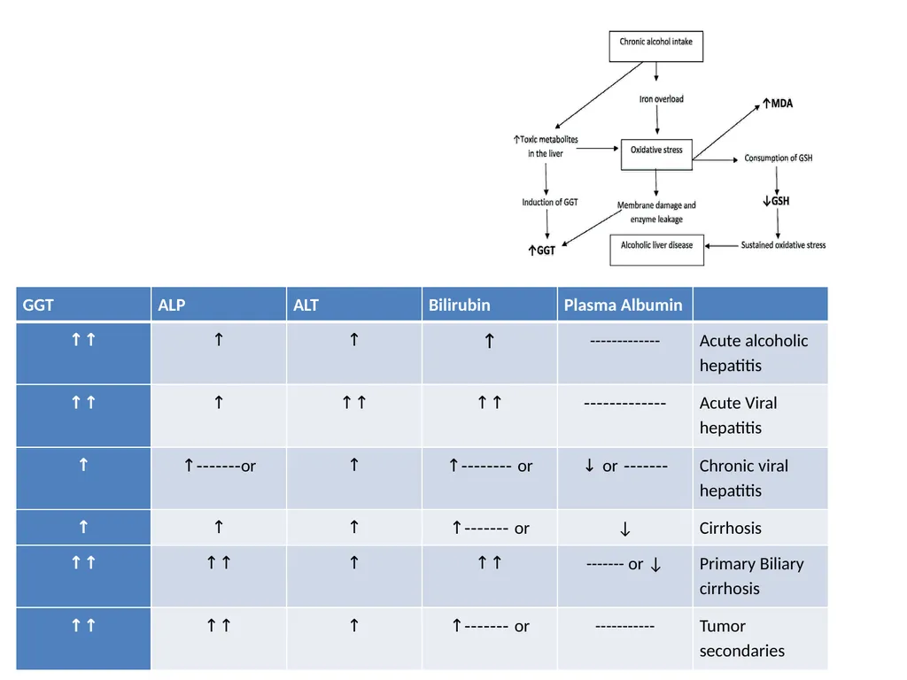
1- Induction of GGT synthesis by these cells (originate primarily from hepatobiliary system) occurs without cell damage: by alcohol or drugs as anticonvulsants.
2- Biliary obstruction:
a- GGT is markedly increased with obstruction jaundice (5-30 folds).
b- Increased earlier (more sensitive) than ALP.
c- Persists longer than ALP.
3- Viral , toxic & alcohol hepatitis:
Increased is only 2-5 folds (less sensitive than ALT&AST).
4- Primary and secondary liver tumors ( GGT is elevated earlier than other enzymes in liver neoplasm) The Decreased Value Of Gamma-Glutamyl Transferase (GGT) in Hypothyroidism.
While the Normal Values Of Gamma-Glutamyl Transfers
Bone disorder and bone growth. 2.Pregnancy. 3.Skeletal muscle disease.
Renal failure.
GGT is the choice of enzyme in the case of Alcoholism.
Very good at monitoring liver disease. Its return to normal in liver disease is an excellent indicator.<br>
GGT correlates with alkaline phosphatase in obstructive jaundice and metastatic liver infiltration, In such a case, GGT is more sensitive and specific.
GGT is not raised in bone disease, while GGT and alkaline phosphatase will be raised in liver disease.
The Functions Of The Gamma-Glutamyl-Transferase (GGT)
GGT transports amino acids across the cell membrane.
GGT is a sensitive indicator of liver disease, especially hepatobiliary obstruction.
GGT is not raised in bone, muscle, and hemolytic anemia. Causes of increased blood GGT:
1- Induction of GGT synthesis by these cells (originate primarily from hepatobiliary system) occurs without cell damage: by alcohol or drugs as anticonvulsants.
2- Biliary obstruction:
a- GGT is markedly increased with obstruction jaundice (5-30 folds).
b- Increased earlier (more sensitive) than ALP.
c- Persists longer than ALP.
3- Viral , toxic & alcohol hepatitis:
Increased is only 2-5 folds (less sensitive than ALT&AST).
4- Primary and secondary liver tumors ( GGT is elevated earlier than other enzymes in liver neoplasm) The Decreased Value Of Gamma-Glutamyl Transferase (GGT) in Hypothyroidism.
While the Normal Values Of Gamma-Glutamyl Transfers
Bone disorder and bone growth. 2.Pregnancy. 3.Skeletal muscle disease.
Renal failure.
GGT is the choice of enzyme in the case of Alcoholism.
Very good at monitoring liver disease. Its return to normal in liver disease is an excellent indicator.<br>
05
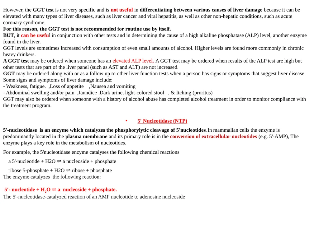
However, the GGT test is not very specific and is not useful in differentiating between various causes of liver damage because it can be elevated with many types of liver diseases, such as liver cancer and viral hepatitis, as well as other non-hepatic conditions, such as acute coronary syndrome.
For this reason, the GGT test is not recommended for routine use by itself.
BUT, it can be useful in conjunction with other tests and in determining the cause of a high alkaline phosphatase (ALP) level, another enzyme found in the liver.
GGT levels are sometimes increased with consumption of even small amounts of alcohol. Higher levels are found more commonly in chronic heavy drinkers.
A GGT test may be ordered when someone has an elevated ALP level. A GGT test may be ordered when results of the ALP test are high but other tests that are part of the liver panel (such as AST and ALT) are not increased.
GGT may be ordered along with or as a follow up to other liver function tests when a person has signs or symptoms that suggest liver disease. Some signs and symptoms of liver damage include:
- Weakness, fatigue. ,Loss of appetite ,Nausea and vomiting
- Abdominal swelling and/or pain ,Jaundice ,Dark urine, light-colored stool , & Itching (pruritus)
GGT may also be ordered when someone with a history of alcohol abuse has completed alcohol treatment in order to monitor compliance with the treatment program.
5′ Nucleotidase (NTP)
5'-nucleotidase is an enzyme which catalyzes the phosphorylytic cleavage of 5'nucleotides.In mammalian cells the enzyme is predominantly located in the plasma membrane and its primary role is in the conversion of extracellular nucleotides (e.g. 5'-AMP), The enzyme plays a key role in the metabolism of nucleotides.
For example, the 5'nucleotidase enzyme catalyses the following chemical reactions
a 5'-nucleotide + H2O ⇌ a nucleoside + phosphate
ribose 5-phosphate + H2O ⇌ ribose + phosphate
The enzyme catalyzes the following reaction:
5′- nucleotide + H2O ⇌ a nucleoside + phosphate.
The 5′-nucleotidase-catalyzed reaction of an AMP nucleotide to adenosine nucleoside<br>
For this reason, the GGT test is not recommended for routine use by itself.
BUT, it can be useful in conjunction with other tests and in determining the cause of a high alkaline phosphatase (ALP) level, another enzyme found in the liver.
GGT levels are sometimes increased with consumption of even small amounts of alcohol. Higher levels are found more commonly in chronic heavy drinkers.
A GGT test may be ordered when someone has an elevated ALP level. A GGT test may be ordered when results of the ALP test are high but other tests that are part of the liver panel (such as AST and ALT) are not increased.
GGT may be ordered along with or as a follow up to other liver function tests when a person has signs or symptoms that suggest liver disease. Some signs and symptoms of liver damage include:
- Weakness, fatigue. ,Loss of appetite ,Nausea and vomiting
- Abdominal swelling and/or pain ,Jaundice ,Dark urine, light-colored stool , & Itching (pruritus)
GGT may also be ordered when someone with a history of alcohol abuse has completed alcohol treatment in order to monitor compliance with the treatment program.
5′ Nucleotidase (NTP)
5'-nucleotidase is an enzyme which catalyzes the phosphorylytic cleavage of 5'nucleotides.In mammalian cells the enzyme is predominantly located in the plasma membrane and its primary role is in the conversion of extracellular nucleotides (e.g. 5'-AMP), The enzyme plays a key role in the metabolism of nucleotides.
For example, the 5'nucleotidase enzyme catalyses the following chemical reactions
a 5'-nucleotide + H2O ⇌ a nucleoside + phosphate
ribose 5-phosphate + H2O ⇌ ribose + phosphate
The enzyme catalyzes the following reaction:
5′- nucleotide + H2O ⇌ a nucleoside + phosphate.
The 5′-nucleotidase-catalyzed reaction of an AMP nucleotide to adenosine nucleoside<br>
06
The combined assays of serum 5'nucleotisase and alkaline phosphatase (ALP) activities are extremely helpful in differential diagnosis since serum 5'nucleotidase activity is increased in obstructive hepatobiliary disorders, whereas serum ALP activity is generally increased in both categories of diseases.
The test is used to determine if elevated protein levels are due to skeletal damage or liver damage.
Elevated levels may indicate cholestasis, destruction of liver cells, hepatitis (liver inflammation), liver ischemia, a liver tumor, or use of liver-damaging drugs.
Ceruloplasmin
Ceruloplasmin is α2- globulin, and glycoprotein (7.5% carbohydrate) which is made by the liver.
Ceruloplasmin contains 95%of the total copper and gives it a blue color. This is an acute-phase protein and it has enzymatic activity (peroxidase activity).
Ceruloplasmin functions:
Ceruloplasmin’s physiological role is reduction and oxidation (Redox).
This may be an oxidant or antioxidant, depending upon the factor, such as the presence of free Ferric (Fe+++) ions and ferritin-binding sites.
Ceruloplasmin is essential for maintaining the ionic state of iron, in particular, oxidizing Fe++ to Fe+++ and preventing the formation of toxic iron products.
Copper is absorbed from the gastrointestinal system in food.
Excess copper is excreted into the bile by the Ceruloplasmin, which is produced by the liver.
In the case of deficiency of Ceruloplasmin, copper accumulates in the body.
Ceruloplasmin is a copper-containing protein that accounts for more than 95% of the copper found in the plasma.
Copper is bound to Ceruloplasmin (α-2 globulin) with ferroxidase activity.
Copper is also transported in plasma loosely bound to albumin(A small fraction of the copper is complexed with amino acids.)
Excretion of copper occurs mainly in the bile and a small fraction in the urine (<40 µg/day).
Wilson’s disease is characterized by the inability of the liver to make a normal quantity of Ceruloplasmin. Ceruloplasmin electrophoresis<br>
The test is used to determine if elevated protein levels are due to skeletal damage or liver damage.
Elevated levels may indicate cholestasis, destruction of liver cells, hepatitis (liver inflammation), liver ischemia, a liver tumor, or use of liver-damaging drugs.
Ceruloplasmin
Ceruloplasmin is α2- globulin, and glycoprotein (7.5% carbohydrate) which is made by the liver.
Ceruloplasmin contains 95%of the total copper and gives it a blue color. This is an acute-phase protein and it has enzymatic activity (peroxidase activity).
Ceruloplasmin functions:
Ceruloplasmin’s physiological role is reduction and oxidation (Redox).
This may be an oxidant or antioxidant, depending upon the factor, such as the presence of free Ferric (Fe+++) ions and ferritin-binding sites.
Ceruloplasmin is essential for maintaining the ionic state of iron, in particular, oxidizing Fe++ to Fe+++ and preventing the formation of toxic iron products.
Copper is absorbed from the gastrointestinal system in food.
Excess copper is excreted into the bile by the Ceruloplasmin, which is produced by the liver.
In the case of deficiency of Ceruloplasmin, copper accumulates in the body.
Ceruloplasmin is a copper-containing protein that accounts for more than 95% of the copper found in the plasma.
Copper is bound to Ceruloplasmin (α-2 globulin) with ferroxidase activity.
Copper is also transported in plasma loosely bound to albumin(A small fraction of the copper is complexed with amino acids.)
Excretion of copper occurs mainly in the bile and a small fraction in the urine (<40 µg/day).
Wilson’s disease is characterized by the inability of the liver to make a normal quantity of Ceruloplasmin. Ceruloplasmin electrophoresis<br>
07
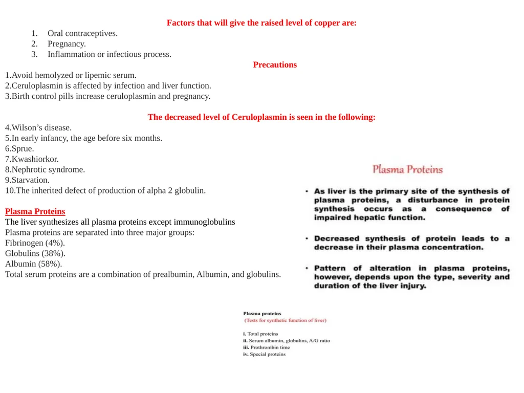
Factors that will give the raised level of copper are:
Oral contraceptives.
Pregnancy.
Inflammation or infectious process.
Precautions
Avoid hemolyzed or lipemic serum.
Ceruloplasmin is affected by infection and liver function.
Birth control pills increase ceruloplasmin and pregnancy.
The decreased level of Ceruloplasmin is seen in the following:
Wilson’s disease.
In early infancy, the age before six months.
Sprue.
Kwashiorkor.
Nephrotic syndrome.
Starvation.
The inherited defect of production of alpha 2 globulin.
Plasma Proteins The liver synthesizes all plasma proteins except immunoglobulins Plasma proteins are separated into three major groups: Fibrinogen (4%). Globulins (38%). Albumin (58%). Total serum proteins are a combination of prealbumin, Albumin, and globulins.<br>
Oral contraceptives.
Pregnancy.
Inflammation or infectious process.
Precautions
Avoid hemolyzed or lipemic serum.
Ceruloplasmin is affected by infection and liver function.
Birth control pills increase ceruloplasmin and pregnancy.
The decreased level of Ceruloplasmin is seen in the following:
Wilson’s disease.
In early infancy, the age before six months.
Sprue.
Kwashiorkor.
Nephrotic syndrome.
Starvation.
The inherited defect of production of alpha 2 globulin.
Plasma Proteins The liver synthesizes all plasma proteins except immunoglobulins Plasma proteins are separated into three major groups: Fibrinogen (4%). Globulins (38%). Albumin (58%). Total serum proteins are a combination of prealbumin, Albumin, and globulins.<br>
08
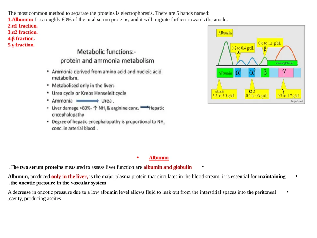
The most common method to separate the proteins is electrophoresis. There are 5 bands named:
Albumin: It is roughly 60% of the total serum proteins, and it will migrate farthest towards the anode.
α1 fraction.
α2 fraction.
β fraction.
γ fraction. Albumin
The two serum proteins measured to assess liver function are albumin and globulin.
Albumin, produced only in the liver, is the major plasma protein that circulates in the blood stream, it is essential for maintaining the oncotic pressure in the vascular system.
A decrease in oncotic pressure due to a low albumin level allows fluid to leak out from the interstitial spaces into the peritoneal cavity, producing ascites.<br>
Albumin: It is roughly 60% of the total serum proteins, and it will migrate farthest towards the anode.
α1 fraction.
α2 fraction.
β fraction.
γ fraction. Albumin
The two serum proteins measured to assess liver function are albumin and globulin.
Albumin, produced only in the liver, is the major plasma protein that circulates in the blood stream, it is essential for maintaining the oncotic pressure in the vascular system.
A decrease in oncotic pressure due to a low albumin level allows fluid to leak out from the interstitial spaces into the peritoneal cavity, producing ascites.<br>
09
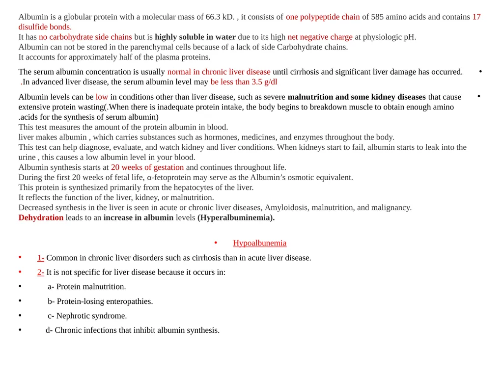
Albumin is a globular protein with a molecular mass of 66.3 kD. , it consists of one polypeptide chain of 585 amino acids and contains 17 disulfide bonds.
It has no carbohydrate side chains but is highly soluble in water due to its high net negative charge at physiologic pH.
Albumin can not be stored in the parenchymal cells because of a lack of side Carbohydrate chains.
It accounts for approximately half of the plasma proteins.
The serum albumin concentration is usually normal in chronic liver disease until cirrhosis and significant liver damage has occurred. In advanced liver disease, the serum albumin level may be less than 3.5 g/dl.
Albumin levels can be low in conditions other than liver disease, such as severe malnutrition and some kidney diseases that cause extensive protein wasting(.When there is inadequate protein intake, the body begins to breakdown muscle to obtain enough amino acids for the synthesis of serum albumin).
This test measures the amount of the protein albumin in blood.
liver makes albumin , which carries substances such as hormones, medicines, and enzymes throughout the body.
This test can help diagnose, evaluate, and watch kidney and liver conditions. When kidneys start to fail, albumin starts to leak into the urine , this causes a low albumin level in your blood.
Albumin synthesis starts at 20 weeks of gestation and continues throughout life.
During the first 20 weeks of fetal life, α-fetoprotein may serve as the Albumin’s osmotic equivalent.
This protein is synthesized primarily from the hepatocytes of the liver.
It reflects the function of the liver, kidney, or malnutrition.
Decreased synthesis in the liver is seen in acute or chronic liver diseases, Amyloidosis, malnutrition, and malignancy.
Dehydration leads to an increase in albumin levels (Hyperalbuminemia).
Hypoalbunemia
1- Common in chronic liver disorders such as cirrhosis than in acute liver disease.
2- It is not specific for liver disease because it occurs in:
a- Protein malnutrition.
b- Protein-losing enteropathies.
c- Nephrotic syndrome.
d- Chronic infections that inhibit albumin synthesis.<br>
It has no carbohydrate side chains but is highly soluble in water due to its high net negative charge at physiologic pH.
Albumin can not be stored in the parenchymal cells because of a lack of side Carbohydrate chains.
It accounts for approximately half of the plasma proteins.
The serum albumin concentration is usually normal in chronic liver disease until cirrhosis and significant liver damage has occurred. In advanced liver disease, the serum albumin level may be less than 3.5 g/dl.
Albumin levels can be low in conditions other than liver disease, such as severe malnutrition and some kidney diseases that cause extensive protein wasting(.When there is inadequate protein intake, the body begins to breakdown muscle to obtain enough amino acids for the synthesis of serum albumin).
This test measures the amount of the protein albumin in blood.
liver makes albumin , which carries substances such as hormones, medicines, and enzymes throughout the body.
This test can help diagnose, evaluate, and watch kidney and liver conditions. When kidneys start to fail, albumin starts to leak into the urine , this causes a low albumin level in your blood.
Albumin synthesis starts at 20 weeks of gestation and continues throughout life.
During the first 20 weeks of fetal life, α-fetoprotein may serve as the Albumin’s osmotic equivalent.
This protein is synthesized primarily from the hepatocytes of the liver.
It reflects the function of the liver, kidney, or malnutrition.
Decreased synthesis in the liver is seen in acute or chronic liver diseases, Amyloidosis, malnutrition, and malignancy.
Dehydration leads to an increase in albumin levels (Hyperalbuminemia).
Hypoalbunemia
1- Common in chronic liver disorders such as cirrhosis than in acute liver disease.
2- It is not specific for liver disease because it occurs in:
a- Protein malnutrition.
b- Protein-losing enteropathies.
c- Nephrotic syndrome.
d- Chronic infections that inhibit albumin synthesis.<br>
10
Hypoalbuminemia:
It is when the albumin level is lower than normal, this may be due to various factors like:
Impaired syntheses of the Albumin from the liver or decreased intake of the proteins.
Increased catabolism due to inflammation or tissue damage.
Due to malabsorption or malnutrition, leading to decreased absorption of the amino acids.
There is an increased loss of Albumin in the urine in conditions like nephrotic syndrome, chronic glomerulonephritis, diabetes mellitus, and SLE.
Protein loss in case of burn or protein-losing-enteropathy.
In the case of ascites, where there is high pressure in the portal system, it drives the Albumin into the peritoneal cavity.
When the albumin level is <2.0 g/L, it will lead to edema formation. This usually occurs when the albumin loss is through the urine or feces.
Serum electrophoresis shows a low albumin spike.
Mechanism of decrease in the albumin synthesis:
Decreased synthesis may be seen in:
Injury to the hepatocytes.
Decreased protein intake, like malnutrition or starvation.
If there is impaired absorption of the protein products like in sprue.
Extensive loss of the Albumin seen in:
In nephrotic syndrome, there is extensive loss of protein in the urine.
There is loos of protein in the extensive burns or exfoliative dermatitis.
In protein-losing intestinal diseases (protein-losing enteropathies).
Urinary loss:
As Albumin is relatively small and globular, a significant amount is filtered into the glomerular urine. Then the majority is reabsorbed by the proximal tubular cells.
Normal urine contains 20 mg of Albumin per gram of creatinine.
Excretion above this level is seen in:
Increased glomerular filtration.
Tubular damage.
The albumin/globulin ratio (A/G) is normally found = >1.0.
A/G ratio <1.0 is usually seen in liver diseases.<br>
It is when the albumin level is lower than normal, this may be due to various factors like:
Impaired syntheses of the Albumin from the liver or decreased intake of the proteins.
Increased catabolism due to inflammation or tissue damage.
Due to malabsorption or malnutrition, leading to decreased absorption of the amino acids.
There is an increased loss of Albumin in the urine in conditions like nephrotic syndrome, chronic glomerulonephritis, diabetes mellitus, and SLE.
Protein loss in case of burn or protein-losing-enteropathy.
In the case of ascites, where there is high pressure in the portal system, it drives the Albumin into the peritoneal cavity.
When the albumin level is <2.0 g/L, it will lead to edema formation. This usually occurs when the albumin loss is through the urine or feces.
Serum electrophoresis shows a low albumin spike.
Mechanism of decrease in the albumin synthesis:
Decreased synthesis may be seen in:
Injury to the hepatocytes.
Decreased protein intake, like malnutrition or starvation.
If there is impaired absorption of the protein products like in sprue.
Extensive loss of the Albumin seen in:
In nephrotic syndrome, there is extensive loss of protein in the urine.
There is loos of protein in the extensive burns or exfoliative dermatitis.
In protein-losing intestinal diseases (protein-losing enteropathies).
Urinary loss:
As Albumin is relatively small and globular, a significant amount is filtered into the glomerular urine. Then the majority is reabsorbed by the proximal tubular cells.
Normal urine contains 20 mg of Albumin per gram of creatinine.
Excretion above this level is seen in:
Increased glomerular filtration.
Tubular damage.
The albumin/globulin ratio (A/G) is normally found = >1.0.
A/G ratio <1.0 is usually seen in liver diseases.<br>
11
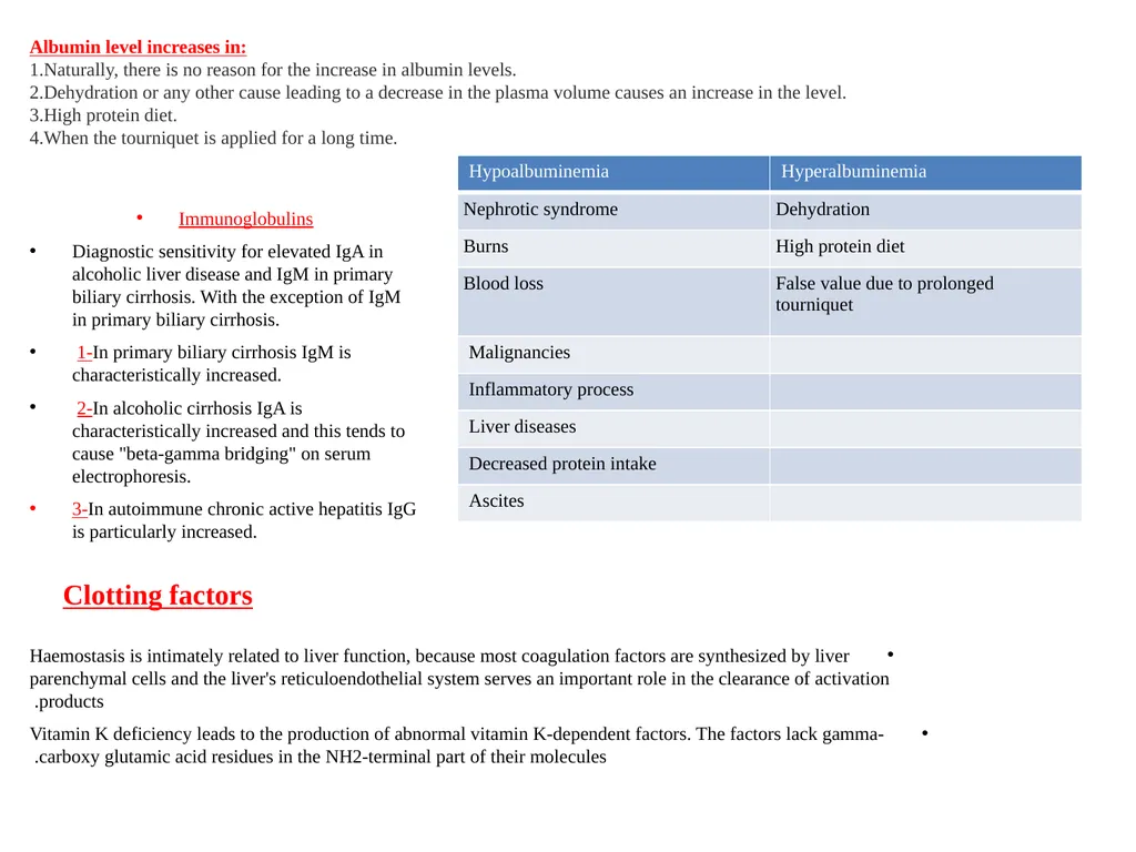
Albumin level increases in:
Naturally, there is no reason for the increase in albumin levels.
Dehydration or any other cause leading to a decrease in the plasma volume causes an increase in the level.
High protein diet.
When the tourniquet is applied for a long time. Immunoglobulins
Diagnostic sensitivity for elevated IgA in alcoholic liver disease and IgM in primary biliary cirrhosis. With the exception of IgM in primary biliary cirrhosis.
1-In primary biliary cirrhosis IgM is characteristically increased.
2-In alcoholic cirrhosis IgA is characteristically increased and this tends to cause "beta-gamma bridging" on serum electrophoresis.
3-In autoimmune chronic active hepatitis IgG is particularly increased. Clotting factors Haemostasis is intimately related to liver function, because most coagulation factors are synthesized by liver parenchymal cells and the liver's reticuloendothelial system serves an important role in the clearance of activation products.
Vitamin K deficiency leads to the production of abnormal vitamin K-dependent factors. The factors lack gamma-carboxy glutamic acid residues in the NH2-terminal part of their molecules.<br>
Naturally, there is no reason for the increase in albumin levels.
Dehydration or any other cause leading to a decrease in the plasma volume causes an increase in the level.
High protein diet.
When the tourniquet is applied for a long time. Immunoglobulins
Diagnostic sensitivity for elevated IgA in alcoholic liver disease and IgM in primary biliary cirrhosis. With the exception of IgM in primary biliary cirrhosis.
1-In primary biliary cirrhosis IgM is characteristically increased.
2-In alcoholic cirrhosis IgA is characteristically increased and this tends to cause "beta-gamma bridging" on serum electrophoresis.
3-In autoimmune chronic active hepatitis IgG is particularly increased. Clotting factors Haemostasis is intimately related to liver function, because most coagulation factors are synthesized by liver parenchymal cells and the liver's reticuloendothelial system serves an important role in the clearance of activation products.
Vitamin K deficiency leads to the production of abnormal vitamin K-dependent factors. The factors lack gamma-carboxy glutamic acid residues in the NH2-terminal part of their molecules.<br>
12
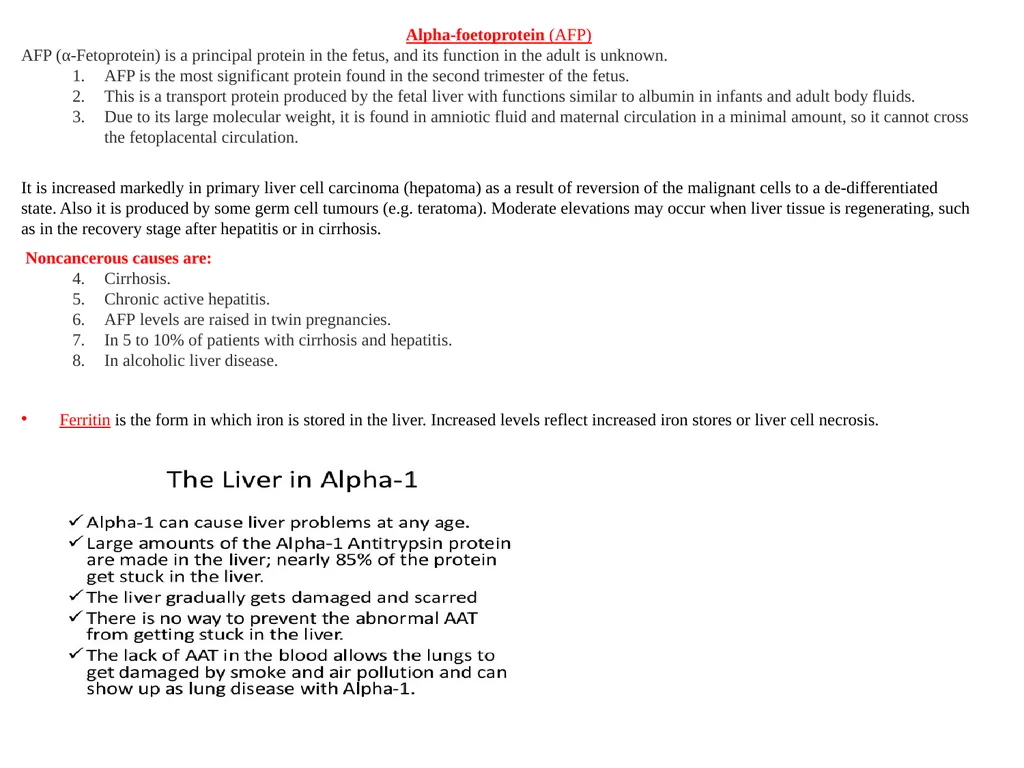
Alpha-foetoprotein (AFP)
AFP (α-Fetoprotein) is a principal protein in the fetus, and its function in the adult is unknown.
AFP is the most significant protein found in the second trimester of the fetus.
This is a transport protein produced by the fetal liver with functions similar to albumin in infants and adult body fluids.
Due to its large molecular weight, it is found in amniotic fluid and maternal circulation in a minimal amount, so it cannot cross the fetoplacental circulation.
It is increased markedly in primary liver cell carcinoma (hepatoma) as a result of reversion of the malignant cells to a de-differentiated state. Also it is produced by some germ cell tumours (e.g. teratoma). Moderate elevations may occur when liver tissue is regenerating, such as in the recovery stage after hepatitis or in cirrhosis.
Noncancerous causes are:
Cirrhosis.
Chronic active hepatitis.
AFP levels are raised in twin pregnancies.
In 5 to 10% of patients with cirrhosis and hepatitis.
In alcoholic liver disease.
Ferritin is the form in which iron is stored in the liver. Increased levels reflect increased iron stores or liver cell necrosis.<br>
AFP (α-Fetoprotein) is a principal protein in the fetus, and its function in the adult is unknown.
AFP is the most significant protein found in the second trimester of the fetus.
This is a transport protein produced by the fetal liver with functions similar to albumin in infants and adult body fluids.
Due to its large molecular weight, it is found in amniotic fluid and maternal circulation in a minimal amount, so it cannot cross the fetoplacental circulation.
It is increased markedly in primary liver cell carcinoma (hepatoma) as a result of reversion of the malignant cells to a de-differentiated state. Also it is produced by some germ cell tumours (e.g. teratoma). Moderate elevations may occur when liver tissue is regenerating, such as in the recovery stage after hepatitis or in cirrhosis.
Noncancerous causes are:
Cirrhosis.
Chronic active hepatitis.
AFP levels are raised in twin pregnancies.
In 5 to 10% of patients with cirrhosis and hepatitis.
In alcoholic liver disease.
Ferritin is the form in which iron is stored in the liver. Increased levels reflect increased iron stores or liver cell necrosis.<br>
13
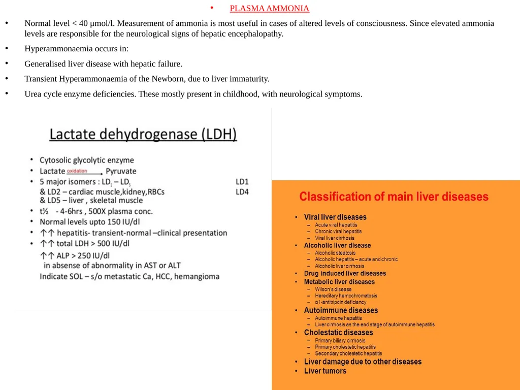
PLASMA AMMONIA
Normal level < 40 μmol/l. Measurement of ammonia is most useful in cases of altered levels of consciousness. Since elevated ammonia levels are responsible for the neurological signs of hepatic encephalopathy.
Hyperammonaemia occurs in:
Generalised liver disease with hepatic failure.
Transient Hyperammonaemia of the Newborn, due to liver immaturity.
Urea cycle enzyme deficiencies. These mostly present in childhood, with neurological symptoms.<br>
Normal level < 40 μmol/l. Measurement of ammonia is most useful in cases of altered levels of consciousness. Since elevated ammonia levels are responsible for the neurological signs of hepatic encephalopathy.
Hyperammonaemia occurs in:
Generalised liver disease with hepatic failure.
Transient Hyperammonaemia of the Newborn, due to liver immaturity.
Urea cycle enzyme deficiencies. These mostly present in childhood, with neurological symptoms.<br>