
MUSCULOSKELETAL INJURIES LESSON -20 BY JITENDER
OM
Published · 58 slides · 0 views

1 / 1







Description
MUSCULOSKELETAL INJURIES LESSON -20 BY JITENDER YADAV INSPPH OBJECTIVES Upon completion of this lesson, you will be able to: Define an open fracture and closed fracture, and list four signs and symptoms. Define a dislocation, a sprain, and
Related Topics
Share
Embed code
Download this presentation From Below
"MUSCULOSKELETAL INJURIES LESSON -20 BY JITENDER" is the property of its rightful owner. Permission is granted to download and print the materials on this website for personal, non-commercial use only, and to display it on your personal computer provided you do not modify the materials and that you retain all copyright notices contained in the materials. By downloading content from our website, you accept the terms of this agreement.
Presentation Transcript
01
MUSCULOSKELETAL INJURIES LESSON -20 BY
JITENDER YADAV
INSP/PH<br>
JITENDER YADAV
INSP/PH<br>
02
OBJECTIVES Upon completion of this lesson, you will be able to:
Define an open fracture and closed fracture, and list four signs and symptoms.
Define a dislocation, a sprain, and a strain and list four signs and symptoms.
Give two reasons for immobilizing a fracture, a sprain or a strain on a patient.
Demonstrate the pre-hospital treatment of fractures and dislocations of the extremities, hips and shoulder.<br>
Define an open fracture and closed fracture, and list four signs and symptoms.
Define a dislocation, a sprain, and a strain and list four signs and symptoms.
Give two reasons for immobilizing a fracture, a sprain or a strain on a patient.
Demonstrate the pre-hospital treatment of fractures and dislocations of the extremities, hips and shoulder.<br>
03
THE SKELETAL SYSTEM The adult skeleton is composed of 206 bones. The human skeleton consists of two main divisions, the axial skeleton and the appendicular skeleton.
Functions of the skeletal system:-
Provides a framework for the body.
Protects vital organs.
Provides for body movement.
Produces red blood cells.<br>
Functions of the skeletal system:-
Provides a framework for the body.
Protects vital organs.
Provides for body movement.
Produces red blood cells.<br>
04
JOINTS (ARTICULATIONS) Joints are bone ends that fit into each other.
There are several types of joints:-
Immovable joints such as in the skull.
Slightly movable joints such as the spine.
Freely movable joints such as the elbow or knee joints (hinge) or the hip joint (ball and socket).<br>
There are several types of joints:-
Immovable joints such as in the skull.
Slightly movable joints such as the spine.
Freely movable joints such as the elbow or knee joints (hinge) or the hip joint (ball and socket).<br>
05
LIGAMENTS AND TENDONS Ligaments connect and hold bones together at the joints.
Tendons attach the skeletal muscles to the bone. These muscles control the movement of the joints.<br>
Tendons attach the skeletal muscles to the bone. These muscles control the movement of the joints.<br>
06
FRACTURES, DISLOCATIONS AND SPRAINS FRACTURES : Any break in the continuity of a bone.
Fractures can be open or closed.
Closed Fracture: One in which the overlying skin is intact. Proper splinting helps prevent closed fracture from becoming open fracture. Cont…<br>
Fractures can be open or closed.
Closed Fracture: One in which the overlying skin is intact. Proper splinting helps prevent closed fracture from becoming open fracture. Cont…<br>
07
Open fracture: One in which the skin has been broken or torn either from the inside by the injured bone, or from the outside by the object that caused the penetrating wound with the associated bone injury. The bone may or may not protrude through the wound. Open fractures are serious because of risk of contamination and risk of infection is greater.<br>
08
NOTE : Treat life-threatening injuries first. It is impossible to rule out a fracture through a physical exam of the patient. Many sprains and dislocations present signs and symptoms similar to a fracture.<br>
09
DISLOCATIONS : Injury in which a bone is moved out of its normal position in a joint and remains that way.
A dislocation sometimes causes the tearing of ligaments and soft tissues if stretched far beyond the normal range of motion. The shoulder, elbow, fingers, hips, and ankles are the joints most frequently affected. Signs and symptoms of dislocation are similar to those of a fracture.<br>
A dislocation sometimes causes the tearing of ligaments and soft tissues if stretched far beyond the normal range of motion. The shoulder, elbow, fingers, hips, and ankles are the joints most frequently affected. Signs and symptoms of dislocation are similar to those of a fracture.<br>
10
SPRAIN : Injury in which ligaments are stretched or partially torn, commonly associated with joint injuries.
Do not confuse a sprain with a strain, which involves muscle injury.
STRAIN: Injury in which a muscle, or a muscle and tendon, are over-extended.
Dislocation, fracture, strain and sprain may all be present in an injury.<br>
Do not confuse a sprain with a strain, which involves muscle injury.
STRAIN: Injury in which a muscle, or a muscle and tendon, are over-extended.
Dislocation, fracture, strain and sprain may all be present in an injury.<br>
11
Signs and Symptoms of a
Musculoskeletal Injury Deformity or angulation: compare with opposite limb.
Pain and tenderness upon palpation or movement.
Crepitus (grating) – a sound or feeling of broken bone ends rubbing together.
Swelling.
Bruising or discoloration.
Exposed bone ends. Cont…<br>
Pain and tenderness upon palpation or movement.
Crepitus (grating) – a sound or feeling of broken bone ends rubbing together.
Swelling.
Bruising or discoloration.
Exposed bone ends. Cont…<br>
12
Joint locked in position – reduced motor ability or reduced ability to move a joint.
Numbness and paralysis – may occur distal to site of injury caused by bone pressing on a nerve.
Compromised circulation distal to injury evidenced by alteration in skin color, temperature, pulse or capillary refill.
NEVER INTENTIONALLY INDUCE CREPITUS. This may cause or aggravate soft tissue injury.<br>
Numbness and paralysis – may occur distal to site of injury caused by bone pressing on a nerve.
Compromised circulation distal to injury evidenced by alteration in skin color, temperature, pulse or capillary refill.
NEVER INTENTIONALLY INDUCE CREPITUS. This may cause or aggravate soft tissue injury.<br>
13
SPLINTING Applying a device to stabilize any painful, swollen or deformed body part.
The primary objective of splinting is to prevent further movement of body parts. For any splint to be effective, it must immobilize adjacent joints and bone ends.<br>
The primary objective of splinting is to prevent further movement of body parts. For any splint to be effective, it must immobilize adjacent joints and bone ends.<br>
14
Reasons for splinting include:
To prevent motion of bone fragments or dislocated joints.
To reduce pain and suffering.
To minimize damage to soft tissues (for example, nerves, arteries , veins and muscle).
To prevent a closed fracture from becoming an open fracture.
To minimize blood loss or shock.<br>
To prevent motion of bone fragments or dislocated joints.
To reduce pain and suffering.
To minimize damage to soft tissues (for example, nerves, arteries , veins and muscle).
To prevent a closed fracture from becoming an open fracture.
To minimize blood loss or shock.<br>
15
TYPES OF SPLINTS Effective splinting may require some ingenuity. Though you may carry many types of splinting devices, many situations will require you to improvise.<br>
16
FIVE BASIC TYPES OF SPLINTS:
Rigid splint: Requires limb to be in anatomical position. Ideal for long-bone injuries (for example cardboard, wood).
Conforming splint: Can be molded to different angles or surrounds the extremity (for example air or vacuum splints). Cont…<br>
Rigid splint: Requires limb to be in anatomical position. Ideal for long-bone injuries (for example cardboard, wood).
Conforming splint: Can be molded to different angles or surrounds the extremity (for example air or vacuum splints). Cont…<br>
17
Traction splint: Used specifically for femur fractures.
Sling and swathe: Two triangular bandages used to hold an injured arm in place against the body.
Improvised splints: a book, cardboard, pillow or blanket etc.<br>
Sling and swathe: Two triangular bandages used to hold an injured arm in place against the body.
Improvised splints: a book, cardboard, pillow or blanket etc.<br>
18
GENERAL RULES FOR SPLINTING Regardless of the method of splinting, general rules apply to all types of immobilization, as follows:
Always communicate your plans with your patient if possible.
Before immobilizing an injured extremity, expose and control bleeding.
Always cut away clothing around the injury site before immobilizing the joint. Remove all jewelry from the site and below it. Cont…<br>
Always communicate your plans with your patient if possible.
Before immobilizing an injured extremity, expose and control bleeding.
Always cut away clothing around the injury site before immobilizing the joint. Remove all jewelry from the site and below it. Cont…<br>
19
Assess P.M.S. (pulse, motor function and sensation).
If limb is severely deformed or distal circulation is compromised (cyanosis distal to fracture site or no distal pulse), align the bone with gentle traction (pulling). If pain or crepitus worsens, discontinue.
Do not attempt to push protruding bone ends back into place. However, when realigning, they may slip back into place. Make a note if this occurs. Cont…<br>
If limb is severely deformed or distal circulation is compromised (cyanosis distal to fracture site or no distal pulse), align the bone with gentle traction (pulling). If pain or crepitus worsens, discontinue.
Do not attempt to push protruding bone ends back into place. However, when realigning, they may slip back into place. Make a note if this occurs. Cont…<br>
20
For patient comfort and proper immobilization, pad voids between the body and the splint, since many rigid splints do not conform to body curves.
Pad a splint before applying it.
If a joint is injured, immobilize it and the bones above and below. Cont…<br>
Pad a splint before applying it.
If a joint is injured, immobilize it and the bones above and below. Cont…<br>
21
AVOID TUNNEL VISION : Do not over-splint the patient — in multisystem trauma patients, do not be distracted from life-threatening injuries by the gross appearance of non-critical injuries.
Securing the patient to a long backboard supports and splints every bone and joint in one step without wasting time.
Be flexible while splinting. Keep in mind patient’s comfort and principles of splinting.<br>
Securing the patient to a long backboard supports and splints every bone and joint in one step without wasting time.
Be flexible while splinting. Keep in mind patient’s comfort and principles of splinting.<br>
22
Pre-hospital Treatment for Suspected Fractures, Dislocations or Sprains Examining involves use of your senses and skills of inspection (looking), palpation (feeling) and auscultation (listening). Use universal precautions and secure the scene.
Perform initial assessment.
Identify and treat life-threatening problems.
Do not be distracted by dramatic-looking injuries.
Remember cervical collar and oxygen, if necessary Cont…<br>
Perform initial assessment.
Identify and treat life-threatening problems.
Do not be distracted by dramatic-looking injuries.
Remember cervical collar and oxygen, if necessary Cont…<br>
23
2) Perform a physical exam. You can use the mnemonic “D.O.T.S.” to guide your exam as you look for signs and symptoms of injuries.
Check for visible deformities. Check all joints and bones through entire length of body.
Check for open injuries, common with extremity injuries.
Palpate for tenderness, which may underlie injury without deformity.
Check for swelling and discoloration. Cont…<br>
Check for visible deformities. Check all joints and bones through entire length of body.
Check for open injuries, common with extremity injuries.
Palpate for tenderness, which may underlie injury without deformity.
Check for swelling and discoloration. Cont…<br>
24
For extremity injuries, always assess for distal pulse, motor function and sensation of (P.M.S.), before and after splinting.
Pulse: Radial in upper extremity injuries, dorsalis pedis (top of foot) or posterior tibial pulse (back of ankles, medially) for lower extremities.
Motor function: check patient’s ability to move, such as wiggling toes or fingers (movement indicates intact nerves).
Sensation: Gently squeeze or pinch the finger of the extremity then the other, asking if patient can feel your touch. Cont…<br>
Pulse: Radial in upper extremity injuries, dorsalis pedis (top of foot) or posterior tibial pulse (back of ankles, medially) for lower extremities.
Motor function: check patient’s ability to move, such as wiggling toes or fingers (movement indicates intact nerves).
Sensation: Gently squeeze or pinch the finger of the extremity then the other, asking if patient can feel your touch. Cont…<br>
25
3) Stabilize the injury. After completing a physical exam, secure injury site providing manual stabilization. Do not release manual stabilization of an injured extremity until it is properly and completely immobilized.
4) Expose the injury. Cut away clothing and remove jewelry before swelling occurs. Cont…<br>
4) Expose the injury. Cut away clothing and remove jewelry before swelling occurs. Cont…<br>
26
5) Treat open wounds and control bleeding. Cover with a clean or sterile dressing, avoid direct pressure over broken bone ends. Use pressure points as needed if bone ends protrude from injury, use caution not to allow bone ends to reenter wound.
6) Prepare your splinting materials. Cont…<br>
6) Prepare your splinting materials. Cont…<br>
27
7) Carefully splint individual injuries.
Measure or adjust the splint into position, maintain manual stabilization as appropriate during splinting until procedure is complete.
Apply and secure to adjacent joints and injury site.
Be careful not to restrict circulation.
8) Reassess pulse, motor function and sensation.
9) Apply cold packs or ice to injury site to reduce pain and swelling.
10) Treat for shock.<br>
Measure or adjust the splint into position, maintain manual stabilization as appropriate during splinting until procedure is complete.
Apply and secure to adjacent joints and injury site.
Be careful not to restrict circulation.
8) Reassess pulse, motor function and sensation.
9) Apply cold packs or ice to injury site to reduce pain and swelling.
10) Treat for shock.<br>
28
Pre-hospital Treatment for Specific
Injuries and Application of Splints IMPORTANT: Always reassess pulse, motor function and sensation before and after splinting.<br>
29
SPLINTING THE UPPER EXTREMITIES 1) Shoulder and clavicle
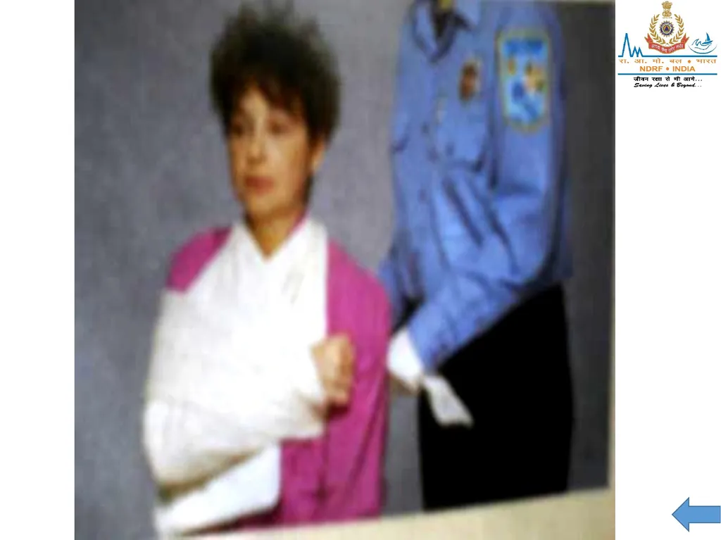
Signs and symptoms: Shoulder appears to be “dropped,” deformity (asymmetry), pain.
Treatment: Apply a sling and swath. Provide any padding necessary to fill void between body and arm.<br>
Signs and symptoms: Shoulder appears to be “dropped,” deformity (asymmetry), pain.
Treatment: Apply a sling and swath. Provide any padding necessary to fill void between body and arm.<br>
30
2) Humorous (Upper Arm) and Shoulder
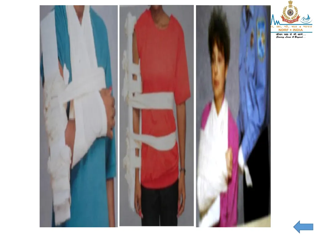
Signs and symptoms: Pain, swelling, deformity.
Treatment: Rigid splint to outside of the arm, pad voids, then apply sling and swath. Alternate method could be by using splints on both sides of the arm.<br>
Signs and symptoms: Pain, swelling, deformity.
Treatment: Rigid splint to outside of the arm, pad voids, then apply sling and swath. Alternate method could be by using splints on both sides of the arm.<br>
31
3) Elbow
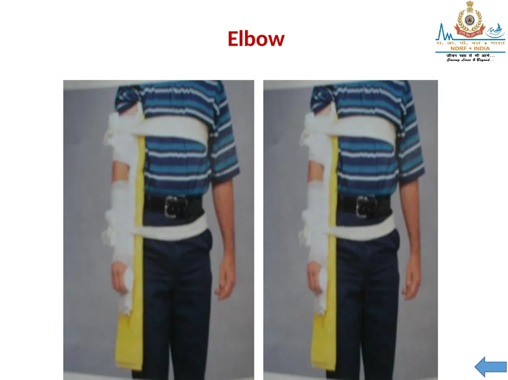
Important: Splint in position found, do not attempt to straighten.
Signs and symptoms: Pain, swelling and deformity.
Treatment:
If arm is bent at elbow, splint with sling & swath. Alternate is pillow or blanket.
If elbow is straight, splint entire arm, armpit to fingertips, both sides.<br>
Important: Splint in position found, do not attempt to straighten.
Signs and symptoms: Pain, swelling and deformity.
Treatment:
If arm is bent at elbow, splint with sling & swath. Alternate is pillow or blanket.
If elbow is straight, splint entire arm, armpit to fingertips, both sides.<br>
32
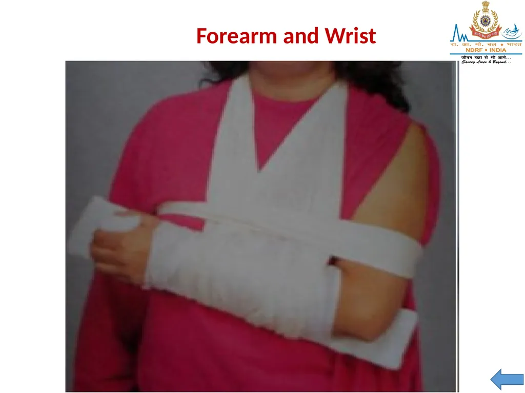
4) Forearm and Wrist
Signs and symptoms: Pain, swelling and deformity.
Treatment: Splint area with arm board, then sling and swath. (Pneumatic splints are an option.)<br>
Signs and symptoms: Pain, swelling and deformity.
Treatment: Splint area with arm board, then sling and swath. (Pneumatic splints are an option.)<br>
33
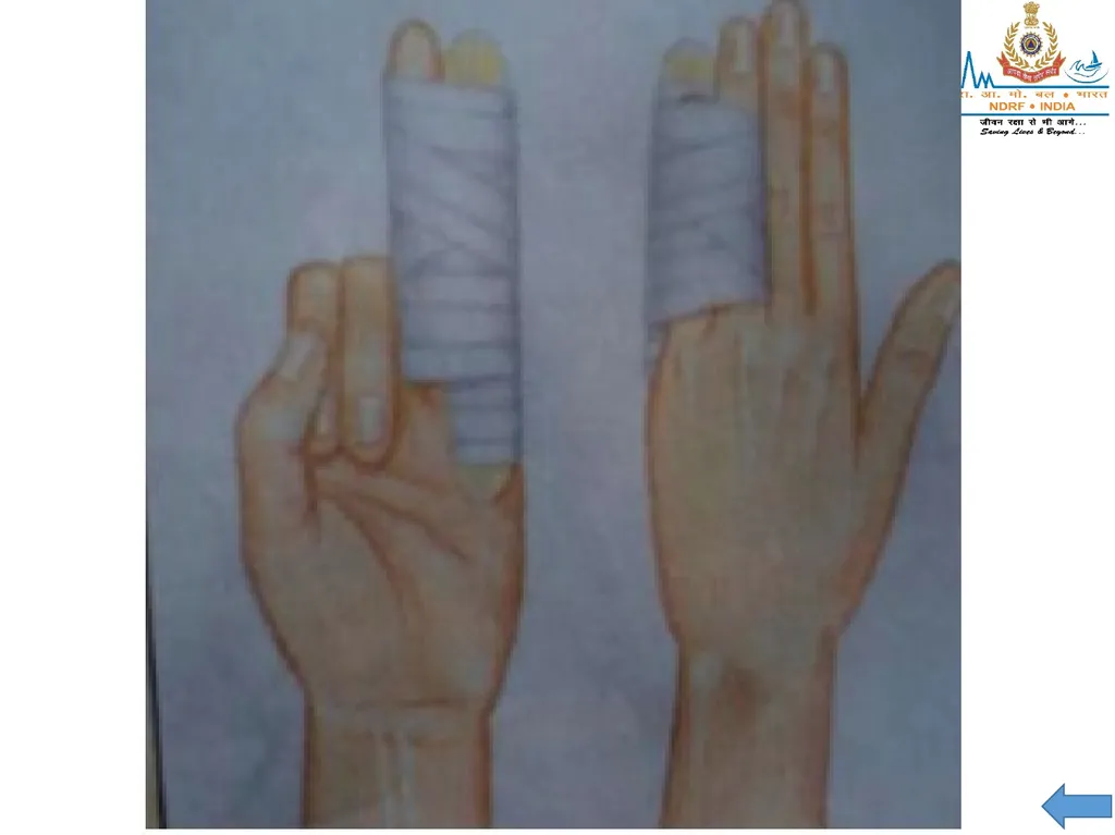
5) Hands and Fingers
Important: Pulse can be checked by capillary refill.
Signs and symptoms: Pain, swelling and deformity.
Treatment:
If one finger is fractured, tape it to an adjacent finger or use tongue depressor to splint.
If more than one finger is fractured, splint the entire hand in the position of function. Place a roll of bandage in palm of hand, or other object, then wrap entire hand and place on arm board.<br>
Important: Pulse can be checked by capillary refill.
Signs and symptoms: Pain, swelling and deformity.
Treatment:
If one finger is fractured, tape it to an adjacent finger or use tongue depressor to splint.
If more than one finger is fractured, splint the entire hand in the position of function. Place a roll of bandage in palm of hand, or other object, then wrap entire hand and place on arm board.<br>
34
SPLINTING THE LOWER EXTREMITIES 1) PELVIS
Pelvic injuries can be life-threatening due to massive blood loss.
Suspect shock.
Any force strong enough to injure the pelvis can also injure the spine. Cont…<br>
Pelvic injuries can be life-threatening due to massive blood loss.
Suspect shock.
Any force strong enough to injure the pelvis can also injure the spine. Cont…<br>
35
Signs and symptoms of pelvic injury:
Pain, especially when pressure is applied to iliac crests or pelvic bones.
Inability to lift legs while lying on back. Cont…<br>
Pain, especially when pressure is applied to iliac crests or pelvic bones.
Inability to lift legs while lying on back. Cont…<br>
36
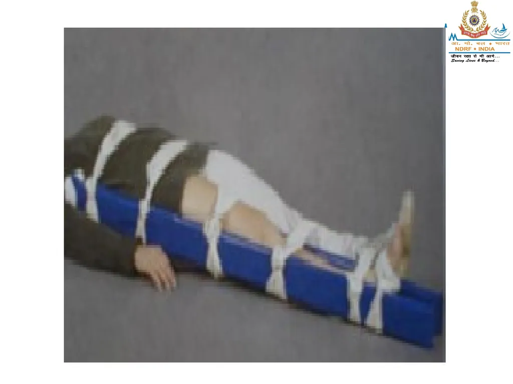
Pre-hospital treatment for pelvic injury:
Minimize patient movement.
Do not log roll or lift with pelvis unsupported.
Place a folded blanket between patient’s legs from groin to feet and bind together with cravats (2 to upper leg, 2 to lower leg).
Place the patient on long backboard.
5) Treat for shock.<br>
Minimize patient movement.
Do not log roll or lift with pelvis unsupported.
Place a folded blanket between patient’s legs from groin to feet and bind together with cravats (2 to upper leg, 2 to lower leg).
Place the patient on long backboard.
5) Treat for shock.<br>
37
2) Hip injuries
With this type of injury, it is difficult to differentiate an upper femur fracture from a hip or pelvic fracture or dislocation. Assess for life threatening injuries as with pelvic injuries.
Signs and symptoms of hip injury
Pain, swelling and discoloration.
Inability to move leg(s).
Possible foot rotation (outward or inward).<br>
With this type of injury, it is difficult to differentiate an upper femur fracture from a hip or pelvic fracture or dislocation. Assess for life threatening injuries as with pelvic injuries.
Signs and symptoms of hip injury
Pain, swelling and discoloration.
Inability to move leg(s).
Possible foot rotation (outward or inward).<br>
38
Pre-hospital treatment for hip injuries
Bind legs together with a folded blanket between patient’s legs.
Support the hip with pillows.
Stabilize patient on long backboard, or use long splints along outer thigh, from foot to armpit with padding; and along the inner thigh, from groin to foot.
Secure with cravats.<br>
Bind legs together with a folded blanket between patient’s legs.
Support the hip with pillows.
Stabilize patient on long backboard, or use long splints along outer thigh, from foot to armpit with padding; and along the inner thigh, from groin to foot.
Secure with cravats.<br>
39
3) Femoral injuries
A femoral fracture can produce massive internal bleeding which may lead to shock. Treat life-threats first.
Signs and symptoms of femoral fracture
Pain (often intense)
Deformity
Rigidity
Shortened limb<br>
A femoral fracture can produce massive internal bleeding which may lead to shock. Treat life-threats first.
Signs and symptoms of femoral fracture
Pain (often intense)
Deformity
Rigidity
Shortened limb<br>
40
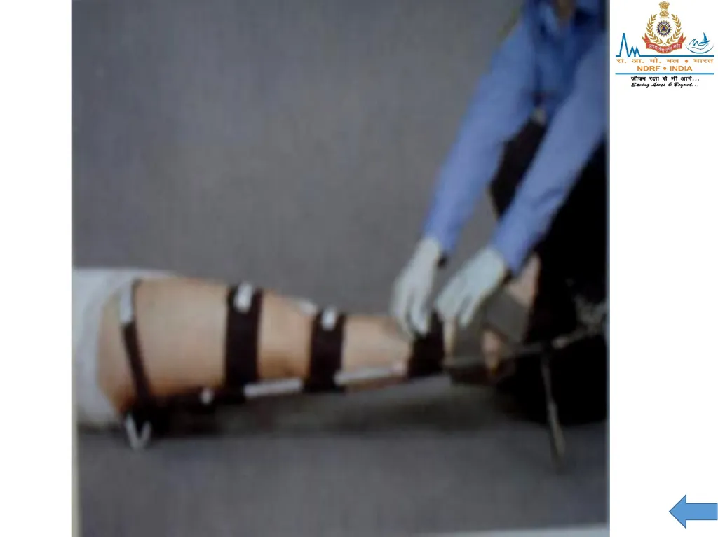
Pre-hospital treatment: If you find the leg in a straight position, use two padded splints one along the inner thigh from groin to the foot, the other along the outer thigh from the armpit to the foot. Secure with cravats.<br>
41
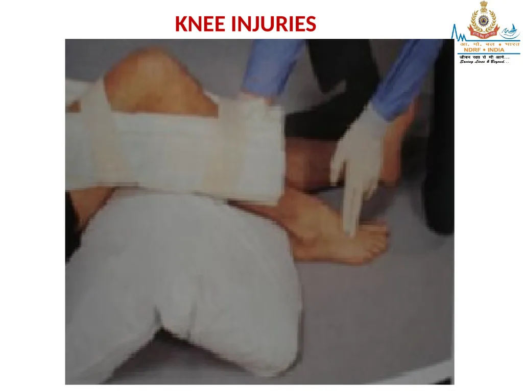
4) Knee injuries
Signs and symptoms: Pain, swelling and deformity.
Bent position: Immobilize in the position found. The bones above and below it should be splinted with short padded boards.
Straight position: Use two padded long splints, the first on the inner thigh from groin to beyond foot. Place the second on the outer thigh, from hip to beyond foot. Secure with cravats.<br>
Signs and symptoms: Pain, swelling and deformity.
Bent position: Immobilize in the position found. The bones above and below it should be splinted with short padded boards.
Straight position: Use two padded long splints, the first on the inner thigh from groin to beyond foot. Place the second on the outer thigh, from hip to beyond foot. Secure with cravats.<br>
42
KNEE INJURIES<br>
43
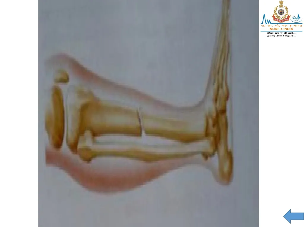
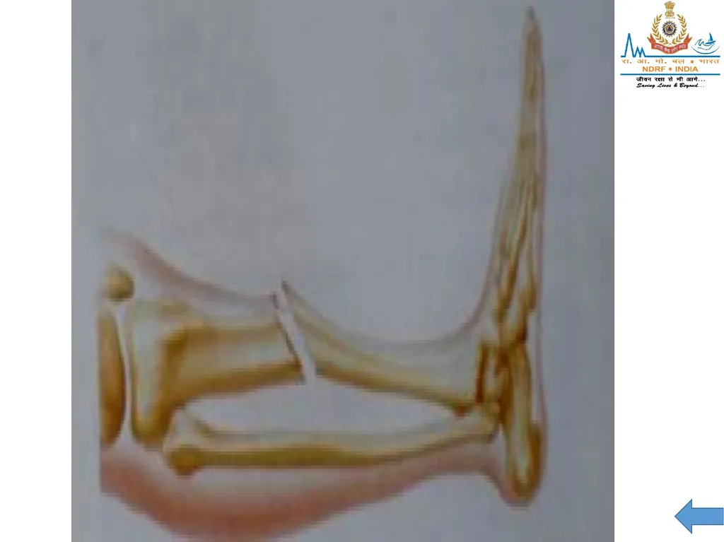
5) Tibia or fibula injury
Signs and symptoms: Pain, swelling and deformity.
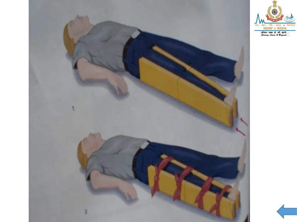
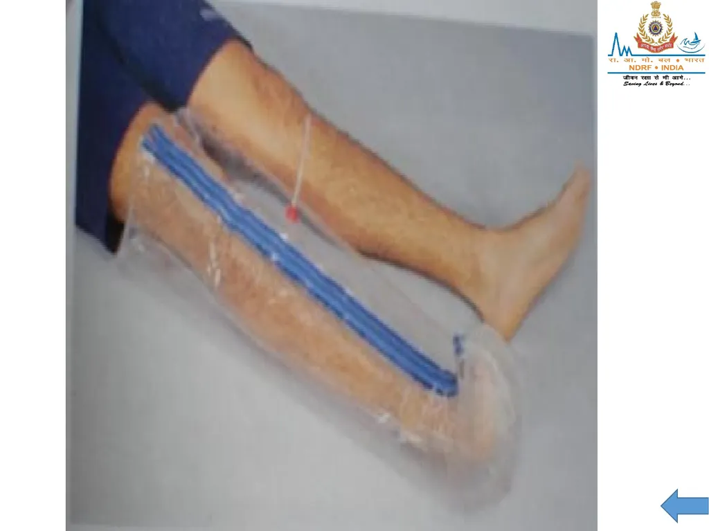
Pre-hospital treatment: Use two padded long splints – groin to foot and thigh to foot. Secure with cravats. Alternative method for a closed injury to the tibia or fibula is to use a circumferential splint or pneumatic splint.<br>
Signs and symptoms: Pain, swelling and deformity.
Pre-hospital treatment: Use two padded long splints – groin to foot and thigh to foot. Secure with cravats. Alternative method for a closed injury to the tibia or fibula is to use a circumferential splint or pneumatic splint.<br>
44
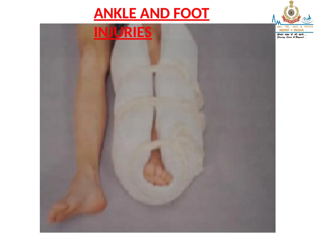
6) Ankle and foot injuries
Signs and symptoms: Pain, swelling and deformity.
Pre-hospital treatment:
Stabilize, remove shoes and socks if possible (expose injury).
Circumferential or formable splint such as a pillow secured with cravats is recommended.
Alternative: Padded boards to mid-thigh.<br>
Signs and symptoms: Pain, swelling and deformity.
Pre-hospital treatment:
Stabilize, remove shoes and socks if possible (expose injury).
Circumferential or formable splint such as a pillow secured with cravats is recommended.
Alternative: Padded boards to mid-thigh.<br>
45
ANKLE AND FOOT INJURIES<br>
46
The axial skeleton consists of 80 bones, including:
• Skull
• Thorax
• Vertebral (spinal) column<br>
• Skull
• Thorax
• Vertebral (spinal) column<br>
47
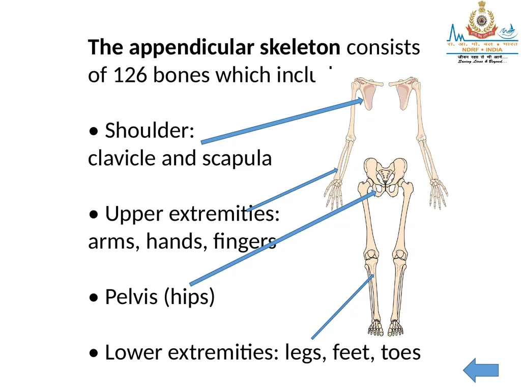
The appendicular skeleton consists of 126 bones which includes:
• Shoulder:
clavicle and scapula
• Upper extremities:
arms, hands, fingers
• Pelvis (hips)
• Lower extremities: legs, feet, toes<br>
• Shoulder:
clavicle and scapula
• Upper extremities:
arms, hands, fingers
• Pelvis (hips)
• Lower extremities: legs, feet, toes<br>
48
Elbow<br>
49
Forearm and Wrist<br>