
Pediatric Trauma Resources: Head and Neck 1

Description: Pediatric Trauma Resources: Head and Neck 1 Disclaimer: This guideline is designed for general use with most patients; providers should use their independent clinical judgment. This guideline is not intended to be a substitute for
Related Topics
Download Presentation
"Pediatric Trauma Resources: Head and Neck 1" is the property of its rightful owner. Permission is granted to download and print the materials on this website for personal, non-commercial use only, and to display it on your personal computer provided you do not modify the materials and that you retain all copyright notices contained in the materials. By downloading content from our website, you accept the terms of this agreement.
Presentation Transcript
slide1. Pediatric Trauma Resources:
Head and Neck 1 Disclaimer: This guideline is designed for general use with most patients; providers should use their independent clinical judgment. This guideline is not intended to be a substitute for professional medical advice, diagnosis, or treatment. Reviewed August 2023<br>
slide2. Disclaimers MMRTAC makes no representations or warranties about the accuracy, reliability, or completeness of the content. Content is provided “as is” and is for informational use only. It is not a substitute for professional medical advice, diagnosis, or treatment. MMRTAC disclaims all warranties, express or implied, statutory or otherwise, including without limitation the implied warranties of merchantability, non-infringement of third parties’ rights, and fitness for a particular purpose.
This content was developed to guide general patient care and may not be suitable for use in all patient care environments. MMRTAC does not endorse, certify, or assess third parties’ competency. You hold all responsibility for your use or nonuse of the content. MMRTAC shall not be liable for claims, losses, or damages arising from or related to any use or misuse of the content. 2 Reviewed August 2023<br>
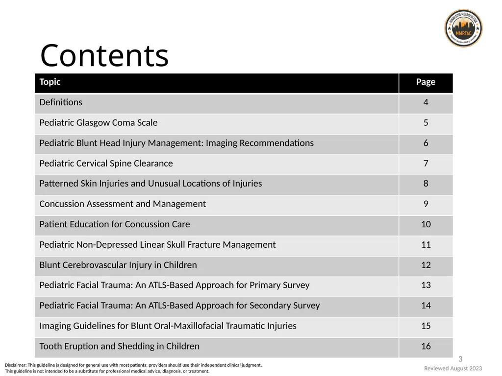
slide3. Contents Reviewed August 2023 3 Disclaimer: This guideline is designed for general use with most patients; providers should use their independent clinical judgment. This guideline is not intended to be a substitute for professional medical advice, diagnosis, or treatment.<br>
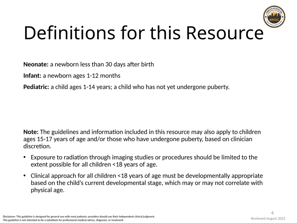
slide4. Definitions for this Resource Neonate: a newborn less than 30 days after birth
Infant: a newborn ages 1-12 months
Pediatric: a child ages 1-14 years; a child who has not yet undergone puberty.
Note: The guidelines and information included in this resource may also apply to children ages 15-17 years of age and/or those who have undergone puberty, based on clinician discretion.
Exposure to radiation through imaging studies or procedures should be limited to the extent possible for all children <18 years of age.
Clinical approach for all children <18 years of age must be developmentally appropriate based on the child’s current developmental stage, which may or may not correlate with physical age. 4 Reviewed August 2023 Disclaimer: This guideline is designed for general use with most patients; providers should use their independent clinical judgment. This guideline is not intended to be a substitute for professional medical advice, diagnosis, or treatment.<br>
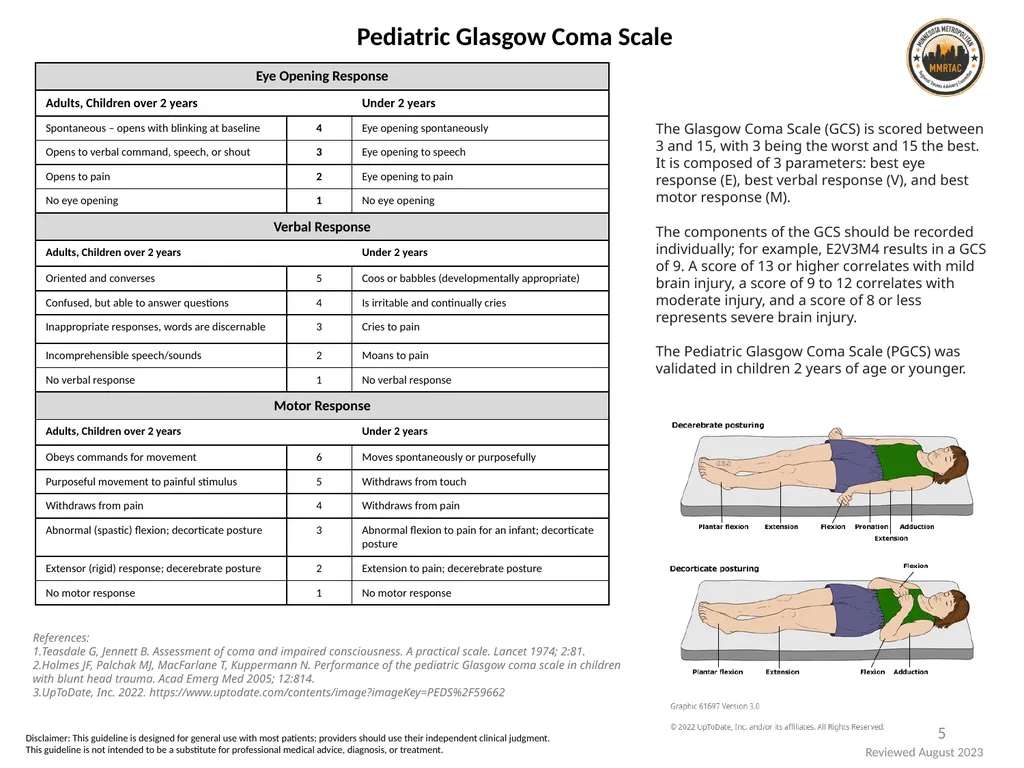
slide5. 5 Pediatric Glasgow Coma Scale References:
Teasdale G, Jennett B. Assessment of coma and impaired consciousness. A practical scale. Lancet 1974; 2:81.
Holmes JF, Palchak MJ, MacFarlane T, Kuppermann N. Performance of the pediatric Glasgow coma scale in children with blunt head trauma. Acad Emerg Med 2005; 12:814.
UpToDate, Inc. 2022. https://www.uptodate.com/contents/image?imageKey=PEDS%2F59662 The Glasgow Coma Scale (GCS) is scored between 3 and 15, with 3 being the worst and 15 the best. It is composed of 3 parameters: best eye response (E), best verbal response (V), and best motor response (M).
The components of the GCS should be recorded individually; for example, E2V3M4 results in a GCS of 9. A score of 13 or higher correlates with mild brain injury, a score of 9 to 12 correlates with moderate injury, and a score of 8 or less represents severe brain injury.
The Pediatric Glasgow Coma Scale (PGCS) was validated in children 2 years of age or younger. Disclaimer: This guideline is designed for general use with most patients; providers should use their independent clinical judgment. This guideline is not intended to be a substitute for professional medical advice, diagnosis, or treatment. Reviewed August 2023<br>
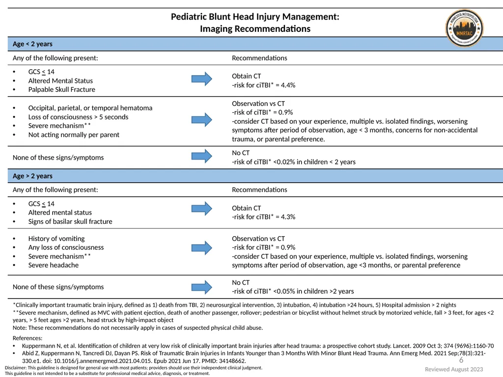
slide6. 6 Disclaimer: This guideline is designed for general use with most patients; providers should use their independent clinical judgment. This guideline is not intended to be a substitute for professional medical advice, diagnosis, or treatment. Reviewed August 2023<br>
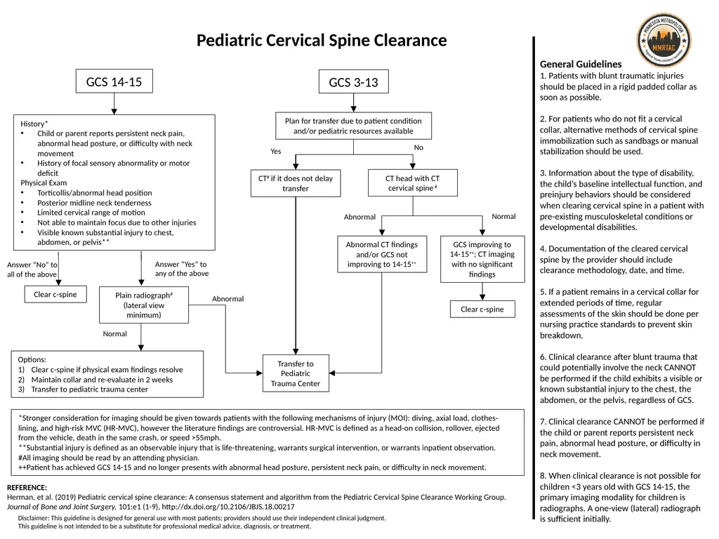
slide7. GCS 14-15 GCS 3-13 History*
Child or parent reports persistent neck pain, abnormal head posture, or difficulty with neck movement
History of focal sensory abnormality or motor deficit
Physical Exam
Torticollis/abnormal head position
Posterior midline neck tenderness
Limited cervical range of motion
Not able to maintain focus due to other injuries
Visible known substantial injury to chest, abdomen, or pelvis** Answer “No” to
all of the above Answer “Yes” to
any of the above Clear c-spine Options:
Clear c-spine if physical exam findings resolve
Maintain collar and re-evaluate in 2 weeks
Transfer to pediatric trauma center Plain radiograph#
(lateral view minimum) Normal Transfer to Pediatric Trauma Center Plan for transfer due to patient condition and/or pediatric resources available Clear c-spine Abnormal Normal Yes No Abnormal CT# if it does not delay transfer CT head with CT cervical spine # *Stronger consideration for imaging should be given towards patients with the following mechanisms of injury (MOI): diving, axial load, clothes-lining, and high-risk MVC (HR-MVC), however the literature findings are controversial. HR-MVC is defined as a head-on collision, rollover, ejected from the vehicle, death in the same crash, or speed >55mph.
**Substantial injury is defined as an observable injury that is life-threatening, warrants surgical intervention, or warrants inpatient observation.
#All imaging should be read by an attending physician.
++Patient has achieved GCS 14-15 and no longer presents with abnormal head posture, persistent neck pain, or difficulty in neck movement. Pediatric Cervical Spine Clearance REFERENCE:
Herman, et al. (2019) Pediatric cervical spine clearance: A consensus statement and algorithm from the Pediatric Cervical Spine Clearance Working Group. Journal of Bone and Joint Surgery, 101:e1 (1-9), http://dx.doi.org/10.2106/JBJS.18.00217 GCS improving to 14-15++; CT imaging with no significant findings Abnormal CT findings and/or GCS not improving to 14-15++ General Guidelines
1. Patients with blunt traumatic injuries should be placed in a rigid padded collar as soon as possible.
2. For patients who do not fit a cervical collar, alternative methods of cervical spine immobilization such as sandbags or manual stabilization should be used.
3. Information about the type of disability, the child’s baseline intellectual function, and preinjury behaviors should be considered when clearing cervical spine in a patient with pre-existing musculoskeletal conditions or developmental disabilities.
4. Documentation of the cleared cervical spine by the provider should include clearance methodology, date, and time.
5. If a patient remains in a cervical collar for extended periods of time, regular assessments of the skin should be done per nursing practice standards to prevent skin breakdown.
6. Clinical clearance after blunt trauma that could potentially involve the neck CANNOT be performed if the child exhibits a visible or known substantial injury to the chest, the abdomen, or the pelvis, regardless of GCS.
7. Clinical clearance CANNOT be performed if the child or parent reports persistent neck pain, abnormal head posture, or difficulty in neck movement.
8. When clinical clearance is not possible for children <3 years old with GCS 14-15, the primary imaging modality for children is radiographs. A one-view (lateral) radiograph is sufficient initially. Disclaimer: This guideline is designed for general use with most patients; providers should use their independent clinical judgment. This guideline is not intended to be a substitute for professional medical advice, diagnosis, or treatment.<br>
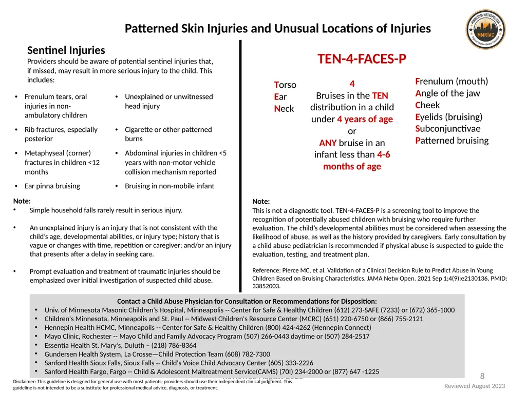
slide8. Patterned Skin Injuries and Unusual Locations of Injuries Note:
This is not a diagnostic tool. TEN-4-FACES-P is a screening tool to improve the recognition of potentially abused children with bruising who require further evaluation. The child’s developmental abilities must be considered when assessing the likelihood of abuse, as well as the history provided by caregivers. Early consultation by a child abuse pediatrician is recommended if physical abuse is suspected to guide the evaluation, testing, and treatment plan.
Reference: Pierce MC, et al. Validation of a Clinical Decision Rule to Predict Abuse in Young Children Based on Bruising Characteristics. JAMA Netw Open. 2021 Sep 1;4(9):e2130136. PMID: 33852003. Sentinel Injuries
Providers should be aware of potential sentinel injuries that, if missed, may result in more serious injury to the child. This includes: Reviewed August 2023 8 Contact a Child Abuse Physician for Consultation or Recommendations for Disposition:
Univ. of Minnesota Masonic Children's Hospital, Minneapolis -- Center for Safe & Healthy Children (612) 273-SAFE (7233) or (672) 365-1000
Children's Minnesota, Minneapolis and St. Paul -- Midwest Children's Resource Center (MCRC) (651) 220-6750 or (866) 755-2121
Hennepin Health HCMC, Minneapolis -- Center for Safe & Healthy Children (800) 424-4262 (Hennepin Connect)
Mayo Clinic, Rochester -- Mayo Child and Family Advocacy Program (507) 266-0443 daytime or (507) 284-2517
Essentia Health St. Mary’s, Duluth – (218) 786-8364
Gundersen Health System, La Crosse—Child Protection Team (608) 782-7300
Sanford Health Sioux Falls, Sioux Falls -- Child's Voice Child Advocacy Center (605) 333-2226
Sanford Health Fargo, Fargo -- Child & Adolescent Maltreatment Service(CAMS) (70I) 234-2000 or (877) 647 -1225 Note:
Simple household falls rarely result in serious injury.
An unexplained injury is an injury that is not consistent with the child’s age, developmental abilities, or injury type; history that is vague or changes with time, repetition or caregiver; and/or an injury that presents after a delay in seeking care.
Prompt evaluation and treatment of traumatic injuries should be emphasized over initial investigation of suspected child abuse. Disclaimer: This guideline is designed for general use with most patients; providers should use their independent clinical judgment. This guideline is not intended to be a substitute for professional medical advice, diagnosis, or treatment. Reviewed August 2023<br>
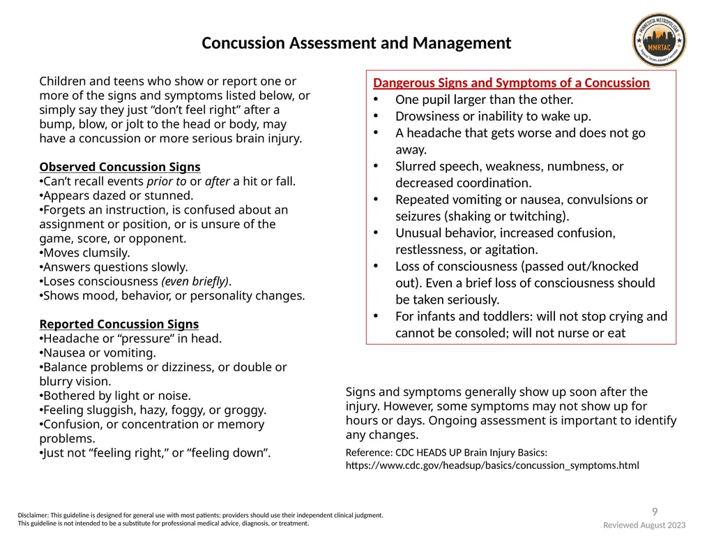
slide9. Concussion Assessment and Management 9 Dangerous Signs and Symptoms of a Concussion
One pupil larger than the other.
Drowsiness or inability to wake up.
A headache that gets worse and does not go away.
Slurred speech, weakness, numbness, or decreased coordination.
Repeated vomiting or nausea, convulsions or seizures (shaking or twitching).
Unusual behavior, increased confusion, restlessness, or agitation.
Loss of consciousness (passed out/knocked out). Even a brief loss of consciousness should be taken seriously.
For infants and toddlers: will not stop crying and cannot be consoled; will not nurse or eat Children and teens who show or report one or more of the signs and symptoms listed below, or simply say they just “don’t feel right” after a bump, blow, or jolt to the head or body, may have a concussion or more serious brain injury.
Observed Concussion Signs
Can’t recall events prior to or after a hit or fall.
Appears dazed or stunned.
Forgets an instruction, is confused about an assignment or position, or is unsure of the game, score, or opponent.
Moves clumsily.
Answers questions slowly.
Loses consciousness (even briefly).
Shows mood, behavior, or personality changes.
Reported Concussion Signs
Headache or “pressure” in head.
Nausea or vomiting.
Balance problems or dizziness, or double or blurry vision.
Bothered by light or noise.
Feeling sluggish, hazy, foggy, or groggy.
Confusion, or concentration or memory problems.
Just not “feeling right,” or “feeling down”. Signs and symptoms generally show up soon after the injury. However, some symptoms may not show up for hours or days. Ongoing assessment is important to identify any changes. Reference: CDC HEADS UP Brain Injury Basics: https://www.cdc.gov/headsup/basics/concussion_symptoms.html Disclaimer: This guideline is designed for general use with most patients; providers should use their independent clinical judgment. This guideline is not intended to be a substitute for professional medical advice, diagnosis, or treatment. Reviewed August 2023<br>
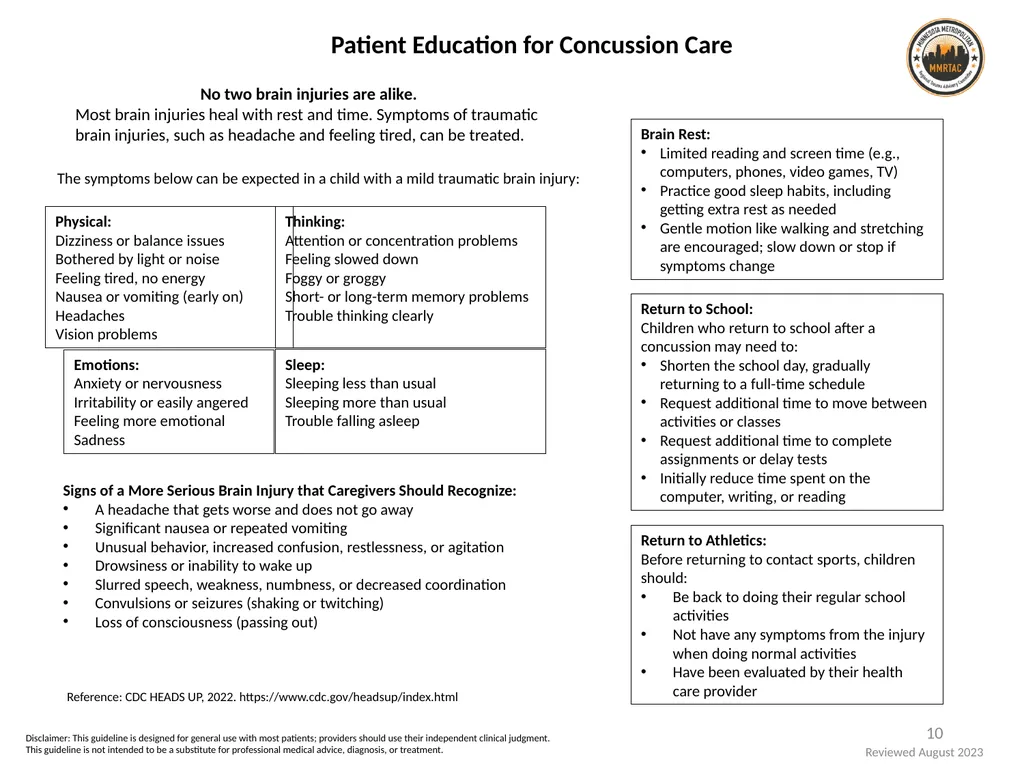
slide10. 10 Brain Rest:
Limited reading and screen time (e.g., computers, phones, video games, TV)
Practice good sleep habits, including getting extra rest as needed
Gentle motion like walking and stretching are encouraged; slow down or stop if symptoms change Patient Education for Concussion Care Signs of a More Serious Brain Injury that Caregivers Should Recognize:
A headache that gets worse and does not go away
Significant nausea or repeated vomiting
Unusual behavior, increased confusion, restlessness, or agitation
Drowsiness or inability to wake up
Slurred speech, weakness, numbness, or decreased coordination
Convulsions or seizures (shaking or twitching)
Loss of consciousness (passing out) Return to Athletics:
Before returning to contact sports, children should:
Be back to doing their regular school activities
Not have any symptoms from the injury when doing normal activities
Have been evaluated by their health care provider No two brain injuries are alike.
Most brain injuries heal with rest and time. Symptoms of traumatic brain injuries, such as headache and feeling tired, can be treated. The symptoms below can be expected in a child with a mild traumatic brain injury: Return to School:
Children who return to school after a concussion may need to:
Shorten the school day, gradually returning to a full-time schedule
Request additional time to move between activities or classes
Request additional time to complete assignments or delay tests
Initially reduce time spent on the computer, writing, or reading Reference: CDC HEADS UP, 2022. https://www.cdc.gov/headsup/index.html Disclaimer: This guideline is designed for general use with most patients; providers should use their independent clinical judgment. This guideline is not intended to be a substitute for professional medical advice, diagnosis, or treatment. Reviewed August 2023<br>
slide11. References:
Hentzen, A. S, Helmer, S.D, et al. Necessity of repeat head computed tomography after isolated skull fracture in the pediatric population. The American Journal of Surgery. 2015; 201, 322-325.
Powell, E.C, Atabaki, S.M, et al. Isolated Linear Skull Fractures in Children With Blunt Head Trauma. Pediatrics. 2015; 135, e851.
Lyons, T.W., Stack, A. M., et al. A QI Initiative to Reduce Hospitalization for Children With Isolated Skull Fractures. Pediatrics. 2016; 137;6;e20153370.
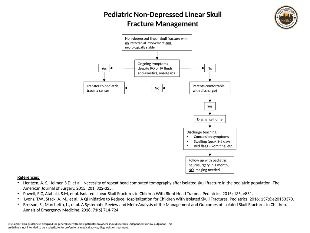
Bressan, S., Marchetto, L., et al. A Systematic Review and Meta-Analysis of the Management and Outcomes of Isolated Skull Fractures in Children. Annals of Emergency Medicine. 2018; 71(6) 714-724 Non-depressed linear skull fracture with no intracranial involvement and
neurologically stable Ongoing symptoms despite PO or IV fluids, anti-emetics, analgesics Yes No Parents comfortable with discharge? No Transfer to pediatric trauma center Yes Follow up with pediatric neurosurgery in 1 month,
NO imaging needed Discharge home Discharge teaching:
Concussion symptoms
Swelling (peak 3-5 days)
Red flags – vomiting, etc. Pediatric Non-Depressed Linear Skull Fracture Management Disclaimer: This guideline is designed for general use with most patients; providers should use their independent clinical judgment. This guideline is not intended to be a substitute for professional medical advice, diagnosis, or treatment.<br>
slide12. 12 Blunt Cerebrovascular Injury in Children References:
Herbert JP, Venkataraman SS, Turkmani AH, Zhu L, Kerr ML, Patel RP, Ugalde IT, Fletcher SA, Sandberg DI, Cox CS, Kitagawa RS, Day AL, Shah MN. Pediatric blunt cerebrovascular injury: the McGovern screening score. J Neurosurg Pediatr. 2018 Jun;21(6):639-649. doi: 10.3171/2017.12.PEDS17498. Epub 2018 Mar 16. PMID: 29547069.
Ravindra VM, Riva-Cambrin J, Sivakumar W, Metzger RR, Bollo RJ. Risk factors for traumatic blunt cerebrovascular injury diagnosed by computed tomography angiography in the pediatric population: a retrospective cohort study. J Neurosurg Pediatr. 2015 Jun;15(6):599-606. doi: 10.3171/2014.11.PEDS14397. Epub 2015 Mar 6. PMID: 25745952.
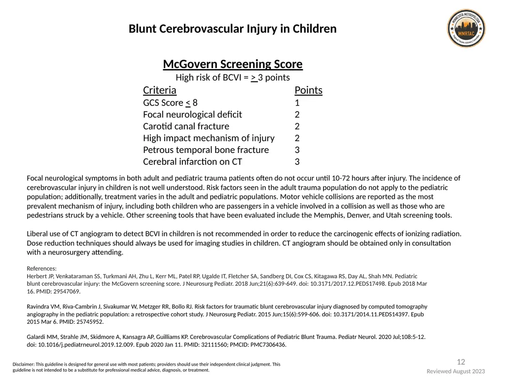
Galardi MM, Strahle JM, Skidmore A, Kansagra AP, Guilliams KP. Cerebrovascular Complications of Pediatric Blunt Trauma. Pediatr Neurol. 2020 Jul;108:5-12. doi: 10.1016/j.pediatrneurol.2019.12.009. Epub 2020 Jan 11. PMID: 32111560; PMCID: PMC7306436. Focal neurological symptoms in both adult and pediatric trauma patients often do not occur until 10-72 hours after injury. The incidence of cerebrovascular injury in children is not well understood. Risk factors seen in the adult trauma population do not apply to the pediatric population; additionally, treatment varies in the adult and pediatric populations. Motor vehicle collisions are reported as the most prevalent mechanism of injury, including both children who are passengers in a vehicle involved in a collision as well as those who are pedestrians struck by a vehicle. Other screening tools that have been evaluated include the Memphis, Denver, and Utah screening tools.
Liberal use of CT angiogram to detect BCVI in children is not recommended in order to reduce the carcinogenic effects of ionizing radiation. Dose reduction techniques should always be used for imaging studies in children. CT angiogram should be obtained only in consultation with a neurosurgery attending. McGovern Screening Score
High risk of BCVI = > 3 points
Criteria Points
GCS Score < 8 1
Focal neurological deficit 2
Carotid canal fracture 2
High impact mechanism of injury 2
Petrous temporal bone fracture 3
Cerebral infarction on CT 3 Disclaimer: This guideline is designed for general use with most patients; providers should use their independent clinical judgment. This guideline is not intended to be a substitute for professional medical advice, diagnosis, or treatment. Reviewed August 2023<br>
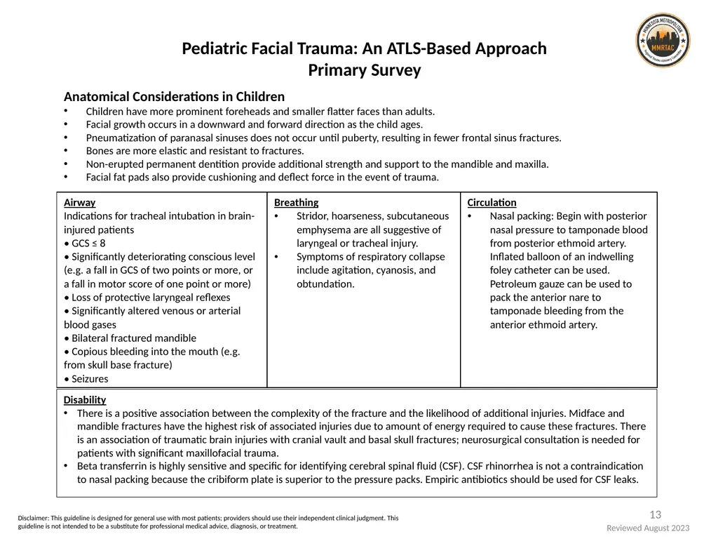
slide13. 13 Pediatric Facial Trauma: An ATLS-Based Approach
Primary Survey Anatomical Considerations in Children
Children have more prominent foreheads and smaller flatter faces than adults.
Facial growth occurs in a downward and forward direction as the child ages.
Pneumatization of paranasal sinuses does not occur until puberty, resulting in fewer frontal sinus fractures.
Bones are more elastic and resistant to fractures.
Non-erupted permanent dentition provide additional strength and support to the mandible and maxilla.
Facial fat pads also provide cushioning and deflect force in the event of trauma. Disability
There is a positive association between the complexity of the fracture and the likelihood of additional injuries. Midface and mandible fractures have the highest risk of associated injuries due to amount of energy required to cause these fractures. There is an association of traumatic brain injuries with cranial vault and basal skull fractures; neurosurgical consultation is needed for patients with significant maxillofacial trauma.
Beta transferrin is highly sensitive and specific for identifying cerebral spinal fluid (CSF). CSF rhinorrhea is not a contraindication to nasal packing because the cribiform plate is superior to the pressure packs. Empiric antibiotics should be used for CSF leaks. Disclaimer: This guideline is designed for general use with most patients; providers should use their independent clinical judgment. This guideline is not intended to be a substitute for professional medical advice, diagnosis, or treatment. Reviewed August 2023<br>
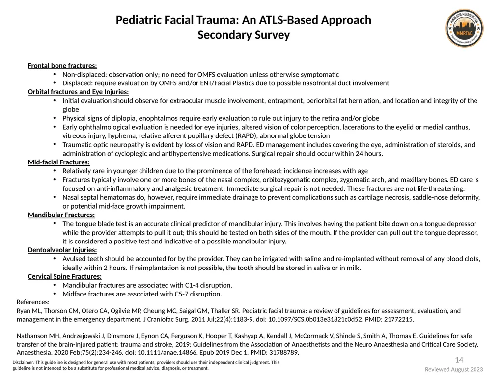
slide14. 14 Frontal bone fractures:
Non-displaced: observation only; no need for OMFS evaluation unless otherwise symptomatic
Displaced: require evaluation by OMFS and/or ENT/Facial Plastics due to possible nasofrontal duct involvement
Orbital fractures and Eye Injuries:
Initial evaluation should observe for extraocular muscle involvement, entrapment, periorbital fat herniation, and location and integrity of the globe
Physical signs of diplopia, enophtalmos require early evaluation to rule out injury to the retina and/or globe
Early ophthalmological evaluation is needed for eye injuries, altered vision of color perception, lacerations to the eyelid or medial canthus, vitreous injury, hyphema, relative afferent pupillary defect (RAPD), abnormal globe tension
Traumatic optic neuropathy is evident by loss of vision and RAPD. ED management includes covering the eye, administration of steroids, and administration of cycloplegic and antihypertensive medications. Surgical repair should occur within 24 hours.
Mid-facial Fractures:
Relatively rare in younger children due to the prominence of the forehead; incidence increases with age
Fractures typically involve one or more bones of the nasal complex, orbitozygomatic complex, zygomatic arch, and maxillary bones. ED care is focused on anti-inflammatory and analgesic treatment. Immediate surgical repair is not needed. These fractures are not life-threatening.
Nasal septal hematomas do, however, require immediate drainage to prevent complications such as cartilage necrosis, saddle-nose deformity, or potential mid-face growth impairment.
Mandibular Fractures:
The tongue blade test is an accurate clinical predictor of mandibular injury. This involves having the patient bite down on a tongue depressor while the provider attempts to pull it out; this should be tested on both sides of the mouth. If the provider can pull out the tongue depressor, it is considered a positive test and indicative of a possible mandibular injury.
Dentoalveolar Injuries:
Avulsed teeth should be accounted for by the provider. They can be irrigated with saline and re-implanted without removal of any blood clots, ideally within 2 hours. If reimplantation is not possible, the tooth should be stored in saliva or in milk.
Cervical Spine Fractures:
Mandibular fractures are associated with C1-4 disruption.
Midface fractures are associated with C5-7 disruption. Pediatric Facial Trauma: An ATLS-Based Approach
Secondary Survey References:
Ryan ML, Thorson CM, Otero CA, Ogilvie MP, Cheung MC, Saigal GM, Thaller SR. Pediatric facial trauma: a review of guidelines for assessment, evaluation, and management in the emergency department. J Craniofac Surg. 2011 Jul;22(4):1183-9. doi: 10.1097/SCS.0b013e31821c0d52. PMID: 21772215.
Nathanson MH, Andrzejowski J, Dinsmore J, Eynon CA, Ferguson K, Hooper T, Kashyap A, Kendall J, McCormack V, Shinde S, Smith A, Thomas E. Guidelines for safe transfer of the brain-injured patient: trauma and stroke, 2019: Guidelines from the Association of Anaesthetists and the Neuro Anaesthesia and Critical Care Society. Anaesthesia. 2020 Feb;75(2):234-246. doi: 10.1111/anae.14866. Epub 2019 Dec 1. PMID: 31788789. Disclaimer: This guideline is designed for general use with most patients; providers should use their independent clinical judgment. This guideline is not intended to be a substitute for professional medical advice, diagnosis, or treatment. Reviewed August 2023<br>
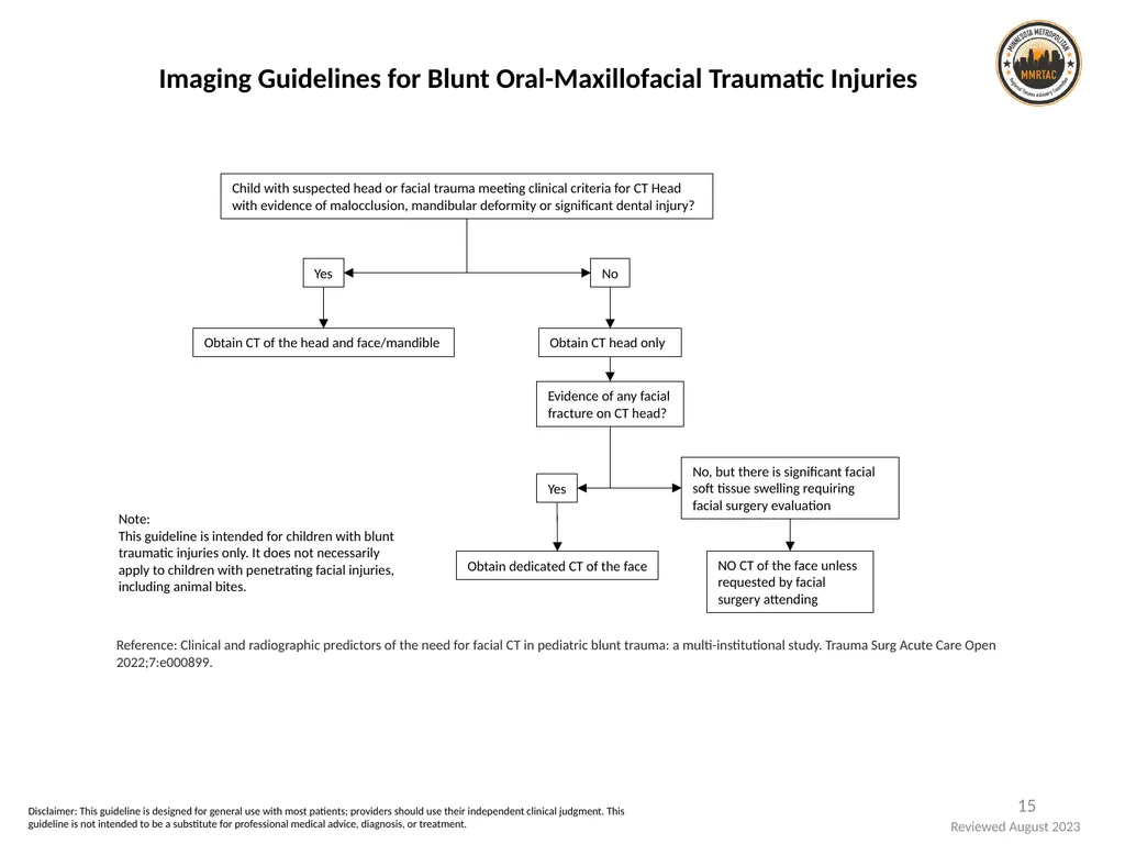
slide15. 15 Imaging Guidelines for Blunt Oral-Maxillofacial Traumatic Injuries Disclaimer: This guideline is designed for general use with most patients; providers should use their independent clinical judgment. This guideline is not intended to be a substitute for professional medical advice, diagnosis, or treatment. Reviewed August 2023<br>
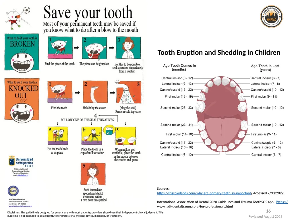
slide16. 16 Sources:
https://friscokidsdds.com/why-are-primary-teeth-so-important/ Accessed 7/30/2022.
International Association of Dental 2020 Guidelines and Trauma ToothSOS app - https://www.iadt-dentaltrauma.org/for-professionals.html Tooth Eruption and Shedding in Children Disclaimer: This guideline is designed for general use with most patients; providers should use their independent clinical judgment. This guideline is not intended to be a substitute for professional medical advice, diagnosis, or treatment. Reviewed August 2023<br>
Head and Neck 1 Disclaimer: This guideline is designed for general use with most patients; providers should use their independent clinical judgment. This guideline is not intended to be a substitute for professional medical advice, diagnosis, or treatment. Reviewed August 2023<br>
slide2. Disclaimers MMRTAC makes no representations or warranties about the accuracy, reliability, or completeness of the content. Content is provided “as is” and is for informational use only. It is not a substitute for professional medical advice, diagnosis, or treatment. MMRTAC disclaims all warranties, express or implied, statutory or otherwise, including without limitation the implied warranties of merchantability, non-infringement of third parties’ rights, and fitness for a particular purpose.
This content was developed to guide general patient care and may not be suitable for use in all patient care environments. MMRTAC does not endorse, certify, or assess third parties’ competency. You hold all responsibility for your use or nonuse of the content. MMRTAC shall not be liable for claims, losses, or damages arising from or related to any use or misuse of the content. 2 Reviewed August 2023<br>
slide3. Contents Reviewed August 2023 3 Disclaimer: This guideline is designed for general use with most patients; providers should use their independent clinical judgment. This guideline is not intended to be a substitute for professional medical advice, diagnosis, or treatment.<br>
slide4. Definitions for this Resource Neonate: a newborn less than 30 days after birth
Infant: a newborn ages 1-12 months
Pediatric: a child ages 1-14 years; a child who has not yet undergone puberty.
Note: The guidelines and information included in this resource may also apply to children ages 15-17 years of age and/or those who have undergone puberty, based on clinician discretion.
Exposure to radiation through imaging studies or procedures should be limited to the extent possible for all children <18 years of age.
Clinical approach for all children <18 years of age must be developmentally appropriate based on the child’s current developmental stage, which may or may not correlate with physical age. 4 Reviewed August 2023 Disclaimer: This guideline is designed for general use with most patients; providers should use their independent clinical judgment. This guideline is not intended to be a substitute for professional medical advice, diagnosis, or treatment.<br>
slide5. 5 Pediatric Glasgow Coma Scale References:
Teasdale G, Jennett B. Assessment of coma and impaired consciousness. A practical scale. Lancet 1974; 2:81.
Holmes JF, Palchak MJ, MacFarlane T, Kuppermann N. Performance of the pediatric Glasgow coma scale in children with blunt head trauma. Acad Emerg Med 2005; 12:814.
UpToDate, Inc. 2022. https://www.uptodate.com/contents/image?imageKey=PEDS%2F59662 The Glasgow Coma Scale (GCS) is scored between 3 and 15, with 3 being the worst and 15 the best. It is composed of 3 parameters: best eye response (E), best verbal response (V), and best motor response (M).
The components of the GCS should be recorded individually; for example, E2V3M4 results in a GCS of 9. A score of 13 or higher correlates with mild brain injury, a score of 9 to 12 correlates with moderate injury, and a score of 8 or less represents severe brain injury.
The Pediatric Glasgow Coma Scale (PGCS) was validated in children 2 years of age or younger. Disclaimer: This guideline is designed for general use with most patients; providers should use their independent clinical judgment. This guideline is not intended to be a substitute for professional medical advice, diagnosis, or treatment. Reviewed August 2023<br>
slide6. 6 Disclaimer: This guideline is designed for general use with most patients; providers should use their independent clinical judgment. This guideline is not intended to be a substitute for professional medical advice, diagnosis, or treatment. Reviewed August 2023<br>
slide7. GCS 14-15 GCS 3-13 History*
Child or parent reports persistent neck pain, abnormal head posture, or difficulty with neck movement
History of focal sensory abnormality or motor deficit
Physical Exam
Torticollis/abnormal head position
Posterior midline neck tenderness
Limited cervical range of motion
Not able to maintain focus due to other injuries
Visible known substantial injury to chest, abdomen, or pelvis** Answer “No” to
all of the above Answer “Yes” to
any of the above Clear c-spine Options:
Clear c-spine if physical exam findings resolve
Maintain collar and re-evaluate in 2 weeks
Transfer to pediatric trauma center Plain radiograph#
(lateral view minimum) Normal Transfer to Pediatric Trauma Center Plan for transfer due to patient condition and/or pediatric resources available Clear c-spine Abnormal Normal Yes No Abnormal CT# if it does not delay transfer CT head with CT cervical spine # *Stronger consideration for imaging should be given towards patients with the following mechanisms of injury (MOI): diving, axial load, clothes-lining, and high-risk MVC (HR-MVC), however the literature findings are controversial. HR-MVC is defined as a head-on collision, rollover, ejected from the vehicle, death in the same crash, or speed >55mph.
**Substantial injury is defined as an observable injury that is life-threatening, warrants surgical intervention, or warrants inpatient observation.
#All imaging should be read by an attending physician.
++Patient has achieved GCS 14-15 and no longer presents with abnormal head posture, persistent neck pain, or difficulty in neck movement. Pediatric Cervical Spine Clearance REFERENCE:
Herman, et al. (2019) Pediatric cervical spine clearance: A consensus statement and algorithm from the Pediatric Cervical Spine Clearance Working Group. Journal of Bone and Joint Surgery, 101:e1 (1-9), http://dx.doi.org/10.2106/JBJS.18.00217 GCS improving to 14-15++; CT imaging with no significant findings Abnormal CT findings and/or GCS not improving to 14-15++ General Guidelines
1. Patients with blunt traumatic injuries should be placed in a rigid padded collar as soon as possible.
2. For patients who do not fit a cervical collar, alternative methods of cervical spine immobilization such as sandbags or manual stabilization should be used.
3. Information about the type of disability, the child’s baseline intellectual function, and preinjury behaviors should be considered when clearing cervical spine in a patient with pre-existing musculoskeletal conditions or developmental disabilities.
4. Documentation of the cleared cervical spine by the provider should include clearance methodology, date, and time.
5. If a patient remains in a cervical collar for extended periods of time, regular assessments of the skin should be done per nursing practice standards to prevent skin breakdown.
6. Clinical clearance after blunt trauma that could potentially involve the neck CANNOT be performed if the child exhibits a visible or known substantial injury to the chest, the abdomen, or the pelvis, regardless of GCS.
7. Clinical clearance CANNOT be performed if the child or parent reports persistent neck pain, abnormal head posture, or difficulty in neck movement.
8. When clinical clearance is not possible for children <3 years old with GCS 14-15, the primary imaging modality for children is radiographs. A one-view (lateral) radiograph is sufficient initially. Disclaimer: This guideline is designed for general use with most patients; providers should use their independent clinical judgment. This guideline is not intended to be a substitute for professional medical advice, diagnosis, or treatment.<br>
slide8. Patterned Skin Injuries and Unusual Locations of Injuries Note:
This is not a diagnostic tool. TEN-4-FACES-P is a screening tool to improve the recognition of potentially abused children with bruising who require further evaluation. The child’s developmental abilities must be considered when assessing the likelihood of abuse, as well as the history provided by caregivers. Early consultation by a child abuse pediatrician is recommended if physical abuse is suspected to guide the evaluation, testing, and treatment plan.
Reference: Pierce MC, et al. Validation of a Clinical Decision Rule to Predict Abuse in Young Children Based on Bruising Characteristics. JAMA Netw Open. 2021 Sep 1;4(9):e2130136. PMID: 33852003. Sentinel Injuries
Providers should be aware of potential sentinel injuries that, if missed, may result in more serious injury to the child. This includes: Reviewed August 2023 8 Contact a Child Abuse Physician for Consultation or Recommendations for Disposition:
Univ. of Minnesota Masonic Children's Hospital, Minneapolis -- Center for Safe & Healthy Children (612) 273-SAFE (7233) or (672) 365-1000
Children's Minnesota, Minneapolis and St. Paul -- Midwest Children's Resource Center (MCRC) (651) 220-6750 or (866) 755-2121
Hennepin Health HCMC, Minneapolis -- Center for Safe & Healthy Children (800) 424-4262 (Hennepin Connect)
Mayo Clinic, Rochester -- Mayo Child and Family Advocacy Program (507) 266-0443 daytime or (507) 284-2517
Essentia Health St. Mary’s, Duluth – (218) 786-8364
Gundersen Health System, La Crosse—Child Protection Team (608) 782-7300
Sanford Health Sioux Falls, Sioux Falls -- Child's Voice Child Advocacy Center (605) 333-2226
Sanford Health Fargo, Fargo -- Child & Adolescent Maltreatment Service(CAMS) (70I) 234-2000 or (877) 647 -1225 Note:
Simple household falls rarely result in serious injury.
An unexplained injury is an injury that is not consistent with the child’s age, developmental abilities, or injury type; history that is vague or changes with time, repetition or caregiver; and/or an injury that presents after a delay in seeking care.
Prompt evaluation and treatment of traumatic injuries should be emphasized over initial investigation of suspected child abuse. Disclaimer: This guideline is designed for general use with most patients; providers should use their independent clinical judgment. This guideline is not intended to be a substitute for professional medical advice, diagnosis, or treatment. Reviewed August 2023<br>
slide9. Concussion Assessment and Management 9 Dangerous Signs and Symptoms of a Concussion
One pupil larger than the other.
Drowsiness or inability to wake up.
A headache that gets worse and does not go away.
Slurred speech, weakness, numbness, or decreased coordination.
Repeated vomiting or nausea, convulsions or seizures (shaking or twitching).
Unusual behavior, increased confusion, restlessness, or agitation.
Loss of consciousness (passed out/knocked out). Even a brief loss of consciousness should be taken seriously.
For infants and toddlers: will not stop crying and cannot be consoled; will not nurse or eat Children and teens who show or report one or more of the signs and symptoms listed below, or simply say they just “don’t feel right” after a bump, blow, or jolt to the head or body, may have a concussion or more serious brain injury.
Observed Concussion Signs
Can’t recall events prior to or after a hit or fall.
Appears dazed or stunned.
Forgets an instruction, is confused about an assignment or position, or is unsure of the game, score, or opponent.
Moves clumsily.
Answers questions slowly.
Loses consciousness (even briefly).
Shows mood, behavior, or personality changes.
Reported Concussion Signs
Headache or “pressure” in head.
Nausea or vomiting.
Balance problems or dizziness, or double or blurry vision.
Bothered by light or noise.
Feeling sluggish, hazy, foggy, or groggy.
Confusion, or concentration or memory problems.
Just not “feeling right,” or “feeling down”. Signs and symptoms generally show up soon after the injury. However, some symptoms may not show up for hours or days. Ongoing assessment is important to identify any changes. Reference: CDC HEADS UP Brain Injury Basics: https://www.cdc.gov/headsup/basics/concussion_symptoms.html Disclaimer: This guideline is designed for general use with most patients; providers should use their independent clinical judgment. This guideline is not intended to be a substitute for professional medical advice, diagnosis, or treatment. Reviewed August 2023<br>
slide10. 10 Brain Rest:
Limited reading and screen time (e.g., computers, phones, video games, TV)
Practice good sleep habits, including getting extra rest as needed
Gentle motion like walking and stretching are encouraged; slow down or stop if symptoms change Patient Education for Concussion Care Signs of a More Serious Brain Injury that Caregivers Should Recognize:
A headache that gets worse and does not go away
Significant nausea or repeated vomiting
Unusual behavior, increased confusion, restlessness, or agitation
Drowsiness or inability to wake up
Slurred speech, weakness, numbness, or decreased coordination
Convulsions or seizures (shaking or twitching)
Loss of consciousness (passing out) Return to Athletics:
Before returning to contact sports, children should:
Be back to doing their regular school activities
Not have any symptoms from the injury when doing normal activities
Have been evaluated by their health care provider No two brain injuries are alike.
Most brain injuries heal with rest and time. Symptoms of traumatic brain injuries, such as headache and feeling tired, can be treated. The symptoms below can be expected in a child with a mild traumatic brain injury: Return to School:
Children who return to school after a concussion may need to:
Shorten the school day, gradually returning to a full-time schedule
Request additional time to move between activities or classes
Request additional time to complete assignments or delay tests
Initially reduce time spent on the computer, writing, or reading Reference: CDC HEADS UP, 2022. https://www.cdc.gov/headsup/index.html Disclaimer: This guideline is designed for general use with most patients; providers should use their independent clinical judgment. This guideline is not intended to be a substitute for professional medical advice, diagnosis, or treatment. Reviewed August 2023<br>
slide11. References:
Hentzen, A. S, Helmer, S.D, et al. Necessity of repeat head computed tomography after isolated skull fracture in the pediatric population. The American Journal of Surgery. 2015; 201, 322-325.
Powell, E.C, Atabaki, S.M, et al. Isolated Linear Skull Fractures in Children With Blunt Head Trauma. Pediatrics. 2015; 135, e851.
Lyons, T.W., Stack, A. M., et al. A QI Initiative to Reduce Hospitalization for Children With Isolated Skull Fractures. Pediatrics. 2016; 137;6;e20153370.
Bressan, S., Marchetto, L., et al. A Systematic Review and Meta-Analysis of the Management and Outcomes of Isolated Skull Fractures in Children. Annals of Emergency Medicine. 2018; 71(6) 714-724 Non-depressed linear skull fracture with no intracranial involvement and
neurologically stable Ongoing symptoms despite PO or IV fluids, anti-emetics, analgesics Yes No Parents comfortable with discharge? No Transfer to pediatric trauma center Yes Follow up with pediatric neurosurgery in 1 month,
NO imaging needed Discharge home Discharge teaching:
Concussion symptoms
Swelling (peak 3-5 days)
Red flags – vomiting, etc. Pediatric Non-Depressed Linear Skull Fracture Management Disclaimer: This guideline is designed for general use with most patients; providers should use their independent clinical judgment. This guideline is not intended to be a substitute for professional medical advice, diagnosis, or treatment.<br>
slide12. 12 Blunt Cerebrovascular Injury in Children References:
Herbert JP, Venkataraman SS, Turkmani AH, Zhu L, Kerr ML, Patel RP, Ugalde IT, Fletcher SA, Sandberg DI, Cox CS, Kitagawa RS, Day AL, Shah MN. Pediatric blunt cerebrovascular injury: the McGovern screening score. J Neurosurg Pediatr. 2018 Jun;21(6):639-649. doi: 10.3171/2017.12.PEDS17498. Epub 2018 Mar 16. PMID: 29547069.
Ravindra VM, Riva-Cambrin J, Sivakumar W, Metzger RR, Bollo RJ. Risk factors for traumatic blunt cerebrovascular injury diagnosed by computed tomography angiography in the pediatric population: a retrospective cohort study. J Neurosurg Pediatr. 2015 Jun;15(6):599-606. doi: 10.3171/2014.11.PEDS14397. Epub 2015 Mar 6. PMID: 25745952.
Galardi MM, Strahle JM, Skidmore A, Kansagra AP, Guilliams KP. Cerebrovascular Complications of Pediatric Blunt Trauma. Pediatr Neurol. 2020 Jul;108:5-12. doi: 10.1016/j.pediatrneurol.2019.12.009. Epub 2020 Jan 11. PMID: 32111560; PMCID: PMC7306436. Focal neurological symptoms in both adult and pediatric trauma patients often do not occur until 10-72 hours after injury. The incidence of cerebrovascular injury in children is not well understood. Risk factors seen in the adult trauma population do not apply to the pediatric population; additionally, treatment varies in the adult and pediatric populations. Motor vehicle collisions are reported as the most prevalent mechanism of injury, including both children who are passengers in a vehicle involved in a collision as well as those who are pedestrians struck by a vehicle. Other screening tools that have been evaluated include the Memphis, Denver, and Utah screening tools.
Liberal use of CT angiogram to detect BCVI in children is not recommended in order to reduce the carcinogenic effects of ionizing radiation. Dose reduction techniques should always be used for imaging studies in children. CT angiogram should be obtained only in consultation with a neurosurgery attending. McGovern Screening Score
High risk of BCVI = > 3 points
Criteria Points
GCS Score < 8 1
Focal neurological deficit 2
Carotid canal fracture 2
High impact mechanism of injury 2
Petrous temporal bone fracture 3
Cerebral infarction on CT 3 Disclaimer: This guideline is designed for general use with most patients; providers should use their independent clinical judgment. This guideline is not intended to be a substitute for professional medical advice, diagnosis, or treatment. Reviewed August 2023<br>
slide13. 13 Pediatric Facial Trauma: An ATLS-Based Approach
Primary Survey Anatomical Considerations in Children
Children have more prominent foreheads and smaller flatter faces than adults.
Facial growth occurs in a downward and forward direction as the child ages.
Pneumatization of paranasal sinuses does not occur until puberty, resulting in fewer frontal sinus fractures.
Bones are more elastic and resistant to fractures.
Non-erupted permanent dentition provide additional strength and support to the mandible and maxilla.
Facial fat pads also provide cushioning and deflect force in the event of trauma. Disability
There is a positive association between the complexity of the fracture and the likelihood of additional injuries. Midface and mandible fractures have the highest risk of associated injuries due to amount of energy required to cause these fractures. There is an association of traumatic brain injuries with cranial vault and basal skull fractures; neurosurgical consultation is needed for patients with significant maxillofacial trauma.
Beta transferrin is highly sensitive and specific for identifying cerebral spinal fluid (CSF). CSF rhinorrhea is not a contraindication to nasal packing because the cribiform plate is superior to the pressure packs. Empiric antibiotics should be used for CSF leaks. Disclaimer: This guideline is designed for general use with most patients; providers should use their independent clinical judgment. This guideline is not intended to be a substitute for professional medical advice, diagnosis, or treatment. Reviewed August 2023<br>
slide14. 14 Frontal bone fractures:
Non-displaced: observation only; no need for OMFS evaluation unless otherwise symptomatic
Displaced: require evaluation by OMFS and/or ENT/Facial Plastics due to possible nasofrontal duct involvement
Orbital fractures and Eye Injuries:
Initial evaluation should observe for extraocular muscle involvement, entrapment, periorbital fat herniation, and location and integrity of the globe
Physical signs of diplopia, enophtalmos require early evaluation to rule out injury to the retina and/or globe
Early ophthalmological evaluation is needed for eye injuries, altered vision of color perception, lacerations to the eyelid or medial canthus, vitreous injury, hyphema, relative afferent pupillary defect (RAPD), abnormal globe tension
Traumatic optic neuropathy is evident by loss of vision and RAPD. ED management includes covering the eye, administration of steroids, and administration of cycloplegic and antihypertensive medications. Surgical repair should occur within 24 hours.
Mid-facial Fractures:
Relatively rare in younger children due to the prominence of the forehead; incidence increases with age
Fractures typically involve one or more bones of the nasal complex, orbitozygomatic complex, zygomatic arch, and maxillary bones. ED care is focused on anti-inflammatory and analgesic treatment. Immediate surgical repair is not needed. These fractures are not life-threatening.
Nasal septal hematomas do, however, require immediate drainage to prevent complications such as cartilage necrosis, saddle-nose deformity, or potential mid-face growth impairment.
Mandibular Fractures:
The tongue blade test is an accurate clinical predictor of mandibular injury. This involves having the patient bite down on a tongue depressor while the provider attempts to pull it out; this should be tested on both sides of the mouth. If the provider can pull out the tongue depressor, it is considered a positive test and indicative of a possible mandibular injury.
Dentoalveolar Injuries:
Avulsed teeth should be accounted for by the provider. They can be irrigated with saline and re-implanted without removal of any blood clots, ideally within 2 hours. If reimplantation is not possible, the tooth should be stored in saliva or in milk.
Cervical Spine Fractures:
Mandibular fractures are associated with C1-4 disruption.
Midface fractures are associated with C5-7 disruption. Pediatric Facial Trauma: An ATLS-Based Approach
Secondary Survey References:
Ryan ML, Thorson CM, Otero CA, Ogilvie MP, Cheung MC, Saigal GM, Thaller SR. Pediatric facial trauma: a review of guidelines for assessment, evaluation, and management in the emergency department. J Craniofac Surg. 2011 Jul;22(4):1183-9. doi: 10.1097/SCS.0b013e31821c0d52. PMID: 21772215.
Nathanson MH, Andrzejowski J, Dinsmore J, Eynon CA, Ferguson K, Hooper T, Kashyap A, Kendall J, McCormack V, Shinde S, Smith A, Thomas E. Guidelines for safe transfer of the brain-injured patient: trauma and stroke, 2019: Guidelines from the Association of Anaesthetists and the Neuro Anaesthesia and Critical Care Society. Anaesthesia. 2020 Feb;75(2):234-246. doi: 10.1111/anae.14866. Epub 2019 Dec 1. PMID: 31788789. Disclaimer: This guideline is designed for general use with most patients; providers should use their independent clinical judgment. This guideline is not intended to be a substitute for professional medical advice, diagnosis, or treatment. Reviewed August 2023<br>
slide15. 15 Imaging Guidelines for Blunt Oral-Maxillofacial Traumatic Injuries Disclaimer: This guideline is designed for general use with most patients; providers should use their independent clinical judgment. This guideline is not intended to be a substitute for professional medical advice, diagnosis, or treatment. Reviewed August 2023<br>
slide16. 16 Sources:
https://friscokidsdds.com/why-are-primary-teeth-so-important/ Accessed 7/30/2022.
International Association of Dental 2020 Guidelines and Trauma ToothSOS app - https://www.iadt-dentaltrauma.org/for-professionals.html Tooth Eruption and Shedding in Children Disclaimer: This guideline is designed for general use with most patients; providers should use their independent clinical judgment. This guideline is not intended to be a substitute for professional medical advice, diagnosis, or treatment. Reviewed August 2023<br>