
Salmonellosis Dr. Bipin Kumar Assistant Professor





Description: Salmonellosis Dr. Bipin Kumar Assistant Professor Department of Veterinary Medicine Bihar Veterinary College, Patna (Bihar Animal Sciences University, Patna) Etiology Salmonella, a rod-shaped gram-negative bacterium is the causative agent
Related Topics
Download Presentation
"Salmonellosis Dr. Bipin Kumar Assistant Professor" is the property of its rightful owner. Permission is granted to download and print the materials on this website for personal, non-commercial use only, and to display it on your personal computer provided you do not modify the materials and that you retain all copyright notices contained in the materials. By downloading content from our website, you accept the terms of this agreement.
Presentation Transcript
slide1. Salmonellosis Dr. Bipin Kumar
Assistant Professor Department of Veterinary Medicine
Bihar Veterinary College, Patna
(Bihar Animal Sciences University, Patna)<br>
slide2. Etiology Salmonella, a rod-shaped gram-negative bacterium is the causative agent of salmonellosis.
Salmonellosis in warm-blooded vertebrates is in most cases associated with serovars of Salmonella enterica.
The most common type of infection is the carrier state.
Clinical disease is characterized by two major syndromes: a systemic septicemia (also termed as typhoid) and an enteritis.
Other less common clinical presentations include abortion, arthritis, respiratory disease, necrosis of extremities, and meningitis. Salmonella spp.
Only a few serotypes produce clinical salmonellosis in healthy animals and typically have a narrow range of host species, a phenomenon termed serovar-host specificity.
Salmonella enterica serovar Typhi (S Typhi) and S Paratyphi produce typhoid in people, S Gallinarum produces a similar disease in poultry, S Abortusovis in sheep, S Choleraesuis in pigs, S Dublin in cattle, etc.<br>
slide3. Young calves, piglets, lambs, and foals may develop both the enteritis and septicemic form.
cattle—S Typhimurium, S Dublin, and S Newport;
sheep and goats—S Typhimurium, S Dublin, S Abortusovis, S Anatum, and S Montevideo;
pigs—S Typhimurium and S Choleraesuis;
horses—S Typhimurium, S Anatum, S Newport, S Enteritidis, and Salmonella serovar IIIa 18:z4:z23; and
poultry—S Enteritidis, S Typhimurium, S Gallinarum, and S Pullorum.<br>
slide4. Transmission Human:
People are often infected when they eat contaminated foods of animal origin such as meat or eggs.
They can also be infected by ingesting organisms in animal feces, either directly or in contaminated food or water.
Directly transmitted human infections are most often acquired from the feces of reptiles, chicks and ducklings. Livestock, dogs, cats, adult poultry and cage birds can also be involved.
Animals:
Salmonella spp. are mainly transmitted by the fecal-oral route.
Vertical transmission occurs in birds, with contamination of the vitelline membrane, albumen and the yolk of eggs.
Salmonella spp. can also be transmitted in utero in mammals.
Animals may also become infected from contaminated feed (including pastures), drinking water, or close contact with infected animal (including humans).
Birds and rodents can spread Salmonella to livestock.
Carnivores are also infected through meat, eggs, and other animal products that are not thoroughly cooked.<br>
slide5. Mechanisms of Pathogenicity
Bacterial products involved in virulence:
Salmonellae owe their pathogenicity largely to their ability to invade tissue and to survive within macrophages.
The Vi antigen is a capsule that affords salmonellae some protection from phagocytosis.
Once phagocytosed, Salmonella inhibits generation of oxidative free radicals and intraphagosomal killing.
Additionally, salmonellae have endotoxic lipopolysaccharide, which is responsible for septic shock in patients with bacteriemia.
Salmonellae that cause enteritis produce at least two enterotoxins that are responsible for many of the clinical signs of enteritis.
The first of these is a small (25-30kD) protein that binds to GM1 gangliosides and cause hypersecretion of fluids and electrolytes by elevating levels of c-AMP.
It appears that both protein kinase C and prostaglandin E2 are involved in this process.
The second enterotoxin is larger (about 100 kD) and is unrelated in structure and mechanism of activity to the first enterotoxin
Salmonella strains that produce enterotoxins have been reported to invade the intestinal wall more effectively and to be more virulent than their non-toxigenic counterparts.<br>
slide6. The Salmonella infection cycle.
• Intestinal infection with salmonellae can follow one of two infection cycle.
One cycle causes enteritis, other causes typhoid
a. Enteritis
Most serotypes cause enteritis, an infection that is limited to the terminal ileum.
The salmonellae invade the intestinal wall and produce enterotoxins that cause nausea, vomiting and diarrhea.
b. Enteric fever (Typhoid): • Two serotypes Typhi and Paratyphi can cause typhoid.
• The salmonella invade the wall of the terminal ileum and than spread to the intestinal lymphatics, where they are phagocytosed by PMNs and macrophages.
• Salmonella phagocytosed by PMNs are killed, but those phagocytosed by macrophages survive and multiply within phagocytic vacuoles.
• Wandering macrophages that contain salmonellae act as “taxi/cabs” that deliver salmonellae to various reticuloendothelial tissues.
• Infected macrophages are eventually destroyed and salmonellae released from lysed macrophages cause septicemia.<br>
slide7. Clinical manifestation Salmonellae often localize in the mucosae of the ileum, caecum and colon, and in the mesenteric lymph nodes of infected animals.
Although most organisms are cleared from the tissues by host defense mechanisms, subclinical infection may persist with shedding of small numbers of salmonellae in the faeces.
Latent infections, in which salmonellae are present in the gall bladder but are not excreted, also occur.
Clinical disease may develop from subclinical and latent infections if affected animals are stressed.
Other factors which determine the clinical outcome of infection include the number of salmonellae ingested, the virulence of the infecting serotype or strain and the susceptibility of the host.
Host susceptibility may be related to immunological status, genetic make- up or age.
Young and debilitated or aged animaIs are particularly susceptible and may develop the septicemic form of the disease.
In most animal species, both enteric and septicemic forms of salmonellosis are recorded.
A number of serotypes have been associated with abortion in farm animals, often without other obvious clinical signs in dams.<br>
slide8. Enteric Salmonellosis
Enterocolitis caused by salmonella organisms can affect most species of farm animals, irrespective of age.
Acute disease is characterized by fever, depression, anorexia and profuse foul-smelling diarrhoea often containing blood, mucus and epithelial casts.
Dehydration and weight loss follow and pregnant animals may abort.
Severely affected young animals become recumbent and may die within a few days of acquiring infection.
On farms with endemic salmonellosis, the milder clinical signs often observed may be attributed to the influence of acquired immunity.
Chronic enterocolitis can follow acute salmonellosis in pigs, cattle and horses.
Intermittent fever, soft faeces and gradual weight loss, leading to emaciation, are common features of this condition.<br>
slide9. Septicaemic Salmonellosis
The septicaemic form can occur in all age groups but is most common in calves, in neonatal foals and in pigs less than four months of age.
Onset of clinical disease is sudden with high fever, depression and recumbency.
If treatment is delayed, many young animals with septicaemic salmonellosis die within 48 hours.
Surviving animals can develop persistent diarrhoea, arthritis, meningitis or pneumonia.
In pigs with septicaemic Salmonella Choleraesuis infection, there is a characteristic bluish discolouration of the ears and snout.
Intercurrent viral infections often predispose to severe clinical forms of the disease.
The close clinical and pathological relationships which have been recognized in animals infected with Salmonella Choleraesuis (hog- cholera bacillus) and classical swine fever virus, either jointly or separately, exemplify both the importance of intercurrent infections and the difficulty of clinically distinguishing the diseases caused by these agents.<br>
slide10. Salmonellosis in poultry
Salmonella Pullorum, Salmonella Gallinarum and Salmonella Enteritidis can infect the ovaries of hens and be transmitted through eggs.
Pullorum disease or bacillary white diarrhoea (Salmonella Pullorum):
Infects young chicks and turkey poults up to 2 to 3 weeks of age.
The mortality rate is high and affected birds huddle under a heat source and are anorexic, depressed and have whitish faecal pasting around their vents.
Characteristic lesions includes whitish nodes throughout the lungs and focal necrosis of liver and spleen.
Fowl typhoid (Salmonella Gallinarum):
Can produce lesions in young chicks and poults similar to those of pullorum disease, but it is also a serious concern in growing and adult poultry
However, in countries where fowl typhoid is endemic, a septicaemic disease of adult birds occurs, often resulting in sudden deaths.
Characteristic findings include an enlarged, friable, bile-stained liver and enlarged spleen.
Paratyphoid is a name given to infections of poultry by non-host-adapted salmonellae such as Salmonella Enteritidis and Salmonella Typhimurium. These infections are often subclinical in laying birds.<br>
slide11. Diagnosis A history of previous outbreaks of the disease may suggest salmonellosis.
At postmortem, enterocolitis with blood-stained luminal contents and enlarged mesenteric lymph nodes are commonly observed.
Specimens for submission should include faeces and blood from live animals.
Intestinal contents and samples from tissue lesions should be submitted from dead animals and abomasal contents from aborted foetuses.
Isolation of salmonellae from blood or parenchymatous organs is deemed to be confirmatory for septicaemic sallmonellosis.
Serological tests such as ELISA and agglutination techniques are of greatest value when used on a herd or flock basis.
A rising antibody titre using paired serum samples is indicative of active infection.
DNA probes can be used to screen large numbers of faecal samples for salmonellae (Maddox and Fales, 1991).
Salmonella isolates then should be sent to a central or reference laboratory for more comprehensive serologic testing and<br>
slide12. Treatment Antibiotic therapy should be based on results of susceptibility testing because R-plasmids coding for multiple resistance are comparatively common in salmonellae.
Oral antimicrobial therapy should be used judiciously for treating enteric salmonellosis because it may disturb the normal intestinal flora, extend the duration of salmonella excretion and increase the probability of drug resistance developing.
In the septicaemic form of the disease, intravenous antibiotic therapy must be used.
Fluid and electrolyte replacement therapy is required to counteract dehydration and shock.<br>
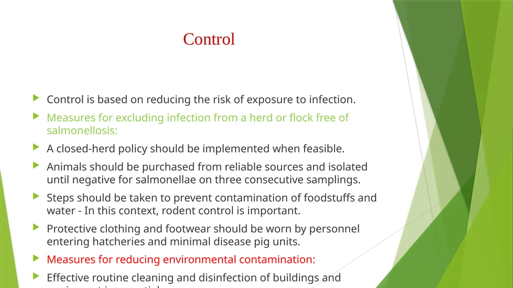
slide13. Control Control is based on reducing the risk of exposure to infection.
Measures for excluding infection from a herd or flock free of salmonellosis:
A closed-herd policy should be implemented when feasible.
Animals should be purchased from reliable sources and isolated until negative for salmonellae on three consecutive samplings.
Steps should be taken to prevent contamination of foodstuffs and water - In this context, rodent control is important.
Protective clothing and footwear should be worn by personnel entering hatcheries and minimal disease pig units.
Measures for reducing environmental contamination:
Effective routine cleaning and disinfection of buildings and equipment is essential.
Overstocking and overcrowding should be avoided.
Slurry should be spread on arable land where possible.<br>
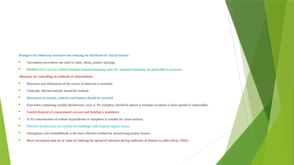
slide14. Strategies for enhancing resistance and reducing the likelihood of clinical disease:
Vaccination procedures are used in cattle, sheep, poultry and pigs.
Modified live vaccines which stimulate humoral immunity and cell- mediated immunity are preferabIe to bacterins.
Measures for controlIing an outbreak of salmonellosis:
Detection and elimination of the source of infection is essential.
Clinically affected animals should be isolated.
Movement of animals, vehicles and humans should be curtailed.
Foot baths containing suitable disinfectant, such as 3% iodophor, should be placed at strategic locations to limit spread of salmonellae.
Careful disposal of contaminated carcases and bedding is mandatory.
A 3% concentration of sodium hypochlorite or iodophors is suitable for clean surfaces.
Phenolic disinfectants are suitable for buildings with residual organic matter.
Fumigation with formaldehyde is the most effective method for disinfecting poultry houses.
Herd vaccination may be of value for limiting the spread of infection during outbreaks of disease in cattle (Wray, 1991).<br>
slide15. Thank You<br>
Assistant Professor Department of Veterinary Medicine
Bihar Veterinary College, Patna
(Bihar Animal Sciences University, Patna)<br>
slide2. Etiology Salmonella, a rod-shaped gram-negative bacterium is the causative agent of salmonellosis.
Salmonellosis in warm-blooded vertebrates is in most cases associated with serovars of Salmonella enterica.
The most common type of infection is the carrier state.
Clinical disease is characterized by two major syndromes: a systemic septicemia (also termed as typhoid) and an enteritis.
Other less common clinical presentations include abortion, arthritis, respiratory disease, necrosis of extremities, and meningitis. Salmonella spp.
Only a few serotypes produce clinical salmonellosis in healthy animals and typically have a narrow range of host species, a phenomenon termed serovar-host specificity.
Salmonella enterica serovar Typhi (S Typhi) and S Paratyphi produce typhoid in people, S Gallinarum produces a similar disease in poultry, S Abortusovis in sheep, S Choleraesuis in pigs, S Dublin in cattle, etc.<br>
slide3. Young calves, piglets, lambs, and foals may develop both the enteritis and septicemic form.
cattle—S Typhimurium, S Dublin, and S Newport;
sheep and goats—S Typhimurium, S Dublin, S Abortusovis, S Anatum, and S Montevideo;
pigs—S Typhimurium and S Choleraesuis;
horses—S Typhimurium, S Anatum, S Newport, S Enteritidis, and Salmonella serovar IIIa 18:z4:z23; and
poultry—S Enteritidis, S Typhimurium, S Gallinarum, and S Pullorum.<br>
slide4. Transmission Human:
People are often infected when they eat contaminated foods of animal origin such as meat or eggs.
They can also be infected by ingesting organisms in animal feces, either directly or in contaminated food or water.
Directly transmitted human infections are most often acquired from the feces of reptiles, chicks and ducklings. Livestock, dogs, cats, adult poultry and cage birds can also be involved.
Animals:
Salmonella spp. are mainly transmitted by the fecal-oral route.
Vertical transmission occurs in birds, with contamination of the vitelline membrane, albumen and the yolk of eggs.
Salmonella spp. can also be transmitted in utero in mammals.
Animals may also become infected from contaminated feed (including pastures), drinking water, or close contact with infected animal (including humans).
Birds and rodents can spread Salmonella to livestock.
Carnivores are also infected through meat, eggs, and other animal products that are not thoroughly cooked.<br>
slide5. Mechanisms of Pathogenicity
Bacterial products involved in virulence:
Salmonellae owe their pathogenicity largely to their ability to invade tissue and to survive within macrophages.
The Vi antigen is a capsule that affords salmonellae some protection from phagocytosis.
Once phagocytosed, Salmonella inhibits generation of oxidative free radicals and intraphagosomal killing.
Additionally, salmonellae have endotoxic lipopolysaccharide, which is responsible for septic shock in patients with bacteriemia.
Salmonellae that cause enteritis produce at least two enterotoxins that are responsible for many of the clinical signs of enteritis.
The first of these is a small (25-30kD) protein that binds to GM1 gangliosides and cause hypersecretion of fluids and electrolytes by elevating levels of c-AMP.
It appears that both protein kinase C and prostaglandin E2 are involved in this process.
The second enterotoxin is larger (about 100 kD) and is unrelated in structure and mechanism of activity to the first enterotoxin
Salmonella strains that produce enterotoxins have been reported to invade the intestinal wall more effectively and to be more virulent than their non-toxigenic counterparts.<br>
slide6. The Salmonella infection cycle.
• Intestinal infection with salmonellae can follow one of two infection cycle.
One cycle causes enteritis, other causes typhoid
a. Enteritis
Most serotypes cause enteritis, an infection that is limited to the terminal ileum.
The salmonellae invade the intestinal wall and produce enterotoxins that cause nausea, vomiting and diarrhea.
b. Enteric fever (Typhoid): • Two serotypes Typhi and Paratyphi can cause typhoid.
• The salmonella invade the wall of the terminal ileum and than spread to the intestinal lymphatics, where they are phagocytosed by PMNs and macrophages.
• Salmonella phagocytosed by PMNs are killed, but those phagocytosed by macrophages survive and multiply within phagocytic vacuoles.
• Wandering macrophages that contain salmonellae act as “taxi/cabs” that deliver salmonellae to various reticuloendothelial tissues.
• Infected macrophages are eventually destroyed and salmonellae released from lysed macrophages cause septicemia.<br>
slide7. Clinical manifestation Salmonellae often localize in the mucosae of the ileum, caecum and colon, and in the mesenteric lymph nodes of infected animals.
Although most organisms are cleared from the tissues by host defense mechanisms, subclinical infection may persist with shedding of small numbers of salmonellae in the faeces.
Latent infections, in which salmonellae are present in the gall bladder but are not excreted, also occur.
Clinical disease may develop from subclinical and latent infections if affected animals are stressed.
Other factors which determine the clinical outcome of infection include the number of salmonellae ingested, the virulence of the infecting serotype or strain and the susceptibility of the host.
Host susceptibility may be related to immunological status, genetic make- up or age.
Young and debilitated or aged animaIs are particularly susceptible and may develop the septicemic form of the disease.
In most animal species, both enteric and septicemic forms of salmonellosis are recorded.
A number of serotypes have been associated with abortion in farm animals, often without other obvious clinical signs in dams.<br>
slide8. Enteric Salmonellosis
Enterocolitis caused by salmonella organisms can affect most species of farm animals, irrespective of age.
Acute disease is characterized by fever, depression, anorexia and profuse foul-smelling diarrhoea often containing blood, mucus and epithelial casts.
Dehydration and weight loss follow and pregnant animals may abort.
Severely affected young animals become recumbent and may die within a few days of acquiring infection.
On farms with endemic salmonellosis, the milder clinical signs often observed may be attributed to the influence of acquired immunity.
Chronic enterocolitis can follow acute salmonellosis in pigs, cattle and horses.
Intermittent fever, soft faeces and gradual weight loss, leading to emaciation, are common features of this condition.<br>
slide9. Septicaemic Salmonellosis
The septicaemic form can occur in all age groups but is most common in calves, in neonatal foals and in pigs less than four months of age.
Onset of clinical disease is sudden with high fever, depression and recumbency.
If treatment is delayed, many young animals with septicaemic salmonellosis die within 48 hours.
Surviving animals can develop persistent diarrhoea, arthritis, meningitis or pneumonia.
In pigs with septicaemic Salmonella Choleraesuis infection, there is a characteristic bluish discolouration of the ears and snout.
Intercurrent viral infections often predispose to severe clinical forms of the disease.
The close clinical and pathological relationships which have been recognized in animals infected with Salmonella Choleraesuis (hog- cholera bacillus) and classical swine fever virus, either jointly or separately, exemplify both the importance of intercurrent infections and the difficulty of clinically distinguishing the diseases caused by these agents.<br>
slide10. Salmonellosis in poultry
Salmonella Pullorum, Salmonella Gallinarum and Salmonella Enteritidis can infect the ovaries of hens and be transmitted through eggs.
Pullorum disease or bacillary white diarrhoea (Salmonella Pullorum):
Infects young chicks and turkey poults up to 2 to 3 weeks of age.
The mortality rate is high and affected birds huddle under a heat source and are anorexic, depressed and have whitish faecal pasting around their vents.
Characteristic lesions includes whitish nodes throughout the lungs and focal necrosis of liver and spleen.
Fowl typhoid (Salmonella Gallinarum):
Can produce lesions in young chicks and poults similar to those of pullorum disease, but it is also a serious concern in growing and adult poultry
However, in countries where fowl typhoid is endemic, a septicaemic disease of adult birds occurs, often resulting in sudden deaths.
Characteristic findings include an enlarged, friable, bile-stained liver and enlarged spleen.
Paratyphoid is a name given to infections of poultry by non-host-adapted salmonellae such as Salmonella Enteritidis and Salmonella Typhimurium. These infections are often subclinical in laying birds.<br>
slide11. Diagnosis A history of previous outbreaks of the disease may suggest salmonellosis.
At postmortem, enterocolitis with blood-stained luminal contents and enlarged mesenteric lymph nodes are commonly observed.
Specimens for submission should include faeces and blood from live animals.
Intestinal contents and samples from tissue lesions should be submitted from dead animals and abomasal contents from aborted foetuses.
Isolation of salmonellae from blood or parenchymatous organs is deemed to be confirmatory for septicaemic sallmonellosis.
Serological tests such as ELISA and agglutination techniques are of greatest value when used on a herd or flock basis.
A rising antibody titre using paired serum samples is indicative of active infection.
DNA probes can be used to screen large numbers of faecal samples for salmonellae (Maddox and Fales, 1991).
Salmonella isolates then should be sent to a central or reference laboratory for more comprehensive serologic testing and<br>
slide12. Treatment Antibiotic therapy should be based on results of susceptibility testing because R-plasmids coding for multiple resistance are comparatively common in salmonellae.
Oral antimicrobial therapy should be used judiciously for treating enteric salmonellosis because it may disturb the normal intestinal flora, extend the duration of salmonella excretion and increase the probability of drug resistance developing.
In the septicaemic form of the disease, intravenous antibiotic therapy must be used.
Fluid and electrolyte replacement therapy is required to counteract dehydration and shock.<br>
slide13. Control Control is based on reducing the risk of exposure to infection.
Measures for excluding infection from a herd or flock free of salmonellosis:
A closed-herd policy should be implemented when feasible.
Animals should be purchased from reliable sources and isolated until negative for salmonellae on three consecutive samplings.
Steps should be taken to prevent contamination of foodstuffs and water - In this context, rodent control is important.
Protective clothing and footwear should be worn by personnel entering hatcheries and minimal disease pig units.
Measures for reducing environmental contamination:
Effective routine cleaning and disinfection of buildings and equipment is essential.
Overstocking and overcrowding should be avoided.
Slurry should be spread on arable land where possible.<br>
slide14. Strategies for enhancing resistance and reducing the likelihood of clinical disease:
Vaccination procedures are used in cattle, sheep, poultry and pigs.
Modified live vaccines which stimulate humoral immunity and cell- mediated immunity are preferabIe to bacterins.
Measures for controlIing an outbreak of salmonellosis:
Detection and elimination of the source of infection is essential.
Clinically affected animals should be isolated.
Movement of animals, vehicles and humans should be curtailed.
Foot baths containing suitable disinfectant, such as 3% iodophor, should be placed at strategic locations to limit spread of salmonellae.
Careful disposal of contaminated carcases and bedding is mandatory.
A 3% concentration of sodium hypochlorite or iodophors is suitable for clean surfaces.
Phenolic disinfectants are suitable for buildings with residual organic matter.
Fumigation with formaldehyde is the most effective method for disinfecting poultry houses.
Herd vaccination may be of value for limiting the spread of infection during outbreaks of disease in cattle (Wray, 1991).<br>
slide15. Thank You<br>