
SCANNING ELECTRON MICROSCOPY, TRANSMISSION
AM
Published · 14 slides · 0 views
1 / 1

Description
SCANNING ELECTRON MICROSCOPY, TRANSMISSION ELECTRON MICROSCOPY AND ATOMIC FORCE MICROSCOPE Dr. B. Kavitha Assistant Professor PG and Research Department of Chemistry C.P.A. College, Bodinayakanur SCANNING ELECTRON MICROSCOPY (SEM) Principle
Related Topics
Share
Embed code
Download this presentation From Below
"SCANNING ELECTRON MICROSCOPY, TRANSMISSION" is the property of its rightful owner. Permission is granted to download and print the materials on this website for personal, non-commercial use only, and to display it on your personal computer provided you do not modify the materials and that you retain all copyright notices contained in the materials. By downloading content from our website, you accept the terms of this agreement.
Presentation Transcript
01
SCANNING ELECTRON MICROSCOPY, TRANSMISSION ELECTRON MICROSCOPY AND ATOMIC FORCE MICROSCOPE Dr. B. Kavitha
Assistant Professor
PG and Research Department of Chemistry
C.P.A. College, Bodinayakanur<br>
Assistant Professor
PG and Research Department of Chemistry
C.P.A. College, Bodinayakanur<br>
02
SCANNING ELECTRON MICROSCOPY (SEM) Principle
In this technique, an electron beam is focused onto sample surface kept in a vacuum by electro-magnetic lenses (since electron possesses dual nature with properties of both particle and wave, hence an electron beam can be focused or condensed like an ordinary light). The beam is then rastered or scanned over the surface of the sample. The scattered electron from the sample is then fed to the detector and then to a cathode ray tube through an amplifier, where the images are formed, which gives the information of the sample Instrumentation
It comprises of a heated filament as a source of electron beam, condenser lenses, aperture, evacuated chamber for placing the sample, electron detector, amplifier, CRT with image forming electronics, etc.
The SEM is an instrument that produces a largely magnified image by using electrons instead of light to form an image. A beam of electrons is produced at the top of the microscope by an electron gun. The electron beam follows a vertical path through the microscope, which is held within a vacuum chamber.<br>
03
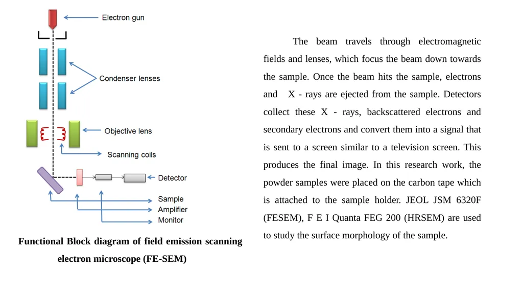
Functional Block diagram of field emission scanning electron microscope (FE-SEM) The beam travels through electromagnetic fields and lenses, which focus the beam down towards the sample. Once the beam hits the sample, electrons and X - rays are ejected from the sample. Detectors collect these X - rays, backscattered electrons and secondary electrons and convert them into a signal that is sent to a screen similar to a television screen. This produces the final image. In this research work, the powder samples were placed on the carbon tape which is attached to the sample holder. JEOL JSM 6320F (FESEM), F E I Quanta FEG 200 (HRSEM) are used to study the surface morphology of the sample.<br>
04
Applications Scanning electron microscopy has been applied to the surface studies of metals, ceramics, polymers, composites and biological materials for both topography as well as compositional analysis.
An extension of this technique is Electron Probe Micro Analysis (EPMA), where the emission of X-rays, from the sample surface, is studied upon exposure to a beam of high energy electrons.
Depending on the type of detectors used this method is classified in to two as: Energy Dispersive Spectrometry (EDS) and Wavelength Dispersive Spectrometry (WDS).
This technique is used extensively in the analysis of metallic and ceramic inclusions, inclusions in polymeric materials and diffusion profiles in electronic components.<br>
05
TRANSMISSION ELECTRON MICROSCOPY (TEM) Principle
In this technique, a beam of high-energy electrons (typically 100 – 400 keV) is collimated by magnetic lenses and allowed to pass through a specimen under high vacuum.
The transmitted beam and a number of diffracted beams can form a resultant diffraction pattern, which is imaged on a fluorescent screen kept below the specimen.
The diffraction pattern gives the information regarding lattice spacing and symmetry of the structure under consideration.
Alternatively, either the transmitted beam or the diffracted beams can be made to form a magnified image of the sample on the viewing screen as bright-and dark field imaging modes respectively. This gives information about the size and shape of the micro-structural constituents of the material.
High - resolution image contains information about the atomic structure of the material. This can be obtained by recombining the transmitted beam and diffracted beams together<br>
06
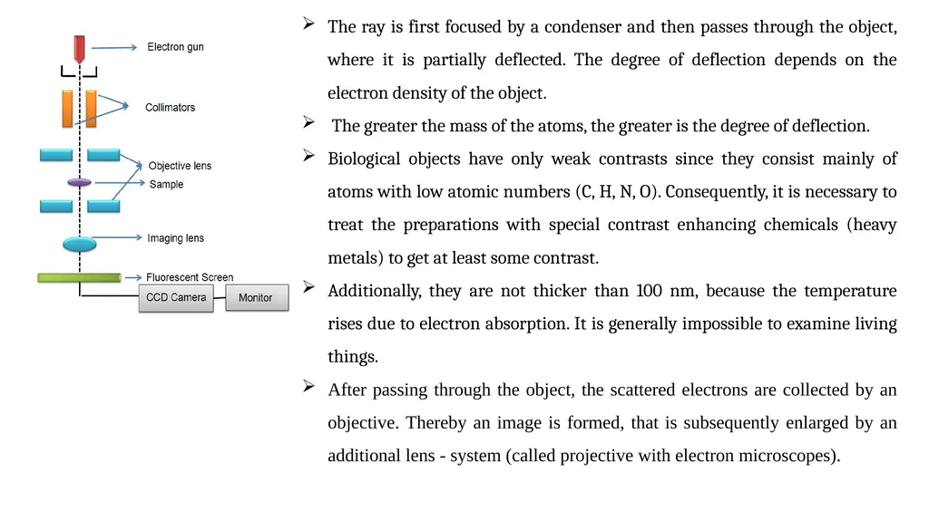
Instrumentation It comprises of a tungsten filament or LaB6 or a field emission gun as source of electron beam, objective lens, imaging lens, CCD camera, monitor, etc.
The ray of electrons is produced by a pin-shaped cathode heated up by current. The electrons are vacuumed up by a high voltage at the anode.
The acceleration voltage is between 50 and 150 kV. The higher it is, the shorter are the electron waves and the higher is the power of resolution, but this factor is hardly ever limiting.
The power of resolution of electron microscopy is usually restrained by the quality of the lens-systems and especially by the technique with which the preparation has been made. Modern gadgets have powers of resolution that range from 0.2 - 0.3 nm.
The accelerated ray of electrons passes a drill-hole at the bottom of the anode. Its following way is analogous to that of a ray of light in a light microscope. The lens-systems consist of electronic coils generating an electromagnetic field.<br>
07
The ray is first focused by a condenser and then passes through the object, where it is partially deflected. The degree of deflection depends on the electron density of the object.
The greater the mass of the atoms, the greater is the degree of deflection.
Biological objects have only weak contrasts since they consist mainly of atoms with low atomic numbers (C, H, N, O). Consequently, it is necessary to treat the preparations with special contrast enhancing chemicals (heavy metals) to get at least some contrast.
Additionally, they are not thicker than 100 nm, because the temperature rises due to electron absorption. It is generally impossible to examine living things.
After passing through the object, the scattered electrons are collected by an objective. Thereby an image is formed, that is subsequently enlarged by an additional lens - system (called projective with electron microscopes).<br>
08
The formed image is made visible on a fluorescent screen or it is documented on photographic material.
Photos taken with electron microscopes are always black and white.
The degree of darkness corresponds to the electron density (differences in atom masses) of the candled preparation. Transmission electron microscopy is used to study the local structures, morphology, dispersion of multi - component polymers, cross sections and crystallization of metallic alloys semiconductors, microstructure of composite materials, etc.
The instrument can be extended to include other detectors like Energy Dispersive Spectrometer (EDS) or Energy Loss Spectrometer (ELS) to study about the local chemistry of the material similar to SEM technique Applications<br>
09
Atomic Force Microscope (AFM) AFM is used to measure surface morphology on atomic scale and obtain material properties.
In 1986 Binnig, Quate and Gerber invented the first Atomic microscope (AFM).
The atomic force microscope is ideal for quantitatively measuring the nanometer scale surface roughness and for visualizing the surface nano-texture on many types of material surfaces including polymer nanocomposites and nanofinished or nanocoated textiles.
It can be operated in air or water.<br>
10
Basic Principle Atomic force microscopy consists of a microscale cantilever with a sharp tip (probe tip radius is in the order of 10 nm) at its end. This tip is used to scan the specimen surface. The tip is brought into proximity of a sample surface. That makes the forces between the tip and the sample leading to a deflection of the cantilever according to Hooke’s law.
F = - KZ
Where, F – Force
K – Stiffness of the lever
Z – Distance of the lever Tips and cantilevers are typically micro fabricated from Silicon (Si) or Silicon nitride (Si3N4). A micro-fabricated cantilever with a sharp tip is deflected by features on a sample surface, much like in a phonograph but on a much smaller scale.
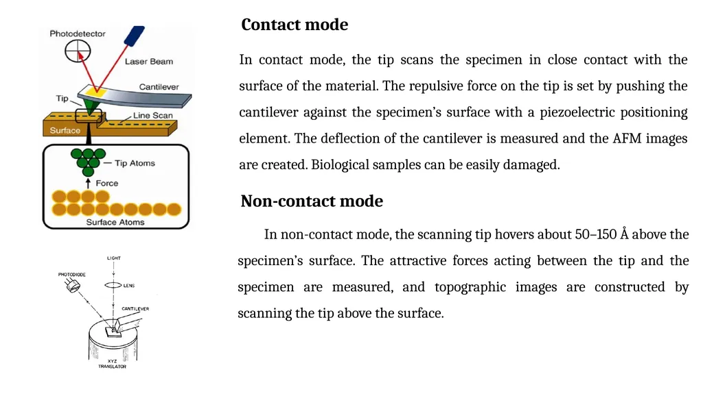
A laser beam reflects off the backside of the cantilever onto a set of photodetectors, allowing the deflection to be measured and assembled into an image of the surface. During scanning, a particular operating parameter is maintained at a constant level, and images are generated through a feedback loop between the optical detection system and the piezoelectric scanners.
There are three scan modes for AFM, namely contact mode, non contact mode and tapping mode.<br>
11
Contact mode In contact mode, the tip scans the specimen in close contact with the surface of the material. The repulsive force on the tip is set by pushing the cantilever against the specimen’s surface with a piezoelectric positioning element. The deflection of the cantilever is measured and the AFM images are created. Biological samples can be easily damaged. Non-contact mode In non-contact mode, the scanning tip hovers about 50–150 Å above the specimen’s surface. The attractive forces acting between the tip and the specimen are measured, and topographic images are constructed by scanning the tip above the surface.<br>
12
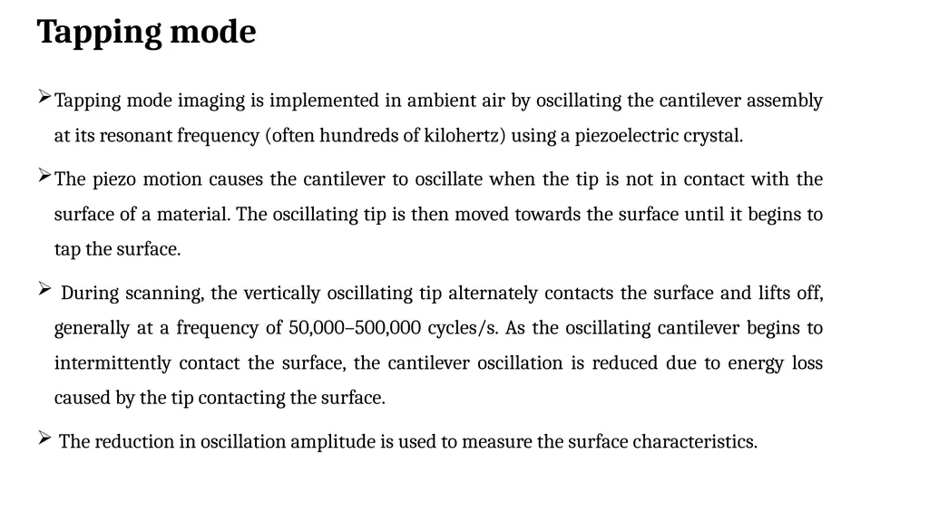
Tapping mode Tapping mode imaging is implemented in ambient air by oscillating the cantilever assembly at its resonant frequency (often hundreds of kilohertz) using a piezoelectric crystal.
The piezo motion causes the cantilever to oscillate when the tip is not in contact with the surface of a material. The oscillating tip is then moved towards the surface until it begins to tap the surface.
During scanning, the vertically oscillating tip alternately contacts the surface and lifts off, generally at a frequency of 50,000–500,000 cycles/s. As the oscillating cantilever begins to intermittently contact the surface, the cantilever oscillation is reduced due to energy loss caused by the tip contacting the surface.
The reduction in oscillation amplitude is used to measure the surface characteristics.<br>
13
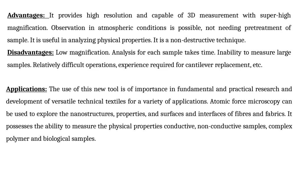
Advantages: It provides high resolution and capable of 3D measurement with super-high magnification. Observation in atmospheric conditions is possible, not needing pretreatment of sample. It is useful in analyzing physical properties. It is a non-destructive technique.
Disadvantages: Low magnification. Analysis for each sample takes time. Inability to measure large samples. Relatively difficult operations, experience required for cantilever replacement, etc. Applications: The use of this new tool is of importance in fundamental and practical research and development of versatile technical textiles for a variety of applications. Atomic force microscopy can be used to explore the nanostructures, properties, and surfaces and interfaces of fibres and fabrics. It possesses the ability to measure the physical properties conductive, non-conductive samples, complex polymer and biological samples.<br>
Disadvantages: Low magnification. Analysis for each sample takes time. Inability to measure large samples. Relatively difficult operations, experience required for cantilever replacement, etc. Applications: The use of this new tool is of importance in fundamental and practical research and development of versatile technical textiles for a variety of applications. Atomic force microscopy can be used to explore the nanostructures, properties, and surfaces and interfaces of fibres and fabrics. It possesses the ability to measure the physical properties conductive, non-conductive samples, complex polymer and biological samples.<br>
14
Thank You<br>