
Shoulder Radiography What to consider when

Description: Shoulder Radiography What to consider when ordering? Image selection should be based on a working diagnosis. Minimum of two orthogonal views. Example an AP view and Scapular Y views. Two views perpendicular to one another. Indications are
Related Topics
Download Presentation
"Shoulder Radiography What to consider when" is the property of its rightful owner. Permission is granted to download and print the materials on this website for personal, non-commercial use only, and to display it on your personal computer provided you do not modify the materials and that you retain all copyright notices contained in the materials. By downloading content from our website, you accept the terms of this agreement.
Presentation Transcript
slide1. Shoulder Radiography<br>
slide2. What to consider when ordering? Image selection should be based on a working diagnosis.
Minimum of two orthogonal views. Example an AP view and Scapular Y views. Two views perpendicular to one another.
Indications are trauma, TTP, restricted ROM, instability…<br>
slide3. Routine AP<br>
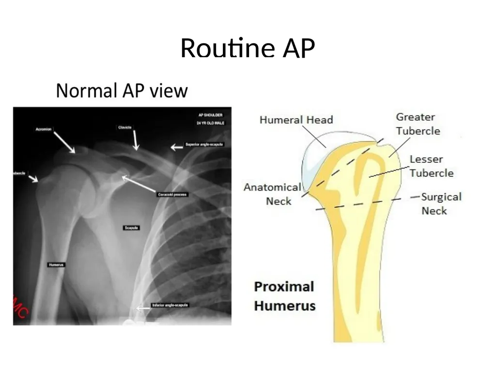
slide4. Routine AP Assess the GH joint, proximal humerus, entire clavicle, AC joint, scapula, and SC joint.
Positioned: GH center of image receptor. Affected arm is in neutral position by patient side.
Its orthogonal view is the lateral Y view.<br>
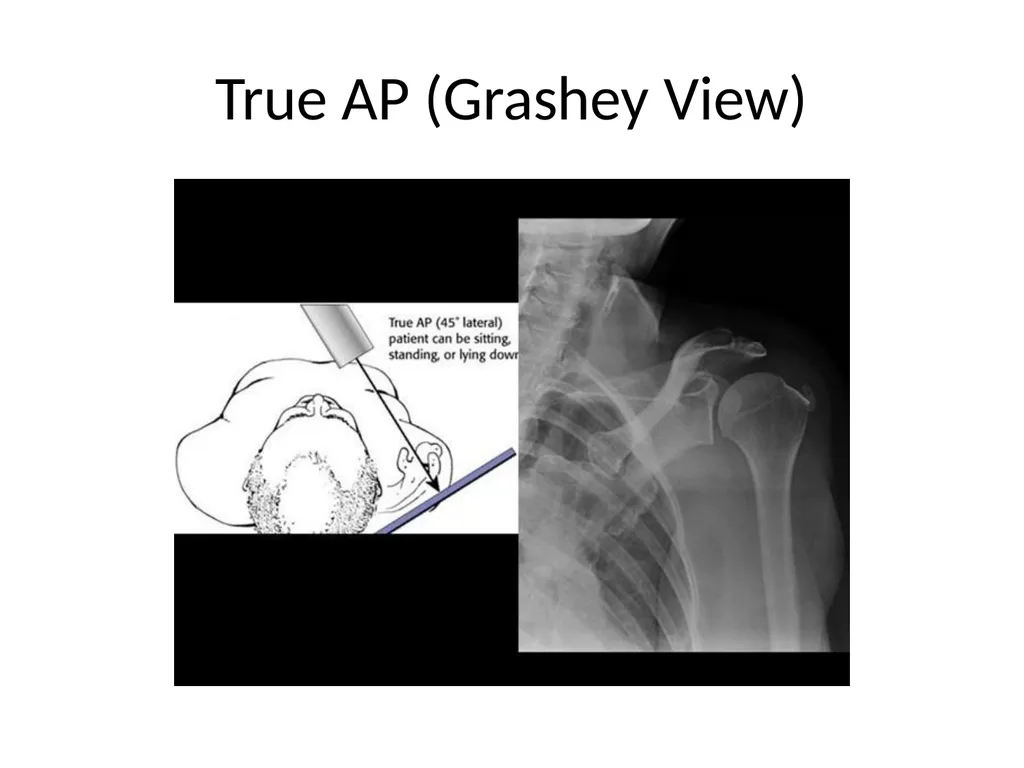
slide5. True AP (Grashey View)<br>
slide6. True AP (Grashey View) Assess GH joint, DJD, proximal migration of humerus… Part of complete trauma series.
Positioned: Patient back against image receptor. GH joint at center of image receptor. Rotate patient 45 degrees toward affected side. Back of hand against thigh (internally rotated).<br>
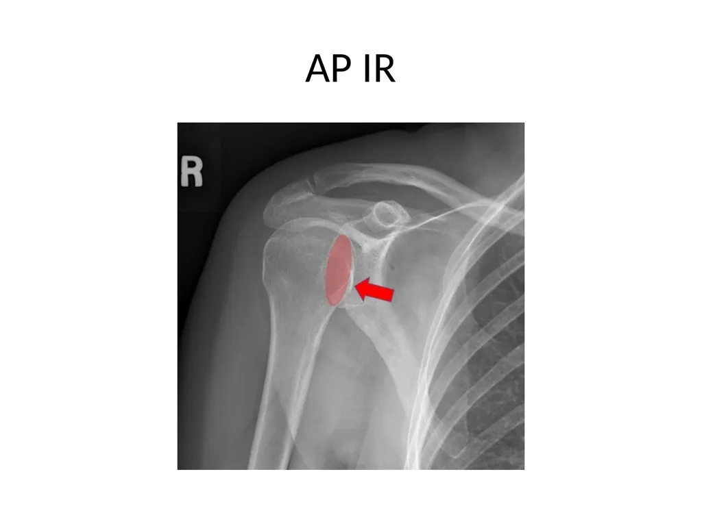
slide7. AP IR<br>
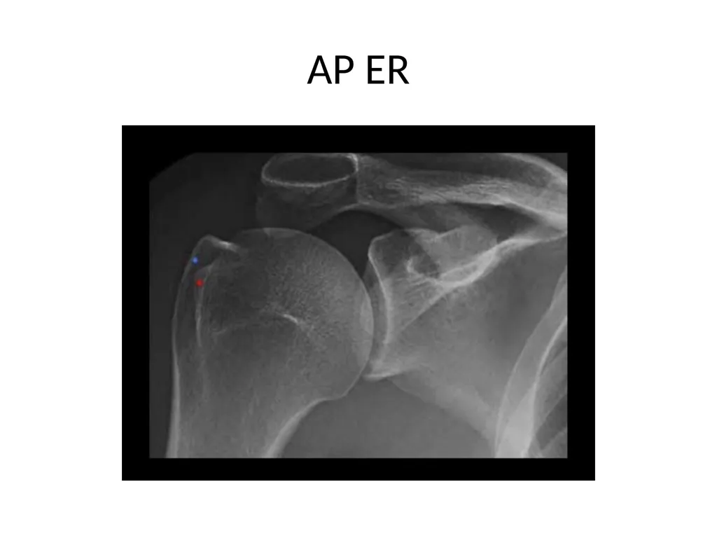
slide8. AP ER<br>
slide9. AP IR/ER Purpose – assess the humeral head for Hill – Sachs lesions.
Positioning: Patients back against image receptor. GH joint center of image receptor.
IR: posterior aspect of hand will be positioned against the hip.
ER: hand will be supinated (facing beam).<br>
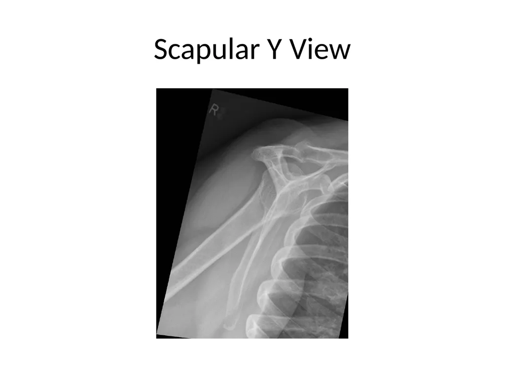
slide10. Scapular Y View<br>
slide11. Scapular Y View Orthogonal view of the AP shoulder view. Profile view of the scapula.
Purpose – assess acromion, coracoid, humeral head relation to glenoid… Part of a complete trauma series.
Positioning: facing the upright detector. Rotate so anterior shoulder is touching detector. Affected hand on patient abdomen (arm flexed). Scapula should be end-on to detector.<br>
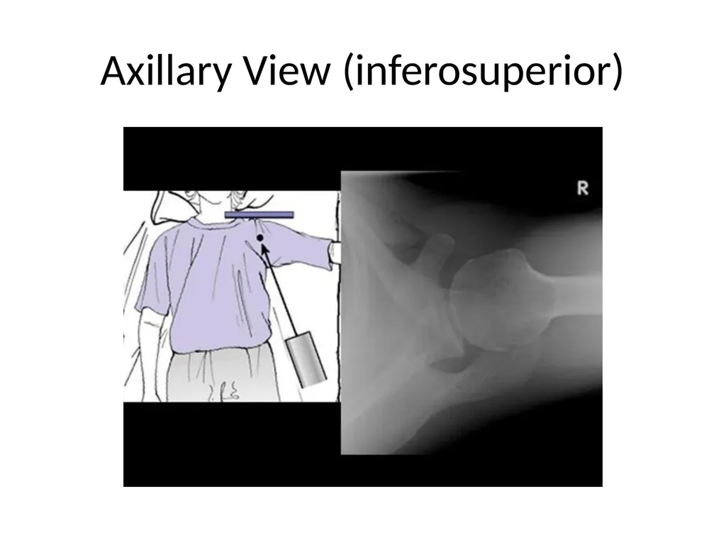
slide12. Axillary View (inferosuperior)<br>
slide13. Axillary View (inferosuperior) Purpose – assess position of humeral head in relation to glenoid. Part of a complete trauma series. Orthogonal view of the AP shoulder.
Positioning: patient supine. Receptor on superior shoulder. Patient has to be able to abduct arm to allow xray tube between arm and body. Arm then externally rotated.<br>
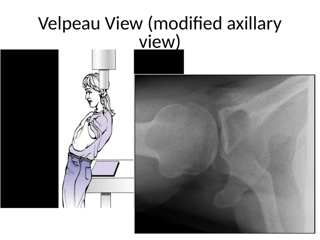
slide14. Velpeau View (modified axillary view)<br>
slide15. Velpeau View Purpose – assess the GH joint. Done in lieu of axillary view for patient who is unable to abduct arm. Orthogonal view to the AP shoulder.
Positioning: Patient sitting. Affected arm beside body. Beam directed superoinferior to receptor.<br>
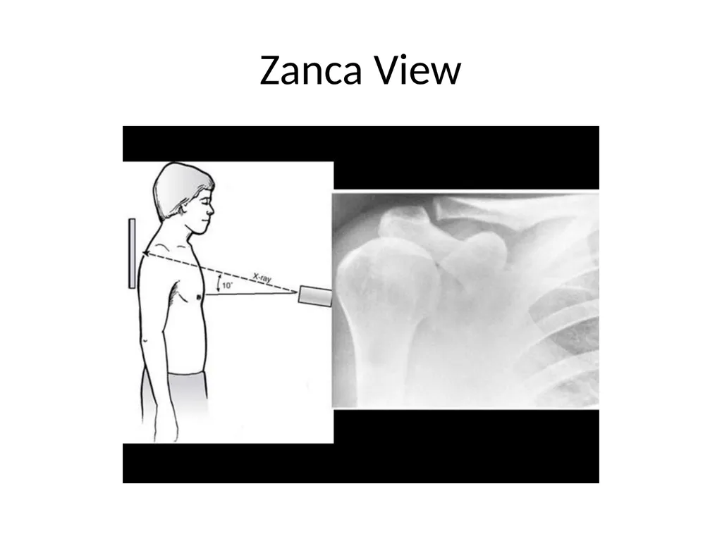
slide16. Zanca View<br>
slide17. Zanca View Purpose – assess distal clavicle and AC joint for separation, distal clavicle fracture, DJD and osteolysis.
Positioning: Back against receptor. AC joint center of image receptor. Arm neutral against body.<br>
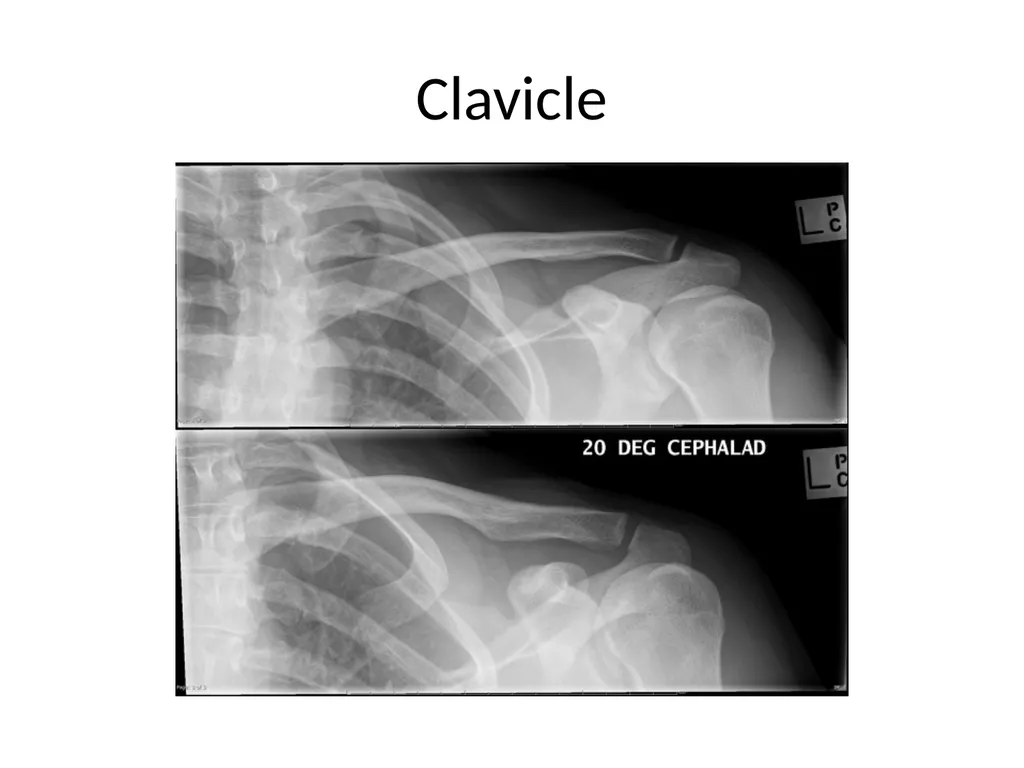
slide18. Clavicle<br>
slide19. Clavicle Purpose – assess clavicle and AC for fracture and AC joint separation.
Positioning: Back against receptor. Clavicle at center of image receptor. Arm neutral against body.<br>
slide20. MRI Useful tool to assess ligaments, tendons, muscle, fibrocartilage, bone marrow…<br>
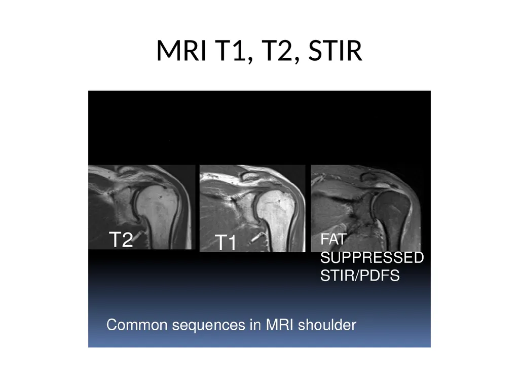
slide21. MRI T1, T2, STIR<br>
slide22. MRI T1, T2, STIR Weighted Sequences:
T1 - Fat is brighter. Bone, fluid, tendon, fibrocartilage are dark. Purpose – assess bone and soft tissue (rotator cuff and capsule).
T2 Fat is suppressed. Fluid and marrow are brighter (revealing inflammation and edema). Bone, ligaments, muscle, fibrocartilage are darker. Purpose – assess bone, soft tissue, muscle atrophy and fatty degeneration.
Short TI Inversion Recovery (STIR) Fat suppressed. Purpose is to distinguish edema from fatty infiltrate, useful in distinguishing acute vs chronic RC tear and identifying occult fractures (bone marrow edema).<br>
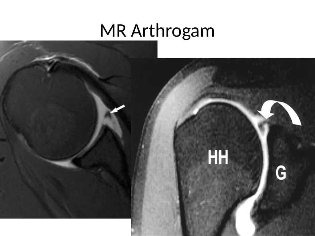
slide23. MR Arthrogam<br>
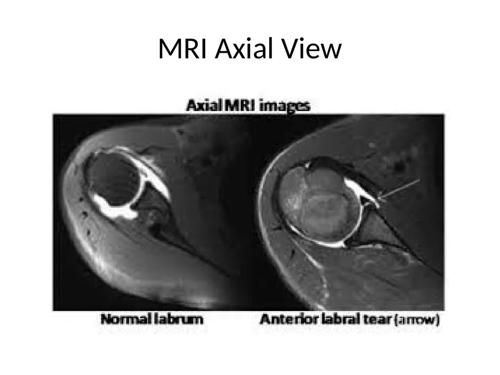
slide24. MRI Axial View<br>
slide25. MR Arthrogam Purpose is to better visualize structures like the labrum and glenohumeral ligaments.
Gadolinium contrast is injected into the joint.
MR Arthrogram may not be needed. Most labral injuries can be seen on MRI.<br>
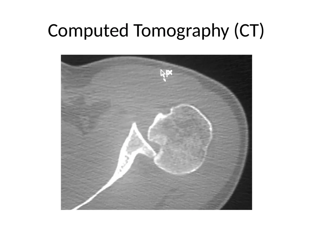
slide26. Computed Tomography (CT)<br>
slide27. CT Provides better bone detail than MRI.
Usually easier and lower cost to obtain.
Down side is added xray exposure for patient.
Useful in surgical planning.<br>
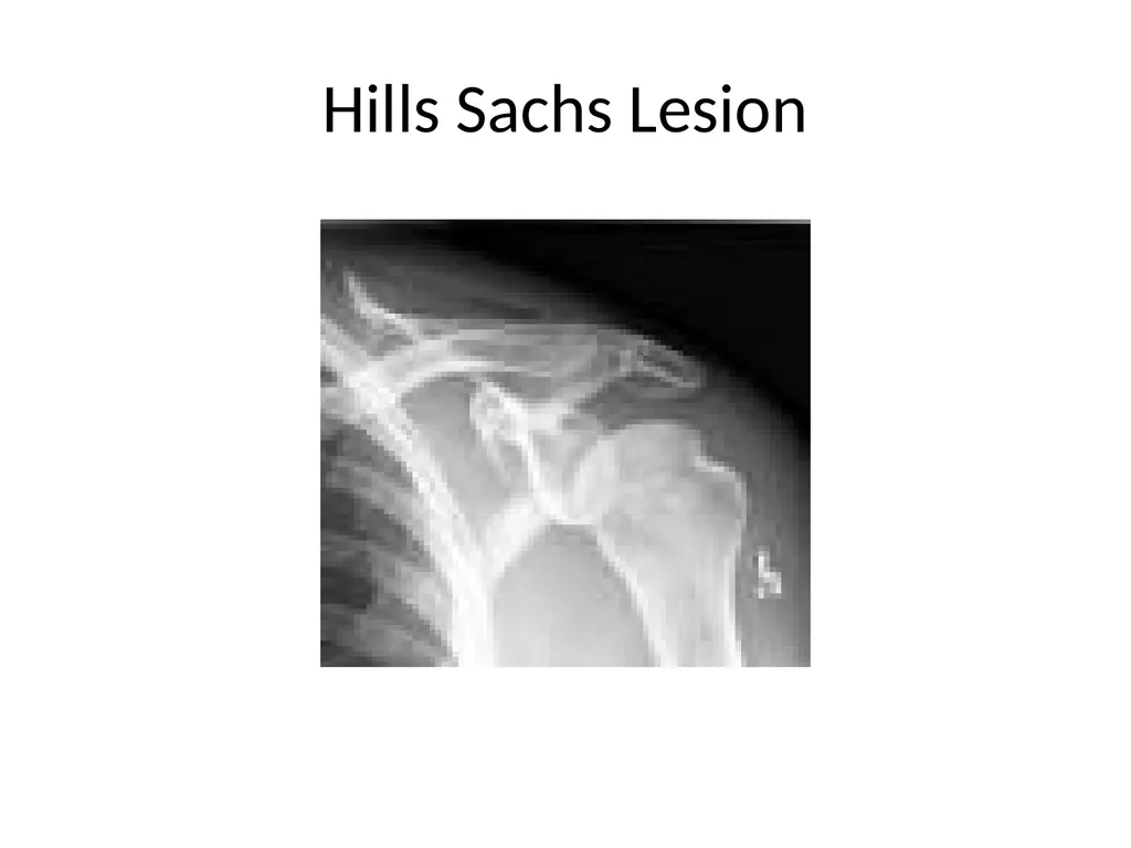
slide28. Hills Sachs Lesion<br>
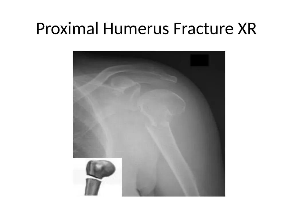
slide29. Proximal Humerus Fracture XR<br>
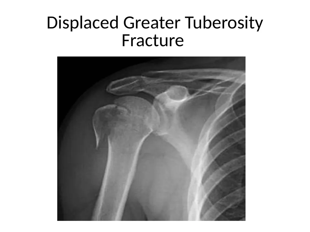
slide30. Displaced Greater Tuberosity Fracture<br>
slide31. Glenohumeral Arthritis XR<br>
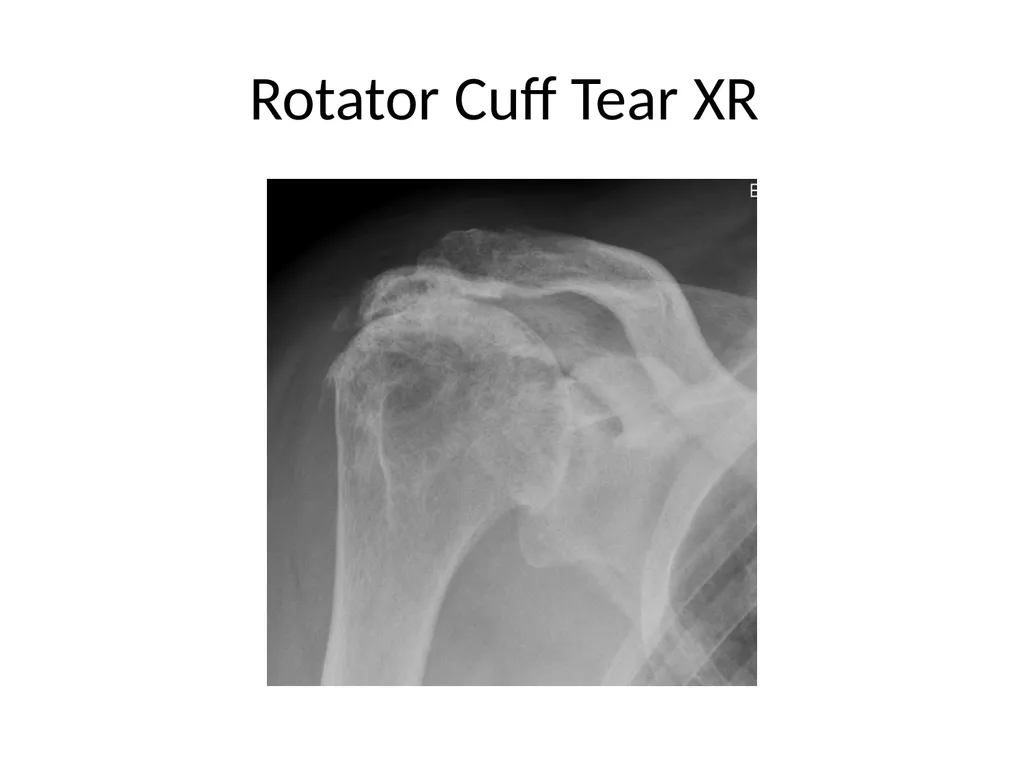
slide32. Rotator Cuff Tear XR<br>
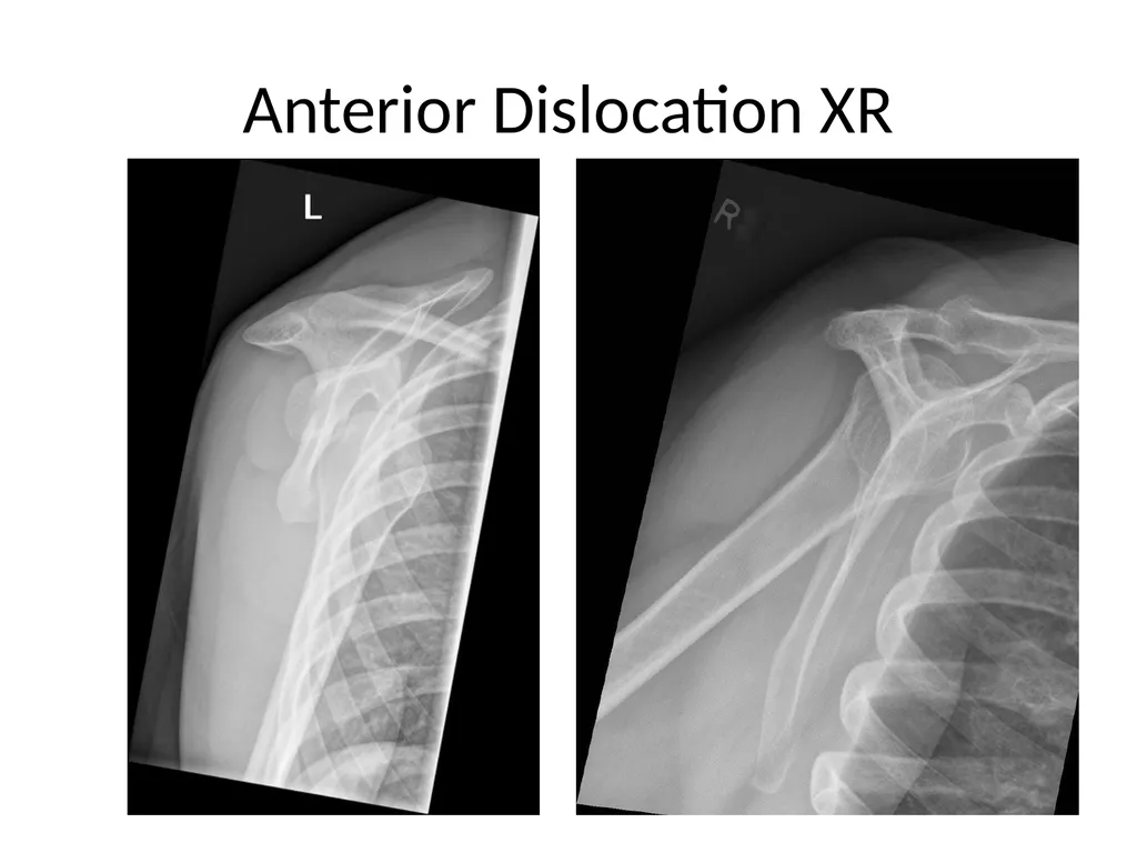
slide33. Anterior Dislocation XR<br>
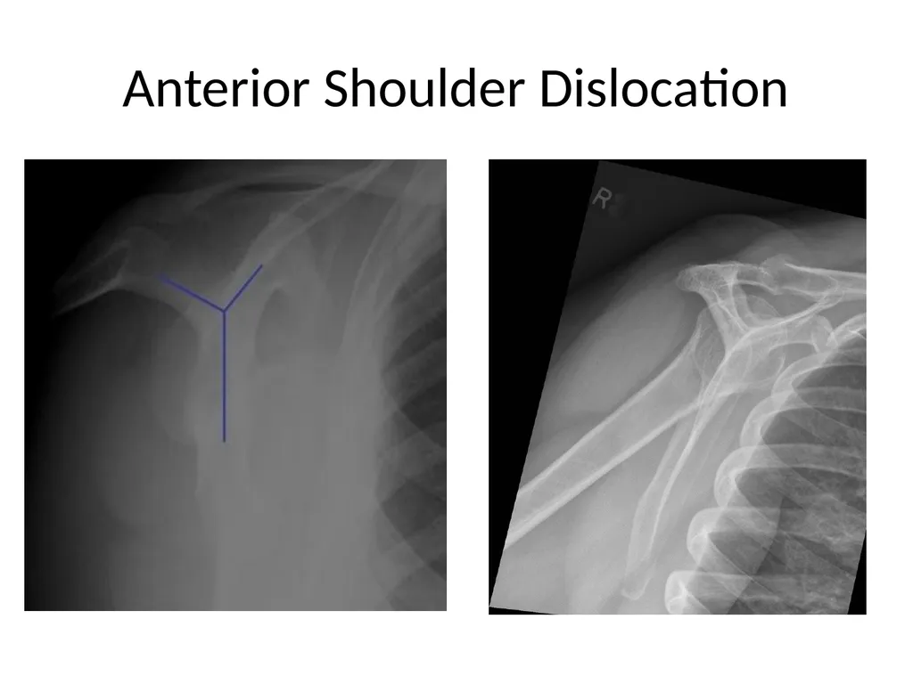
slide34. Anterior Shoulder Dislocation<br>
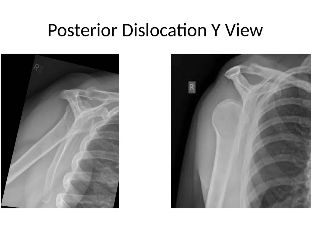
slide35. Posterior Dislocation Y View<br>
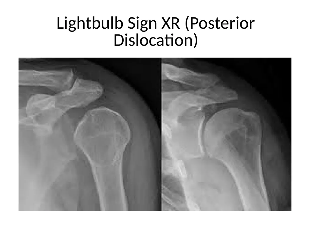
slide36. Lightbulb Sign XR (Posterior Dislocation)<br>
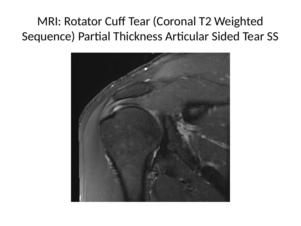
slide37. MRI: Rotator Cuff Tear (Coronal T2 Weighted Sequence) Partial Thickness Articular Sided Tear SS<br>
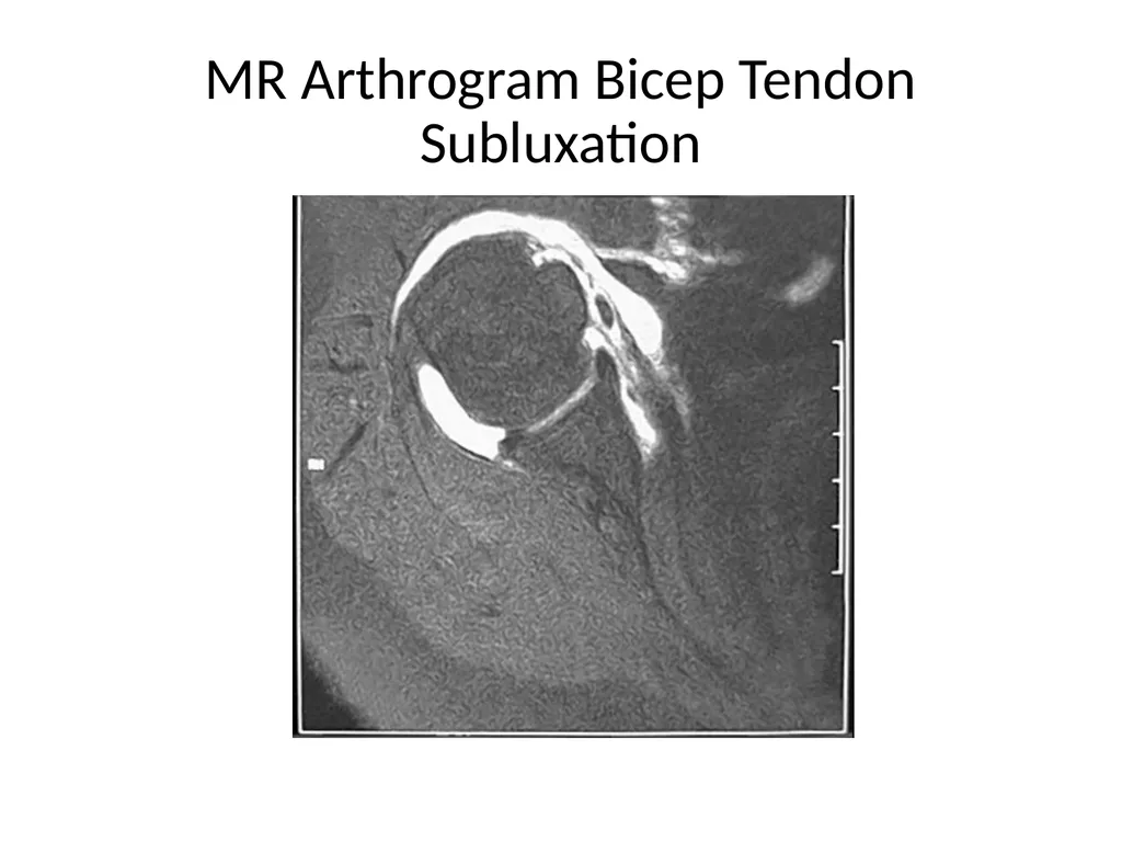
slide38. MR Arthrogram Bicep Tendon Subluxation<br>
slide39. References Orthobullets. (2016) Shoulder Imaging. Retrieved from https://www.orthobullets.com/shoulder-and-elbow/3038/shoulder-imaging?expandLeftMenu=true
Slide Share. (2014) Common Sequences in MRI Shoulder. Retrieved from https://www.slideshare.net/drkrishnakiran/shoulder-slideshare
Radiopedia. (2005-2020) Retrieved from https://radiopedia.org/articles-ap-view-1?lang=us<br>
slide2. What to consider when ordering? Image selection should be based on a working diagnosis.
Minimum of two orthogonal views. Example an AP view and Scapular Y views. Two views perpendicular to one another.
Indications are trauma, TTP, restricted ROM, instability…<br>
slide3. Routine AP<br>
slide4. Routine AP Assess the GH joint, proximal humerus, entire clavicle, AC joint, scapula, and SC joint.
Positioned: GH center of image receptor. Affected arm is in neutral position by patient side.
Its orthogonal view is the lateral Y view.<br>
slide5. True AP (Grashey View)<br>
slide6. True AP (Grashey View) Assess GH joint, DJD, proximal migration of humerus… Part of complete trauma series.
Positioned: Patient back against image receptor. GH joint at center of image receptor. Rotate patient 45 degrees toward affected side. Back of hand against thigh (internally rotated).<br>
slide7. AP IR<br>
slide8. AP ER<br>
slide9. AP IR/ER Purpose – assess the humeral head for Hill – Sachs lesions.
Positioning: Patients back against image receptor. GH joint center of image receptor.
IR: posterior aspect of hand will be positioned against the hip.
ER: hand will be supinated (facing beam).<br>
slide10. Scapular Y View<br>
slide11. Scapular Y View Orthogonal view of the AP shoulder view. Profile view of the scapula.
Purpose – assess acromion, coracoid, humeral head relation to glenoid… Part of a complete trauma series.
Positioning: facing the upright detector. Rotate so anterior shoulder is touching detector. Affected hand on patient abdomen (arm flexed). Scapula should be end-on to detector.<br>
slide12. Axillary View (inferosuperior)<br>
slide13. Axillary View (inferosuperior) Purpose – assess position of humeral head in relation to glenoid. Part of a complete trauma series. Orthogonal view of the AP shoulder.
Positioning: patient supine. Receptor on superior shoulder. Patient has to be able to abduct arm to allow xray tube between arm and body. Arm then externally rotated.<br>
slide14. Velpeau View (modified axillary view)<br>
slide15. Velpeau View Purpose – assess the GH joint. Done in lieu of axillary view for patient who is unable to abduct arm. Orthogonal view to the AP shoulder.
Positioning: Patient sitting. Affected arm beside body. Beam directed superoinferior to receptor.<br>
slide16. Zanca View<br>
slide17. Zanca View Purpose – assess distal clavicle and AC joint for separation, distal clavicle fracture, DJD and osteolysis.
Positioning: Back against receptor. AC joint center of image receptor. Arm neutral against body.<br>
slide18. Clavicle<br>
slide19. Clavicle Purpose – assess clavicle and AC for fracture and AC joint separation.
Positioning: Back against receptor. Clavicle at center of image receptor. Arm neutral against body.<br>
slide20. MRI Useful tool to assess ligaments, tendons, muscle, fibrocartilage, bone marrow…<br>
slide21. MRI T1, T2, STIR<br>
slide22. MRI T1, T2, STIR Weighted Sequences:
T1 - Fat is brighter. Bone, fluid, tendon, fibrocartilage are dark. Purpose – assess bone and soft tissue (rotator cuff and capsule).
T2 Fat is suppressed. Fluid and marrow are brighter (revealing inflammation and edema). Bone, ligaments, muscle, fibrocartilage are darker. Purpose – assess bone, soft tissue, muscle atrophy and fatty degeneration.
Short TI Inversion Recovery (STIR) Fat suppressed. Purpose is to distinguish edema from fatty infiltrate, useful in distinguishing acute vs chronic RC tear and identifying occult fractures (bone marrow edema).<br>
slide23. MR Arthrogam<br>
slide24. MRI Axial View<br>
slide25. MR Arthrogam Purpose is to better visualize structures like the labrum and glenohumeral ligaments.
Gadolinium contrast is injected into the joint.
MR Arthrogram may not be needed. Most labral injuries can be seen on MRI.<br>
slide26. Computed Tomography (CT)<br>
slide27. CT Provides better bone detail than MRI.
Usually easier and lower cost to obtain.
Down side is added xray exposure for patient.
Useful in surgical planning.<br>
slide28. Hills Sachs Lesion<br>
slide29. Proximal Humerus Fracture XR<br>
slide30. Displaced Greater Tuberosity Fracture<br>
slide31. Glenohumeral Arthritis XR<br>
slide32. Rotator Cuff Tear XR<br>
slide33. Anterior Dislocation XR<br>
slide34. Anterior Shoulder Dislocation<br>
slide35. Posterior Dislocation Y View<br>
slide36. Lightbulb Sign XR (Posterior Dislocation)<br>
slide37. MRI: Rotator Cuff Tear (Coronal T2 Weighted Sequence) Partial Thickness Articular Sided Tear SS<br>
slide38. MR Arthrogram Bicep Tendon Subluxation<br>
slide39. References Orthobullets. (2016) Shoulder Imaging. Retrieved from https://www.orthobullets.com/shoulder-and-elbow/3038/shoulder-imaging?expandLeftMenu=true
Slide Share. (2014) Common Sequences in MRI Shoulder. Retrieved from https://www.slideshare.net/drkrishnakiran/shoulder-slideshare
Radiopedia. (2005-2020) Retrieved from https://radiopedia.org/articles-ap-view-1?lang=us<br>