
The Reproductive System and Human Genetics

Description: The Reproductive System and Human Genetics Abdullah A. Alrumayh BSc EMS, MSc Cardiovascular, Lecturer Department of Basic Sciences, PSCEMS http:fac.ksu.edu.saaalrumayh aalrumayhksu.edu.sa Learning Objectives List the essential and
Related Topics
Download Presentation
"The Reproductive System and Human Genetics" is the property of its rightful owner. Permission is granted to download and print the materials on this website for personal, non-commercial use only, and to display it on your personal computer provided you do not modify the materials and that you retain all copyright notices contained in the materials. By downloading content from our website, you accept the terms of this agreement.
Presentation Transcript
slide1. The Reproductive System and Human Genetics Abdullah A. Alrumayh
BSc EMS, MSc Cardiovascular, Lecturer
Department of Basic Sciences, PSCEMS
http://fac.ksu.edu.sa/aalrumayh
aalrumayh@ksu.edu.sa<br>
slide2. Learning Objectives List the essential and accessory reproductive organs of the male and female, describing the general function of each.
Name the hormones necessary for the formation of gametes.
Describe the difference between spermatogenesis and oogenesis.<br>
slide3. Learning Objectives Describe the three layers of the uterine wall.
Briefly describe the life cycle of an oocyte.
Describe the menstrual cycle in terms of changes in hormone levels and the condition of the endometrium.<br>
slide4. Learning Objectives Define the following terms: diploid, haploid, gametes, endometrium, genetic disease, homologous chromosomes, autosomes, sex chromosomes, genes, alleles, genotype, phenotype, homozygous, and heterozygous.
Identify and describe the structures that constitute the external genitalia in both sexes.
Name the parts of a sperm cell.
Beginning with fertilization, describe the major developmental changes during gestation.
Describe the structure and function of the placenta and umbilical cord.
State the length of an average gestation period.<br>
slide5. Learning Objectives Describe the stages of labor.
Describe the difference between fetal circulation/respiration and adult circulation/respiration.
Describe the major changes that take place in an infant at birth.
Discuss the difference between dominant and recessive traits.
List some important genetic diseases found in out-of-hospital care.
Explain how genes can cause disease.<br>
slide6. Introduction The only body system not essential to survival
Male and female reproductive systems are very different.
The reproductive system is linked with genetics.
Certain reproductive organs secrete hormones needed for development and maintenance of secondary sex characteristics and for reproduction.<br>
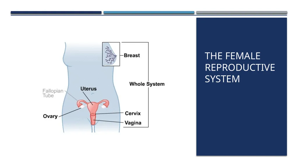
slide7. The Female Reproductive System<br>
slide8. Female reproductive system Oocytes – female sex cells (egg cells) produced and maintained by the female reproductive organs
The female reproductive organs:
Transport oocytes to the site of fertilization
Provide a nurturing environment for the developing fetus
Give birth to a child
Produce female sex hormones<br>
slide9. Produce female sex cells and sex hormones
Ovarian tissue consists of:
Inner medulla
Outer cortex
Suspended in place by:
Mesovarium
Suspensory ligament
Ovarian ligament Ovaries<br>
slide10. Ovaries Three main functions:
Produce oocytes
Immature female gametes (egg or ovum)
Secrete female sex hormones
Including estrogens and progestins
Secrete inhibin
Involved in the feedback control of pituitary follicle-stimulating hormone production<br>
slide11. Ovaries Oogenesis
Maturation process undergone by oocytes
Results in production of an ovum
Ovum
Female gamete or mature egg
During the reproductive years of a woman's life, the pituitary gland releases hormones at roughly monthly intervals that affect the ovaries<br>
slide12. Ovaries Hormones that stimulate one oocyte to undergo meiosis
Follicle-stimulating hormone (FSH)
Luteinizing hormone (LH)
Meiosis
Process of cell division that occurs during the formation of a mature ovum or spermatocyte
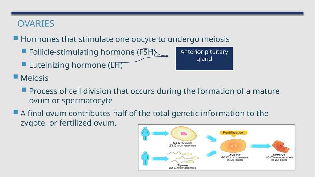
A final ovum contributes half of the total genetic information to the zygote, or fertilized ovum. Anterior pituitary gland<br>
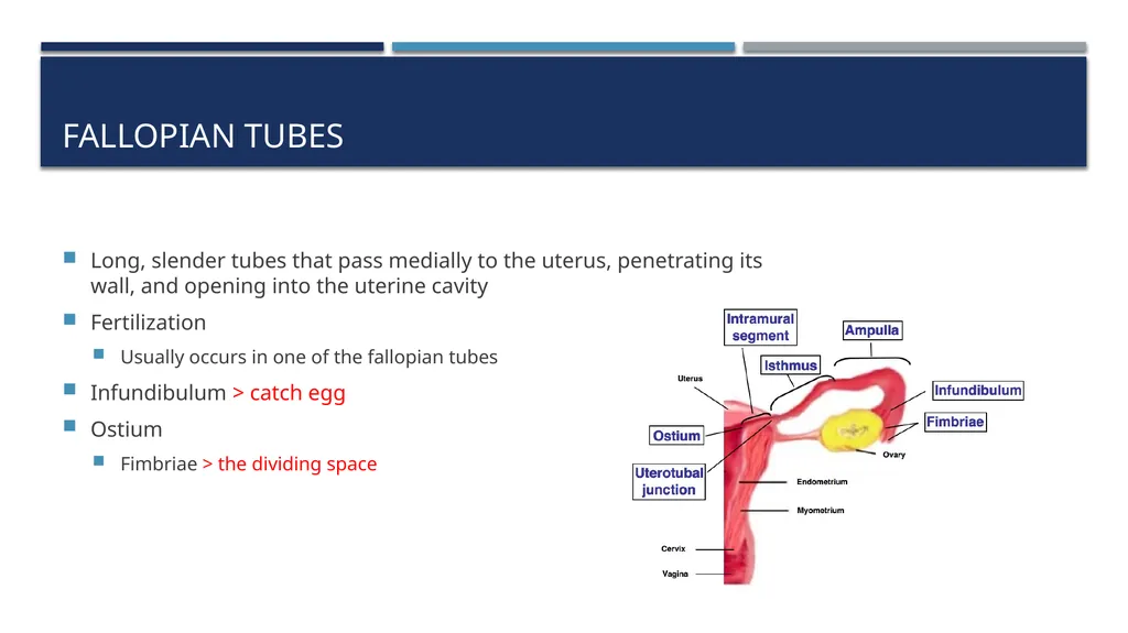
slide13. Fallopian Tubes Long, slender tubes that pass medially to the uterus, penetrating its wall, and opening into the uterine cavity
Fertilization
Usually occurs in one of the fallopian tubes
Infundibulum > catch egg
Ostium
Fimbriae > the dividing space<br>
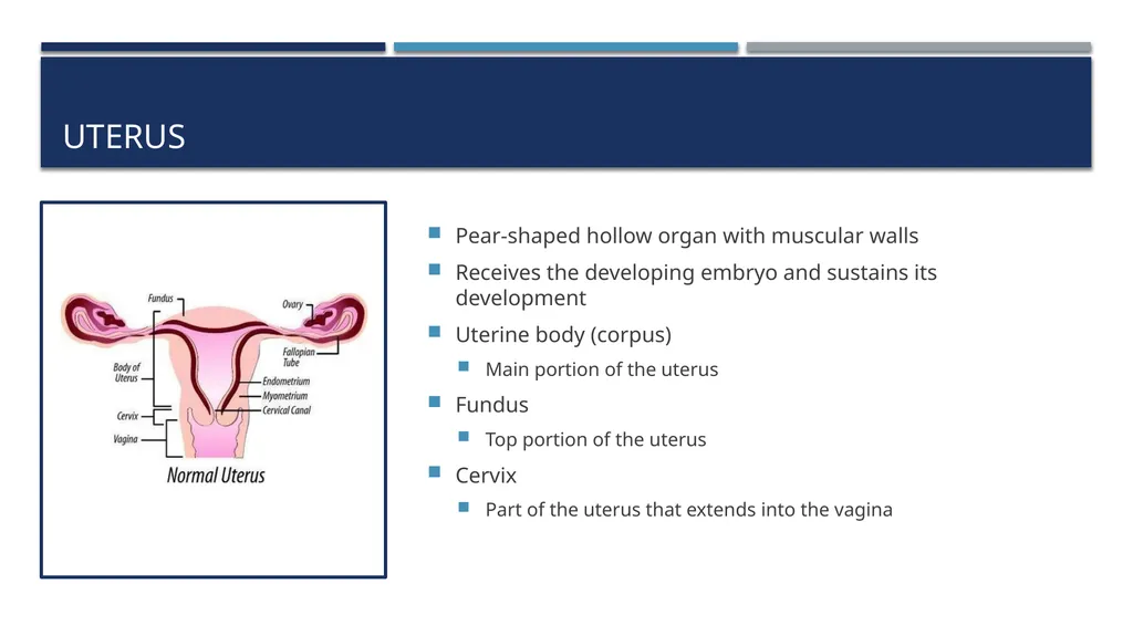
slide14. Uterus Pear-shaped hollow organ with muscular walls
Receives the developing embryo and sustains its development
Uterine body (corpus)
Main portion of the uterus
Fundus
Top portion of the uterus
Cervix
Part of the uterus that extends into the vagina<br>
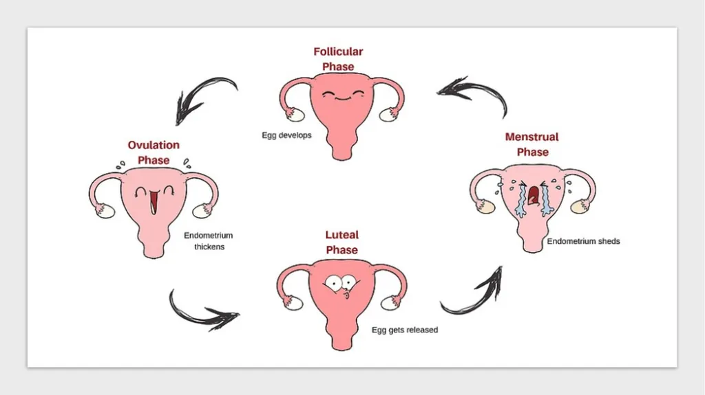
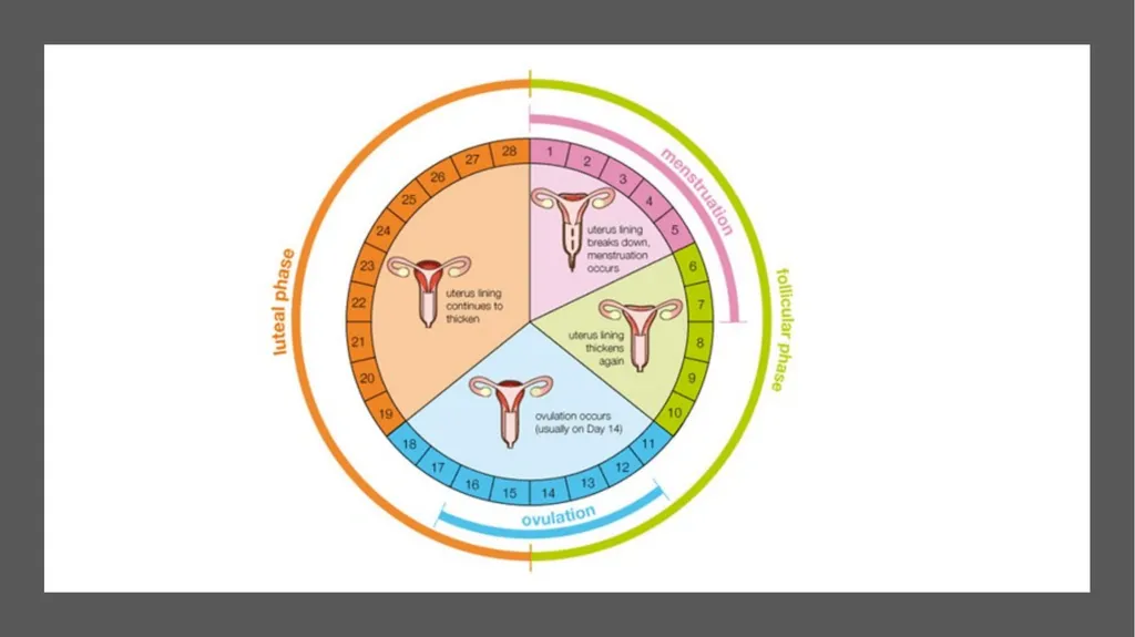
slide15. Menstrual Cycle During the menstrual cycle, an ovum matures, forming a graafian follicle.
Graafian follicle
Developed ovum
At ovulation, this follicle ruptures through the surface of the ovary.
If fertilization occurs, the fertilized egg proceeds through fallopian tube to implant in the uterus.
If fertilization does not occur, a series of hormonal changes causes the corpus luteum to be sloughed, along with the uterine lining. Corpus luteum
Remnants of the graafian follicle<br>
slide16. Menstrual Cycle Recurring cycle
Menarche
Time of first menstrual cycle
Menopause
Ending of menses
During each cycle, the lining of the uterus proliferates in preparation for pregnancy.
Sheds if pregnancy does not occur
Average cycle is 28 days.<br>
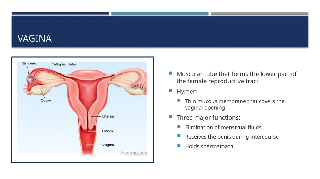
slide19. Vagina Muscular tube that forms the lower part of the female reproductive tract
Hymen
Thin mucous membrane that covers the vaginal opening
Three major functions:
Elimination of menstrual fluids
Receives the penis during intercourse
Holds spermatozoa<br>
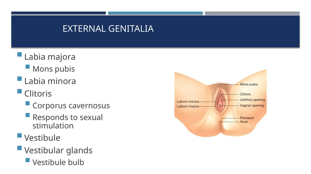
slide20. External Genitalia Labia majora
Mons pubis
Labia minora
Clitoris
Corporus cavernosus
Responds to sexual stimulation
Vestibule
Vestibular glands
Vestibule bulb<br>
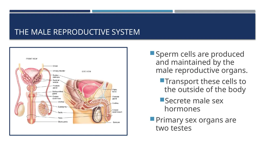
slide21. The Male Reproductive System Sperm cells are produced and maintained by the male reproductive organs.
Transport these cells to the outside of the body
Secrete male sex hormones
Primary sex organs are two testes<br>
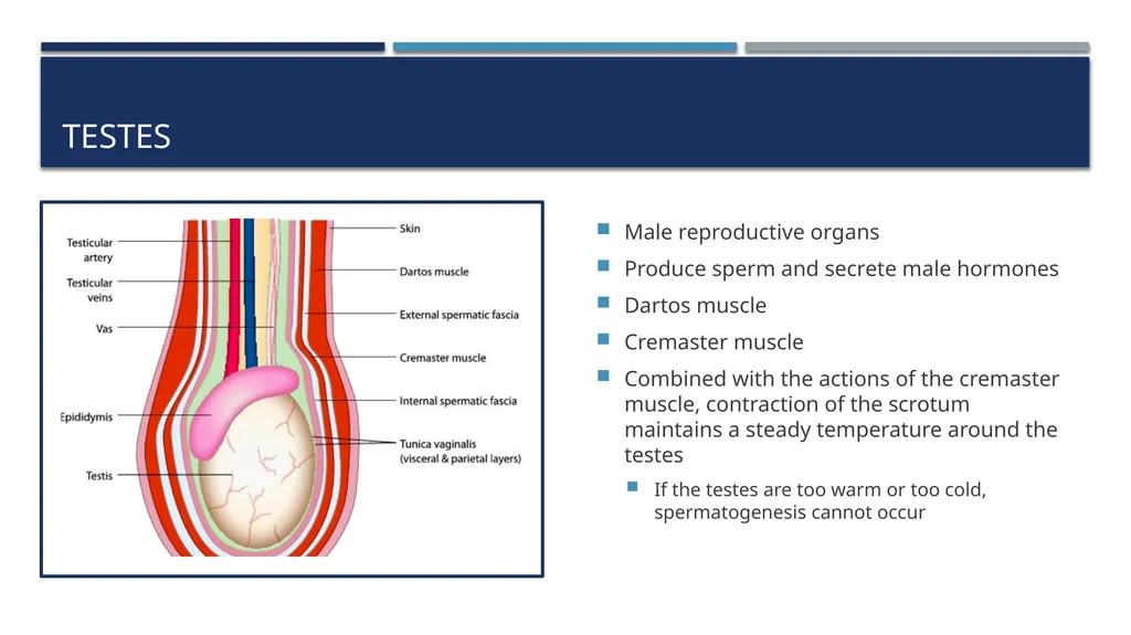
slide22. Testes Male reproductive organs
Produce sperm and secrete male hormones
Dartos muscle
Cremaster muscle
Combined with the actions of the cremaster muscle, contraction of the scrotum maintains a steady temperature around the testes
If the testes are too warm or too cold, spermatogenesis cannot occur<br>
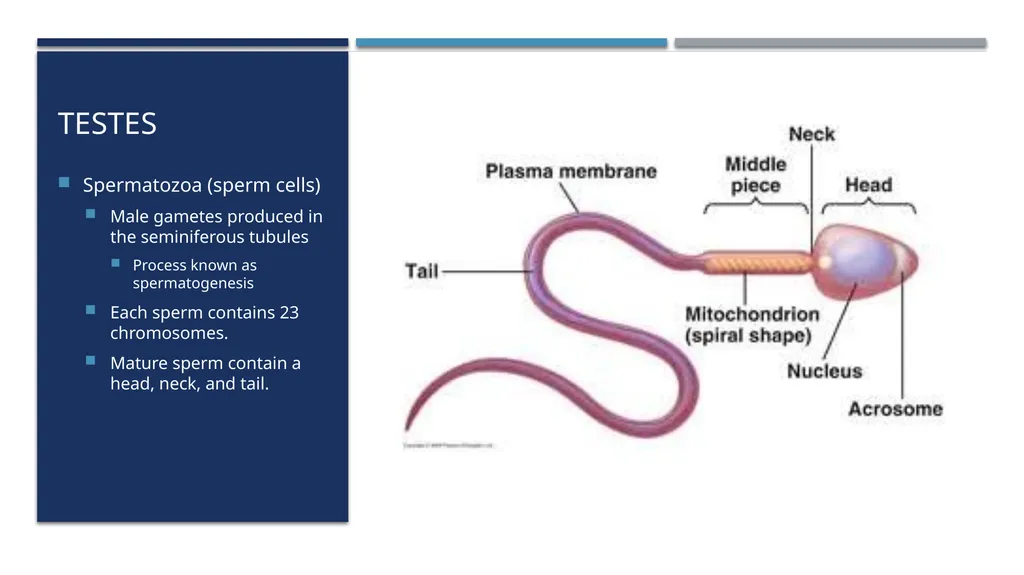
slide23. Testes Spermatozoa (sperm cells)
Male gametes produced in the seminiferous tubules
Process known as spermatogenesis
Each sperm contains 23 chromosomes.
Mature sperm contain a head, neck, and tail.<br>
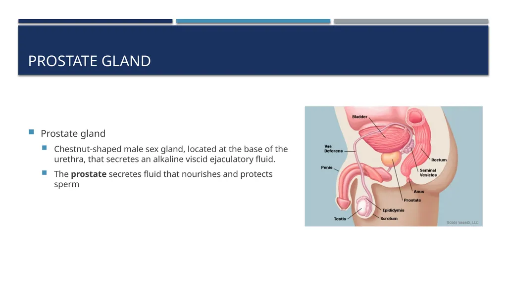
slide24. Prostate gland Prostate gland
Chestnut-shaped male sex gland, located at the base of the urethra, that secretes an alkaline viscid ejaculatory fluid.
The prostate secretes fluid that nourishes and protects sperm<br>
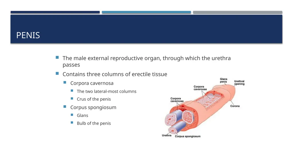
slide25. Penis The male external reproductive organ, through which the urethra passes
Contains three columns of erectile tissue
Corpora cavernosa
The two lateral-most columns
Crus of the penis
Corpus spongiosum
Glans
Bulb of the penis<br>
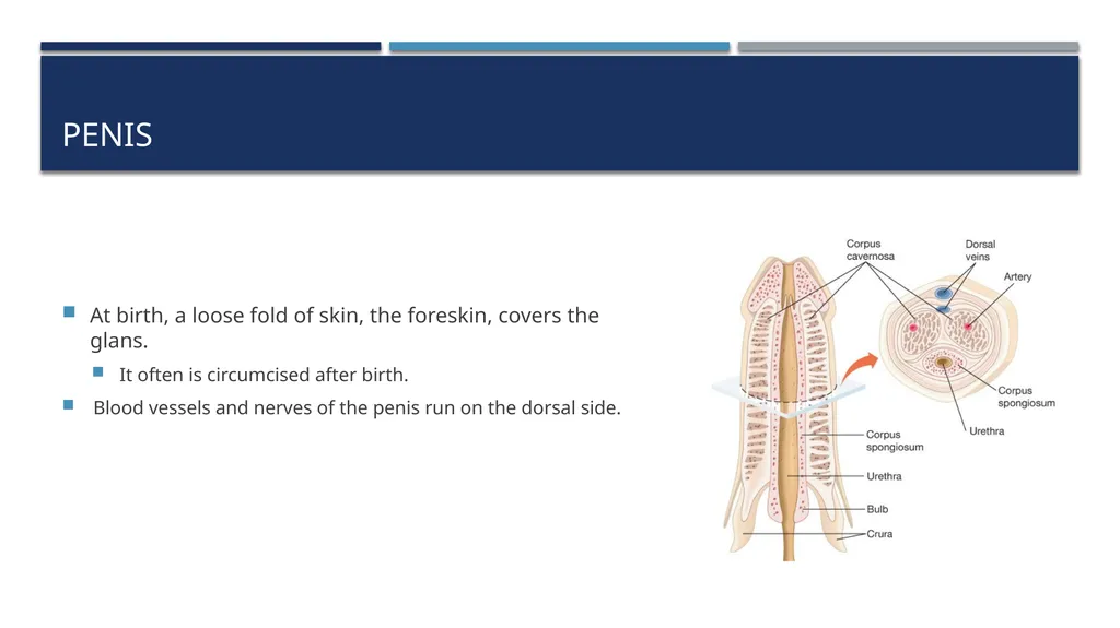
slide26. Penis At birth, a loose fold of skin, the foreskin, covers the glans.
It often is circumcised after birth.
Blood vessels and nerves of the penis run on the dorsal side.<br>
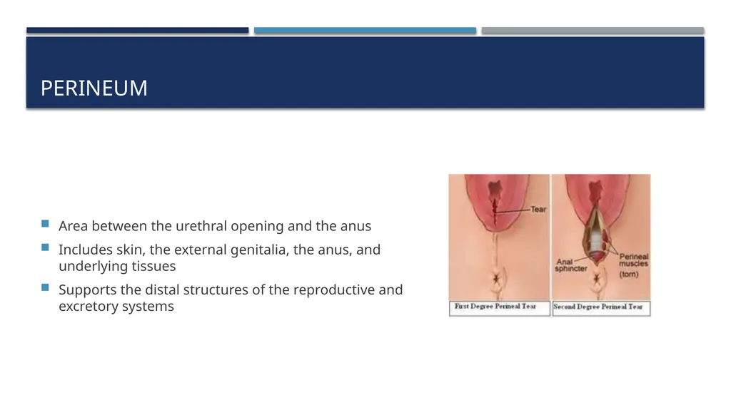
slide27. Perineum Area between the urethral opening and the anus
Includes skin, the external genitalia, the anus, and underlying tissues
Supports the distal structures of the reproductive and excretory systems<br>
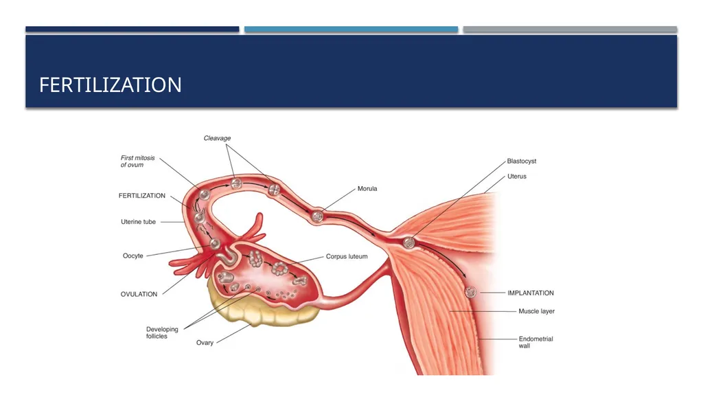
slide28. Pregnancy Fertilization
Occurs when sperm and an ovum meet in the fallopian tube
The zygote that results moves through the fallopian tube toward the uterus.
At the same time, it undergoes progressive cell divisions.
When the zygote contains approximately 32 cells, it usually implants into the uterine wall.<br>
slide29. Fertilization<br>
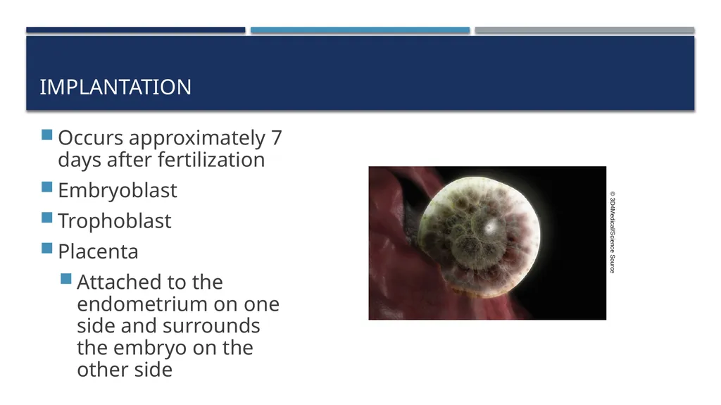
slide30. Implantation Occurs approximately 7 days after fertilization
Embryoblast
Trophoblast
Placenta
Attached to the endometrium on one side and surrounds the embryo on the other side © 3D4Medical/Science Source<br>
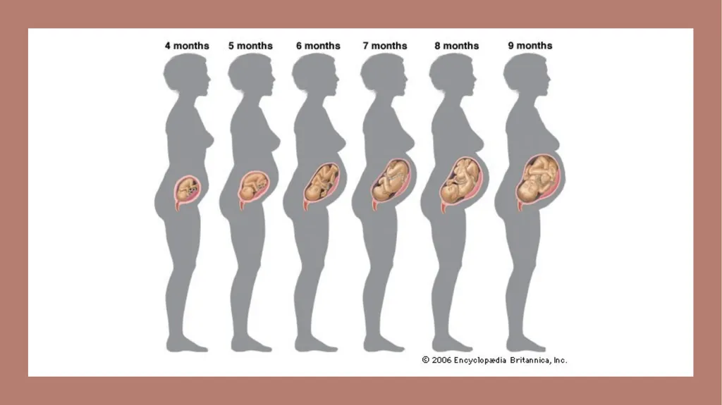
slide31. Gestation Gestation is the process of fetal development
Stimulated by production of the hormone hCG by the trophoblast
Human chorionic gonadotropin (hCG)
Hormone that stimulates the corpus luteum to produce progesterone
Essential for normal continuation of the pregnancy
Process of fetal development following fertilization of an egg
First week of gestation
Fertilized egg moves along the fallopian tube, dividing continuously, and implants into the uterine wall
Normal human gestation period is 266 days<br>
slide32. Gestation Divided into three trimesters
First trimester
Extends to week 12
Major events include:
A positive pregnancy test result
Palpable uterine fundus at the pubic symphysis at week 12
Audible fetal heart tones noted on Doppler ultrasound<br>
slide33. Gestation Second week of gestation
The amniotic membranes develop to form the amniotic sac
The placenta and membranes that surround and protect the developing embryo
Amniotic sac
The sac formed from the amniotic membranes<br>
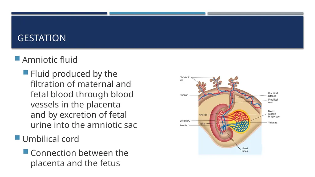
slide34. Amniotic fluid
Fluid produced by the filtration of maternal and fetal blood through blood vessels in the placenta and by excretion of fetal urine into the amniotic sac
Umbilical cord
Connection between the placenta and the fetus Gestation<br>
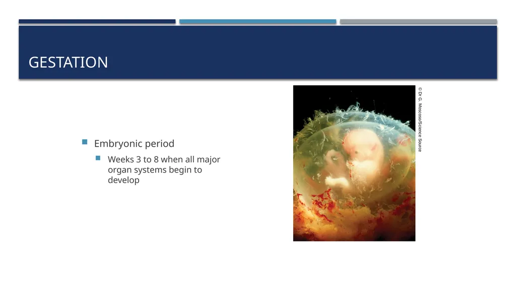
slide35. Gestation Embryonic period
Weeks 3 to 8 when all major organ systems begin to develop © Dr G. Moscoso/Science Source<br>
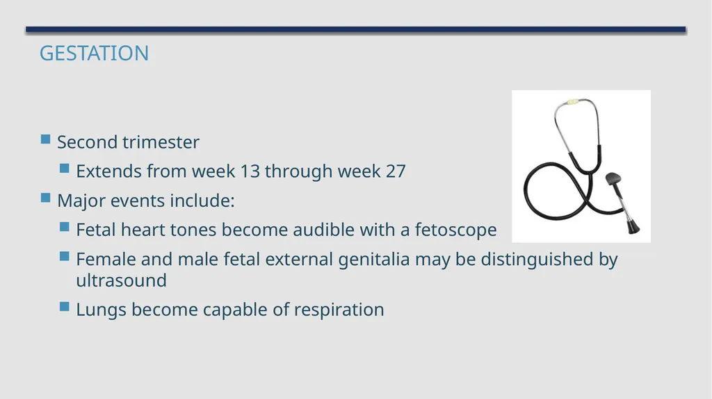
slide36. Gestation Second trimester
Extends from week 13 through week 27
Major events include:
Fetal heart tones become audible with a fetoscope
Female and male fetal external genitalia may be distinguished by ultrasound
Lungs become capable of respiration<br>
slide37. Gestation Third trimester
Extends from week 28 until term, or week 40
Major events include:
The presence of a papillary light reflex in the fetus
Descent of the fetal head to the pelvic inlet
Rupture of the fetal membranes<br>
slide38. Labor Scientists are still uncertain exactly what initiates labor
Likely due to a combination of maternal and fetal stimuli
Consists of three stages:
First stage – uterine wall begins to contract and the cervix dilates
Second stage – birth of the baby occurs
Third stage – placenta is expelled<br>
slide39. The Fetus The lungs are fluid-filled and nonfunctioning
All oxygen and carbon dioxide exchange takes place through the placenta
Fetus is attached to the placenta by the umbilical cord
Oxygenated blood from the mother's circulation
Travels through the placenta, into the fetus
Then back into the placenta
Fetal waste diffuses into the mother's bloodstream for eventual excretion<br>
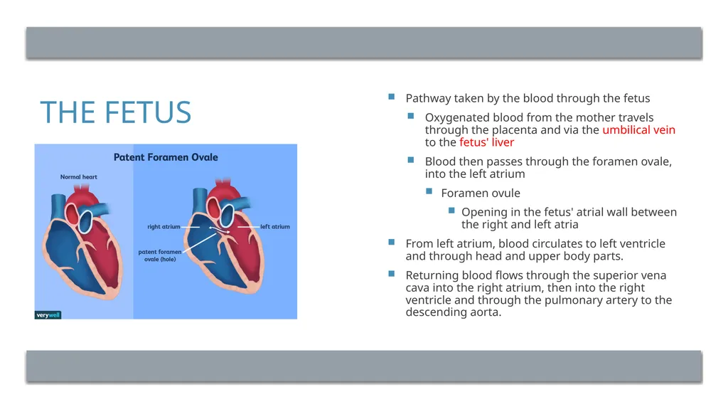
slide40. The Fetus Pathway taken by the blood through the fetus
Oxygenated blood from the mother travels through the placenta and via the umbilical vein to the fetus' liver
Blood then passes through the foramen ovale, into the left atrium
Foramen ovule
Opening in the fetus' atrial wall between the right and left atria
From left atrium, blood circulates to left ventricle and through head and upper body parts.
Returning blood flows through the superior vena cava into the right atrium, then into the right ventricle and through the pulmonary artery to the descending aorta.<br>
slide41. The Fetus From the descending aorta, blood circulates through the lower body parts.
Blood that flows through the umbilical arteries back to the placenta carries the fetal waste.<br>
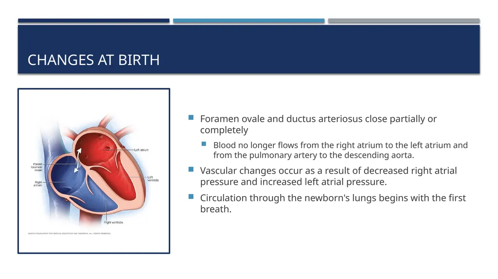
slide42. Changes at Birth Foramen ovale and ductus arteriosus close partially or completely
Blood no longer flows from the right atrium to the left atrium and from the pulmonary artery to the descending aorta.
Vascular changes occur as a result of decreased right atrial pressure and increased left atrial pressure.
Circulation through the newborn's lungs begins with the first breath.<br>
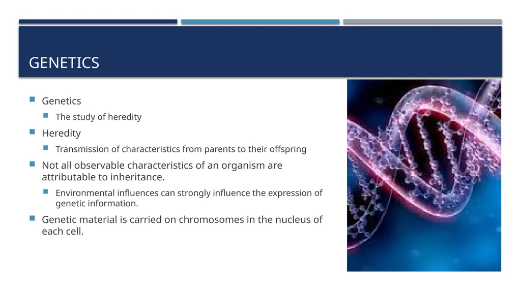
slide44. Genetics Genetics
The study of heredity
Heredity
Transmission of characteristics from parents to their offspring
Not all observable characteristics of an organism are attributable to inheritance.
Environmental influences can strongly influence the expression of genetic information.
Genetic material is carried on chromosomes in the nucleus of each cell.<br>
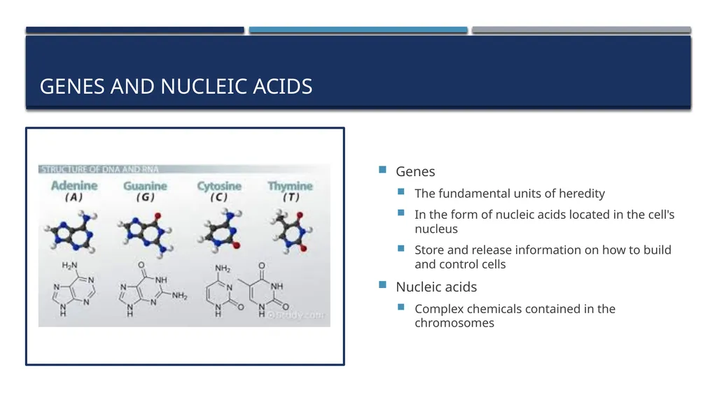
slide45. Genes and Nucleic Acids Genes
The fundamental units of heredity
In the form of nucleic acids located in the cell's nucleus
Store and release information on how to build and control cells
Nucleic acids
Complex chemicals contained in the chromosomes<br>
slide46. Genes and Nucleic Acids Deoxyribonucleic acid (DNA)
Complex chemical that carries the so-called genetic code
Ribonucleic acid (RNA)
Made from DNA in the cell's nucleus
RNA then moves to the cytoplasm of the cell
The central dogma of life
Hypothesis ascribed to Francis Crick
“DNA leads to RNA, which leads to proteins, which leads to cells”<br>
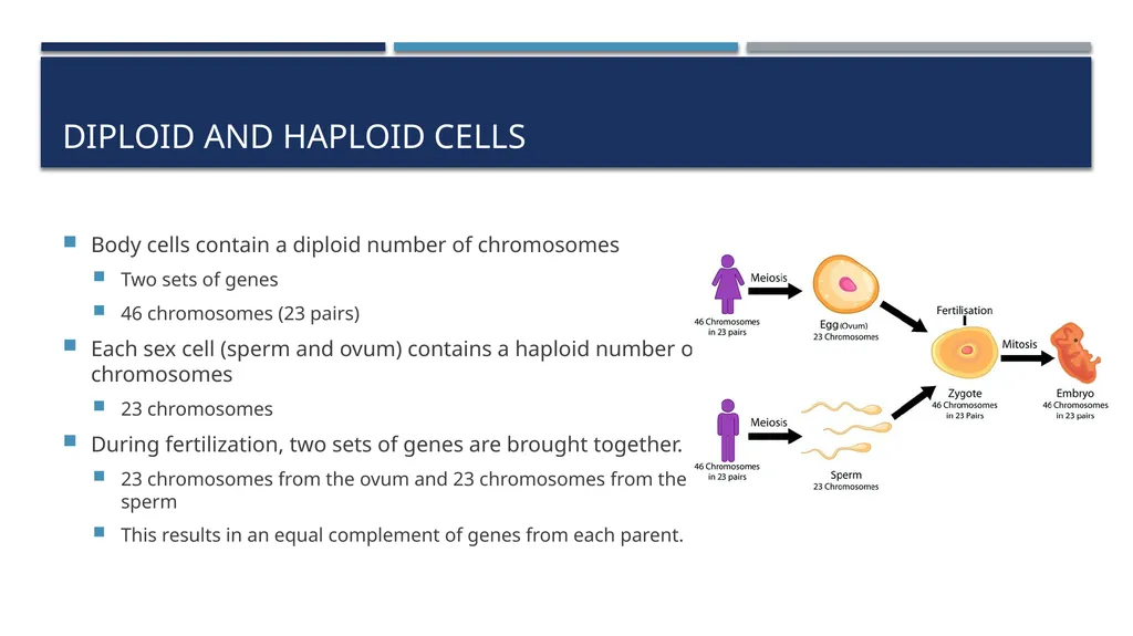
slide47. Diploid and Haploid Cells Body cells contain a diploid number of chromosomes
Two sets of genes
46 chromosomes (23 pairs)
Each sex cell (sperm and ovum) contains a haploid number of chromosomes
23 chromosomes
During fertilization, two sets of genes are brought together.
23 chromosomes from the ovum and 23 chromosomes from the sperm
This results in an equal complement of genes from each parent.<br>
slide48. Autosomal and Sex-Linked Traits Body cells and sex cells contain two different types of chromosomes
Autosomes carry genes that code for numerous body proteins independent of sex
Sex chromosomes code for proteins that determine sex, as well as for other proteins
Two sex chromosomes
X
Y<br>
slide49. Autosomal and Sex-Linked Traits Each ovum contains one X chromosome
Each spermatocyte contains either an X or a Y chromosome
Once fertilized, the zygote can have:
Two X chromosomes (XX) = female embryo
X and Y chromosome (XY) = male embryo<br>
slide50. Autosomal and Sex-Linked Traits Traits are classified by the type of chromosome on which they reside.
Autosomal-linked traits reside in genes located on autosomes.
Sex-linked traits reside in genes located on the sex chromosomes.<br>
slide51. Alleles and Traits Allele
Each single gene contributed by one parent for any particular characteristic
For many inherited features, there may be more than one potential allele.
Each parent contributes one gene (allele) for each inherited characteristic.
Recessive trait
Dominant trait<br>
slide52. Alleles and Traits Letters are used to indicate various alleles
The dominant allele is written in upper case.
The recessive allele is written in lower case.
Each gamete carries one allele.
The fertilized egg has two alleles.
One allele from each parental gamete<br>
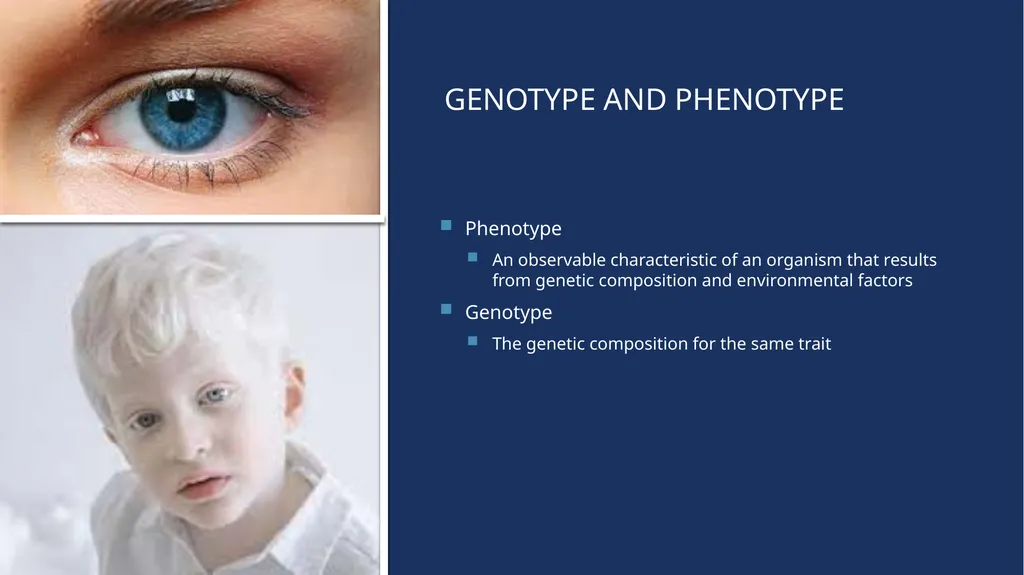
slide53. Genotype and Phenotype Phenotype
An observable characteristic of an organism that results from genetic composition and environmental factors
Genotype
The genetic composition for the same trait<br>
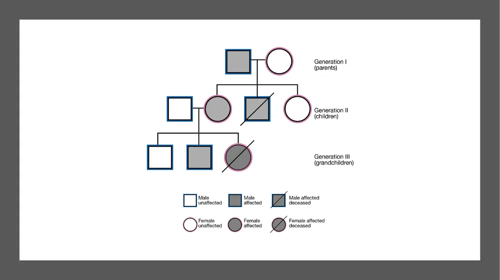
slide54. Genotype and Phenotype Dominant trait
If either or both dominant alleles are present in the genotype, the trait has an observable phenotype
Recessive trait/characteristic
Only visible clinically (have an observable phenotype) when the genotype is composed of two recessive alleles for that particular trait<br>
slide56. Ref. https://www.youtube.com/watch?v=RFDatCchpus&t=2s (Female reproductive)
https://www.youtube.com/watch?v=-XQcnO4iX_U&t=3s (Male reproductive)
https://www.youtube.com/watch?v=SUdAEGXLO-8&t=3s (Sex & fertilization)
https://www.youtube.com/watch?v=BtsSbZ85yiQ&t=3s (pregnancy & development)<br>
BSc EMS, MSc Cardiovascular, Lecturer
Department of Basic Sciences, PSCEMS
http://fac.ksu.edu.sa/aalrumayh
aalrumayh@ksu.edu.sa<br>
slide2. Learning Objectives List the essential and accessory reproductive organs of the male and female, describing the general function of each.
Name the hormones necessary for the formation of gametes.
Describe the difference between spermatogenesis and oogenesis.<br>
slide3. Learning Objectives Describe the three layers of the uterine wall.
Briefly describe the life cycle of an oocyte.
Describe the menstrual cycle in terms of changes in hormone levels and the condition of the endometrium.<br>
slide4. Learning Objectives Define the following terms: diploid, haploid, gametes, endometrium, genetic disease, homologous chromosomes, autosomes, sex chromosomes, genes, alleles, genotype, phenotype, homozygous, and heterozygous.
Identify and describe the structures that constitute the external genitalia in both sexes.
Name the parts of a sperm cell.
Beginning with fertilization, describe the major developmental changes during gestation.
Describe the structure and function of the placenta and umbilical cord.
State the length of an average gestation period.<br>
slide5. Learning Objectives Describe the stages of labor.
Describe the difference between fetal circulation/respiration and adult circulation/respiration.
Describe the major changes that take place in an infant at birth.
Discuss the difference between dominant and recessive traits.
List some important genetic diseases found in out-of-hospital care.
Explain how genes can cause disease.<br>
slide6. Introduction The only body system not essential to survival
Male and female reproductive systems are very different.
The reproductive system is linked with genetics.
Certain reproductive organs secrete hormones needed for development and maintenance of secondary sex characteristics and for reproduction.<br>
slide7. The Female Reproductive System<br>
slide8. Female reproductive system Oocytes – female sex cells (egg cells) produced and maintained by the female reproductive organs
The female reproductive organs:
Transport oocytes to the site of fertilization
Provide a nurturing environment for the developing fetus
Give birth to a child
Produce female sex hormones<br>
slide9. Produce female sex cells and sex hormones
Ovarian tissue consists of:
Inner medulla
Outer cortex
Suspended in place by:
Mesovarium
Suspensory ligament
Ovarian ligament Ovaries<br>
slide10. Ovaries Three main functions:
Produce oocytes
Immature female gametes (egg or ovum)
Secrete female sex hormones
Including estrogens and progestins
Secrete inhibin
Involved in the feedback control of pituitary follicle-stimulating hormone production<br>
slide11. Ovaries Oogenesis
Maturation process undergone by oocytes
Results in production of an ovum
Ovum
Female gamete or mature egg
During the reproductive years of a woman's life, the pituitary gland releases hormones at roughly monthly intervals that affect the ovaries<br>
slide12. Ovaries Hormones that stimulate one oocyte to undergo meiosis
Follicle-stimulating hormone (FSH)
Luteinizing hormone (LH)
Meiosis
Process of cell division that occurs during the formation of a mature ovum or spermatocyte
A final ovum contributes half of the total genetic information to the zygote, or fertilized ovum. Anterior pituitary gland<br>
slide13. Fallopian Tubes Long, slender tubes that pass medially to the uterus, penetrating its wall, and opening into the uterine cavity
Fertilization
Usually occurs in one of the fallopian tubes
Infundibulum > catch egg
Ostium
Fimbriae > the dividing space<br>
slide14. Uterus Pear-shaped hollow organ with muscular walls
Receives the developing embryo and sustains its development
Uterine body (corpus)
Main portion of the uterus
Fundus
Top portion of the uterus
Cervix
Part of the uterus that extends into the vagina<br>
slide15. Menstrual Cycle During the menstrual cycle, an ovum matures, forming a graafian follicle.
Graafian follicle
Developed ovum
At ovulation, this follicle ruptures through the surface of the ovary.
If fertilization occurs, the fertilized egg proceeds through fallopian tube to implant in the uterus.
If fertilization does not occur, a series of hormonal changes causes the corpus luteum to be sloughed, along with the uterine lining. Corpus luteum
Remnants of the graafian follicle<br>
slide16. Menstrual Cycle Recurring cycle
Menarche
Time of first menstrual cycle
Menopause
Ending of menses
During each cycle, the lining of the uterus proliferates in preparation for pregnancy.
Sheds if pregnancy does not occur
Average cycle is 28 days.<br>
slide19. Vagina Muscular tube that forms the lower part of the female reproductive tract
Hymen
Thin mucous membrane that covers the vaginal opening
Three major functions:
Elimination of menstrual fluids
Receives the penis during intercourse
Holds spermatozoa<br>
slide20. External Genitalia Labia majora
Mons pubis
Labia minora
Clitoris
Corporus cavernosus
Responds to sexual stimulation
Vestibule
Vestibular glands
Vestibule bulb<br>
slide21. The Male Reproductive System Sperm cells are produced and maintained by the male reproductive organs.
Transport these cells to the outside of the body
Secrete male sex hormones
Primary sex organs are two testes<br>
slide22. Testes Male reproductive organs
Produce sperm and secrete male hormones
Dartos muscle
Cremaster muscle
Combined with the actions of the cremaster muscle, contraction of the scrotum maintains a steady temperature around the testes
If the testes are too warm or too cold, spermatogenesis cannot occur<br>
slide23. Testes Spermatozoa (sperm cells)
Male gametes produced in the seminiferous tubules
Process known as spermatogenesis
Each sperm contains 23 chromosomes.
Mature sperm contain a head, neck, and tail.<br>
slide24. Prostate gland Prostate gland
Chestnut-shaped male sex gland, located at the base of the urethra, that secretes an alkaline viscid ejaculatory fluid.
The prostate secretes fluid that nourishes and protects sperm<br>
slide25. Penis The male external reproductive organ, through which the urethra passes
Contains three columns of erectile tissue
Corpora cavernosa
The two lateral-most columns
Crus of the penis
Corpus spongiosum
Glans
Bulb of the penis<br>
slide26. Penis At birth, a loose fold of skin, the foreskin, covers the glans.
It often is circumcised after birth.
Blood vessels and nerves of the penis run on the dorsal side.<br>
slide27. Perineum Area between the urethral opening and the anus
Includes skin, the external genitalia, the anus, and underlying tissues
Supports the distal structures of the reproductive and excretory systems<br>
slide28. Pregnancy Fertilization
Occurs when sperm and an ovum meet in the fallopian tube
The zygote that results moves through the fallopian tube toward the uterus.
At the same time, it undergoes progressive cell divisions.
When the zygote contains approximately 32 cells, it usually implants into the uterine wall.<br>
slide29. Fertilization<br>
slide30. Implantation Occurs approximately 7 days after fertilization
Embryoblast
Trophoblast
Placenta
Attached to the endometrium on one side and surrounds the embryo on the other side © 3D4Medical/Science Source<br>
slide31. Gestation Gestation is the process of fetal development
Stimulated by production of the hormone hCG by the trophoblast
Human chorionic gonadotropin (hCG)
Hormone that stimulates the corpus luteum to produce progesterone
Essential for normal continuation of the pregnancy
Process of fetal development following fertilization of an egg
First week of gestation
Fertilized egg moves along the fallopian tube, dividing continuously, and implants into the uterine wall
Normal human gestation period is 266 days<br>
slide32. Gestation Divided into three trimesters
First trimester
Extends to week 12
Major events include:
A positive pregnancy test result
Palpable uterine fundus at the pubic symphysis at week 12
Audible fetal heart tones noted on Doppler ultrasound<br>
slide33. Gestation Second week of gestation
The amniotic membranes develop to form the amniotic sac
The placenta and membranes that surround and protect the developing embryo
Amniotic sac
The sac formed from the amniotic membranes<br>
slide34. Amniotic fluid
Fluid produced by the filtration of maternal and fetal blood through blood vessels in the placenta and by excretion of fetal urine into the amniotic sac
Umbilical cord
Connection between the placenta and the fetus Gestation<br>
slide35. Gestation Embryonic period
Weeks 3 to 8 when all major organ systems begin to develop © Dr G. Moscoso/Science Source<br>
slide36. Gestation Second trimester
Extends from week 13 through week 27
Major events include:
Fetal heart tones become audible with a fetoscope
Female and male fetal external genitalia may be distinguished by ultrasound
Lungs become capable of respiration<br>
slide37. Gestation Third trimester
Extends from week 28 until term, or week 40
Major events include:
The presence of a papillary light reflex in the fetus
Descent of the fetal head to the pelvic inlet
Rupture of the fetal membranes<br>
slide38. Labor Scientists are still uncertain exactly what initiates labor
Likely due to a combination of maternal and fetal stimuli
Consists of three stages:
First stage – uterine wall begins to contract and the cervix dilates
Second stage – birth of the baby occurs
Third stage – placenta is expelled<br>
slide39. The Fetus The lungs are fluid-filled and nonfunctioning
All oxygen and carbon dioxide exchange takes place through the placenta
Fetus is attached to the placenta by the umbilical cord
Oxygenated blood from the mother's circulation
Travels through the placenta, into the fetus
Then back into the placenta
Fetal waste diffuses into the mother's bloodstream for eventual excretion<br>
slide40. The Fetus Pathway taken by the blood through the fetus
Oxygenated blood from the mother travels through the placenta and via the umbilical vein to the fetus' liver
Blood then passes through the foramen ovale, into the left atrium
Foramen ovule
Opening in the fetus' atrial wall between the right and left atria
From left atrium, blood circulates to left ventricle and through head and upper body parts.
Returning blood flows through the superior vena cava into the right atrium, then into the right ventricle and through the pulmonary artery to the descending aorta.<br>
slide41. The Fetus From the descending aorta, blood circulates through the lower body parts.
Blood that flows through the umbilical arteries back to the placenta carries the fetal waste.<br>
slide42. Changes at Birth Foramen ovale and ductus arteriosus close partially or completely
Blood no longer flows from the right atrium to the left atrium and from the pulmonary artery to the descending aorta.
Vascular changes occur as a result of decreased right atrial pressure and increased left atrial pressure.
Circulation through the newborn's lungs begins with the first breath.<br>
slide44. Genetics Genetics
The study of heredity
Heredity
Transmission of characteristics from parents to their offspring
Not all observable characteristics of an organism are attributable to inheritance.
Environmental influences can strongly influence the expression of genetic information.
Genetic material is carried on chromosomes in the nucleus of each cell.<br>
slide45. Genes and Nucleic Acids Genes
The fundamental units of heredity
In the form of nucleic acids located in the cell's nucleus
Store and release information on how to build and control cells
Nucleic acids
Complex chemicals contained in the chromosomes<br>
slide46. Genes and Nucleic Acids Deoxyribonucleic acid (DNA)
Complex chemical that carries the so-called genetic code
Ribonucleic acid (RNA)
Made from DNA in the cell's nucleus
RNA then moves to the cytoplasm of the cell
The central dogma of life
Hypothesis ascribed to Francis Crick
“DNA leads to RNA, which leads to proteins, which leads to cells”<br>
slide47. Diploid and Haploid Cells Body cells contain a diploid number of chromosomes
Two sets of genes
46 chromosomes (23 pairs)
Each sex cell (sperm and ovum) contains a haploid number of chromosomes
23 chromosomes
During fertilization, two sets of genes are brought together.
23 chromosomes from the ovum and 23 chromosomes from the sperm
This results in an equal complement of genes from each parent.<br>
slide48. Autosomal and Sex-Linked Traits Body cells and sex cells contain two different types of chromosomes
Autosomes carry genes that code for numerous body proteins independent of sex
Sex chromosomes code for proteins that determine sex, as well as for other proteins
Two sex chromosomes
X
Y<br>
slide49. Autosomal and Sex-Linked Traits Each ovum contains one X chromosome
Each spermatocyte contains either an X or a Y chromosome
Once fertilized, the zygote can have:
Two X chromosomes (XX) = female embryo
X and Y chromosome (XY) = male embryo<br>
slide50. Autosomal and Sex-Linked Traits Traits are classified by the type of chromosome on which they reside.
Autosomal-linked traits reside in genes located on autosomes.
Sex-linked traits reside in genes located on the sex chromosomes.<br>
slide51. Alleles and Traits Allele
Each single gene contributed by one parent for any particular characteristic
For many inherited features, there may be more than one potential allele.
Each parent contributes one gene (allele) for each inherited characteristic.
Recessive trait
Dominant trait<br>
slide52. Alleles and Traits Letters are used to indicate various alleles
The dominant allele is written in upper case.
The recessive allele is written in lower case.
Each gamete carries one allele.
The fertilized egg has two alleles.
One allele from each parental gamete<br>
slide53. Genotype and Phenotype Phenotype
An observable characteristic of an organism that results from genetic composition and environmental factors
Genotype
The genetic composition for the same trait<br>
slide54. Genotype and Phenotype Dominant trait
If either or both dominant alleles are present in the genotype, the trait has an observable phenotype
Recessive trait/characteristic
Only visible clinically (have an observable phenotype) when the genotype is composed of two recessive alleles for that particular trait<br>
slide56. Ref. https://www.youtube.com/watch?v=RFDatCchpus&t=2s (Female reproductive)
https://www.youtube.com/watch?v=-XQcnO4iX_U&t=3s (Male reproductive)
https://www.youtube.com/watch?v=SUdAEGXLO-8&t=3s (Sex & fertilization)
https://www.youtube.com/watch?v=BtsSbZ85yiQ&t=3s (pregnancy & development)<br>