
This teaching presentation for the ISOM website


Description: This teaching presentation for the ISOM website has been prepared by Tal Marom, MD and Sharon Ovnat Tamir, MD Department of Otolaryngology-Head and Neck Surgery Edith Wolfson Medical Center Sackler Faculty of Medicine Tel Aviv University
Related Topics
Download Presentation
"This teaching presentation for the ISOM website" is the property of its rightful owner. Permission is granted to download and print the materials on this website for personal, non-commercial use only, and to display it on your personal computer provided you do not modify the materials and that you retain all copyright notices contained in the materials. By downloading content from our website, you accept the terms of this agreement.
Presentation Transcript
slide1. This teaching presentation for the ISOM website has been prepared by
Tal Marom, MD and Sharon Ovnat Tamir, MD
Department of Otolaryngology-Head and Neck Surgery
Edith Wolfson Medical Center
Sackler Faculty of Medicine
Tel Aviv University
Holon, Israel Definitions of Otitis Media<br>
slide2. Acknowledgement This presentation is aimed for teaching purposes of students, residents and other allied healthcare workers
Please visit the International Society for Otitis Media website for more resources, www.otitismediasociety.org<br>
slide3. What are the Definitions of Otitis Media ?<br>
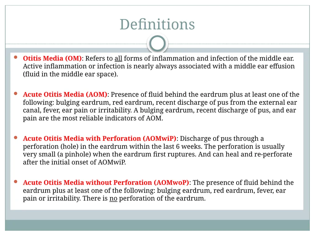
slide4. Definitions Otitis Media (OM): Refers to all forms of inflammation and infection of the middle ear. Active inflammation or infection is nearly always associated with a middle ear effusion (fluid in the middle ear space).
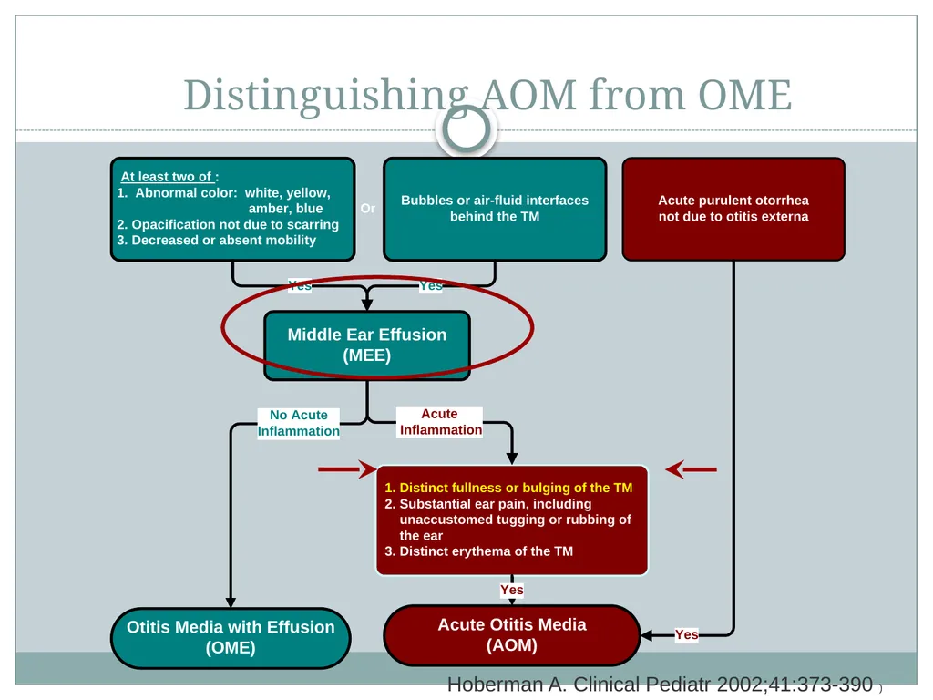
Acute Otitis Media (AOM): Presence of fluid behind the eardrum plus at least one of the following: bulging eardrum, red eardrum, recent discharge of pus from the external ear canal, fever, ear pain or irritability. A bulging eardrum, recent discharge of pus, and ear pain are the most reliable indicators of AOM.
Acute Otitis Media with Perforation (AOMwiP): Discharge of pus through a perforation (hole) in the eardrum within the last 6 weeks. The perforation is usually very small (a pinhole) when the eardrum first ruptures. And can heal and re-perforate after the initial onset of AOMwiP.
Acute Otitis Media without Perforation (AOMwoP): The presence of fluid behind the eardrum plus at least one of the following: bulging eardrum, red eardrum, fever, ear pain or irritability. There is no perforation of the eardrum.<br>
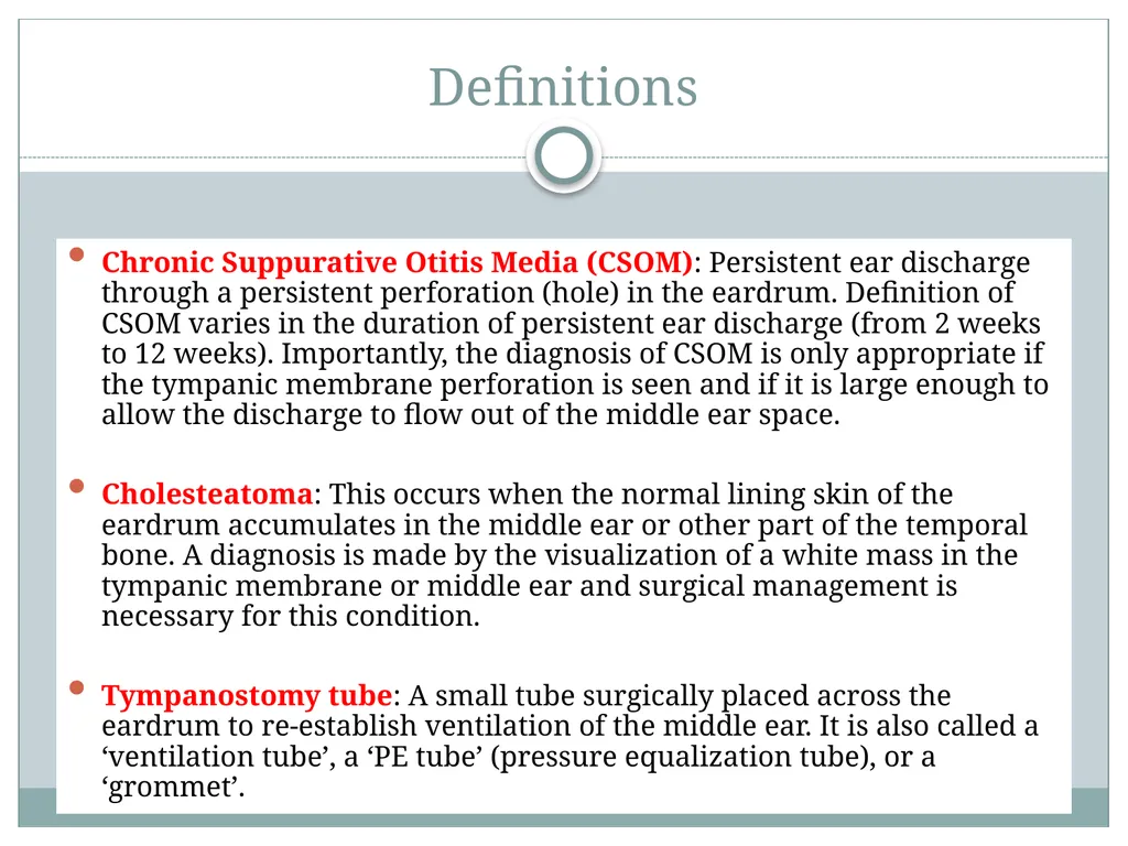
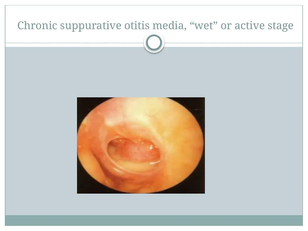
slide5. Definitions Chronic Suppurative Otitis Media (CSOM): Persistent ear discharge through a persistent perforation (hole) in the eardrum. Definition of CSOM varies in the duration of persistent ear discharge (from 2 weeks to 12 weeks). Importantly, the diagnosis of CSOM is only appropriate if the tympanic membrane perforation is seen and if it is large enough to allow the discharge to flow out of the middle ear space.
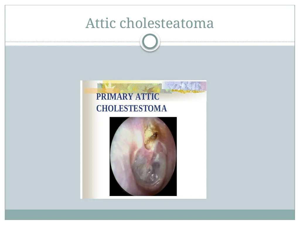
Cholesteatoma: This occurs when the normal lining skin of the eardrum accumulates in the middle ear or other part of the temporal bone. A diagnosis is made by the visualization of a white mass in the tympanic membrane or middle ear and surgical management is necessary for this condition.
Tympanostomy tube: A small tube surgically placed across the eardrum to re-establish ventilation of the middle ear. It is also called a ‘ventilation tube’, a ‘PE tube’ (pressure equalization tube), or a ‘grommet’.<br>
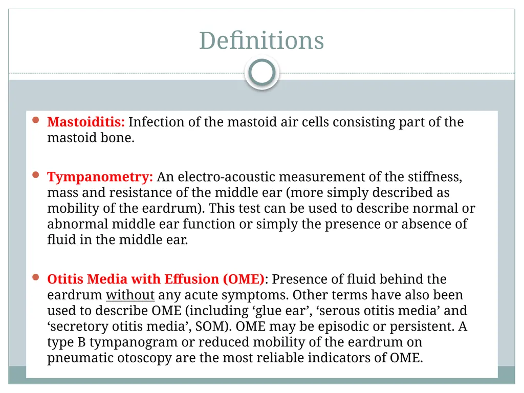
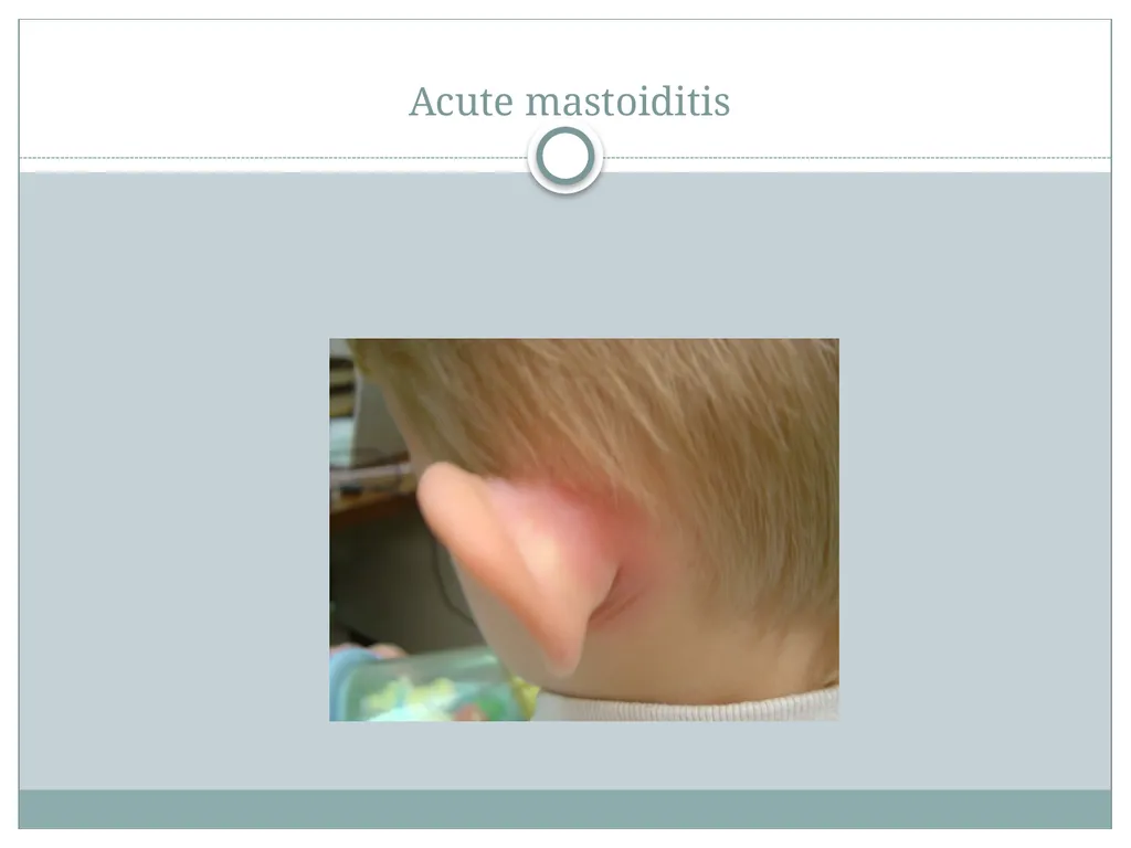
slide6. Definitions Mastoiditis: Infection of the mastoid air cells consisting part of the mastoid bone.
Tympanometry: An electro-acoustic measurement of the stiffness, mass and resistance of the middle ear (more simply described as mobility of the eardrum). This test can be used to describe normal or abnormal middle ear function or simply the presence or absence of fluid in the middle ear.
Otitis Media with Effusion (OME): Presence of fluid behind the eardrum without any acute symptoms. Other terms have also been used to describe OME (including ‘glue ear’, ‘serous otitis media’ and ‘secretory otitis media’, SOM). OME may be episodic or persistent. A type B tympanogram or reduced mobility of the eardrum on pneumatic otoscopy are the most reliable indicators of OME.<br>
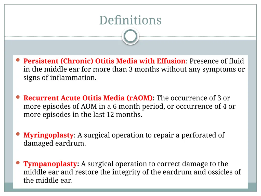
slide7. Definitions Persistent (Chronic) Otitis Media with Effusion: Presence of fluid in the middle ear for more than 3 months without any symptoms or signs of inflammation.
Recurrent Acute Otitis Media (rAOM): The occurrence of 3 or more episodes of AOM in a 6 month period, or occurrence of 4 or more episodes in the last 12 months.
Myringoplasty: A surgical operation to repair a perforated of damaged eardrum.
Tympanoplasty: A surgical operation to correct damage to the middle ear and restore the integrity of the eardrum and ossicles of the middle ear.<br>
slide8. What is the difference between AOM and OME?<br>
slide9. Distinguishing AOM from OME Hoberman A. Clinical Pediatr 2002;41:373-390 )<br>
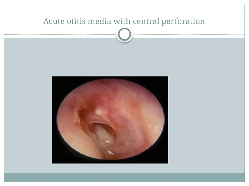
slide10. Acute otitis media with central perforation<br>
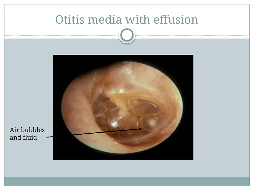
slide11. Otitis media with effusion Air bubbles
and fluid<br>
slide12. Ventilation tube (grommet)<br>
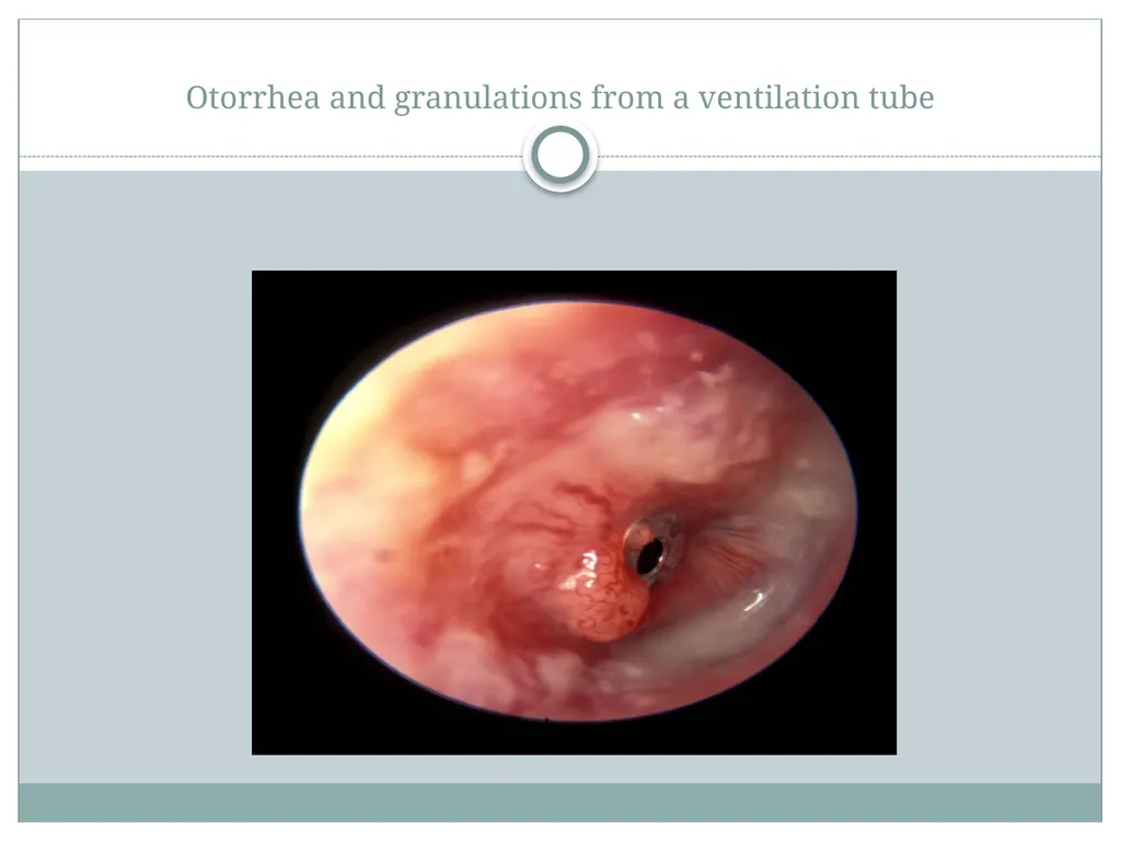
slide13. Otorrhea and granulations from a ventilation tube<br>
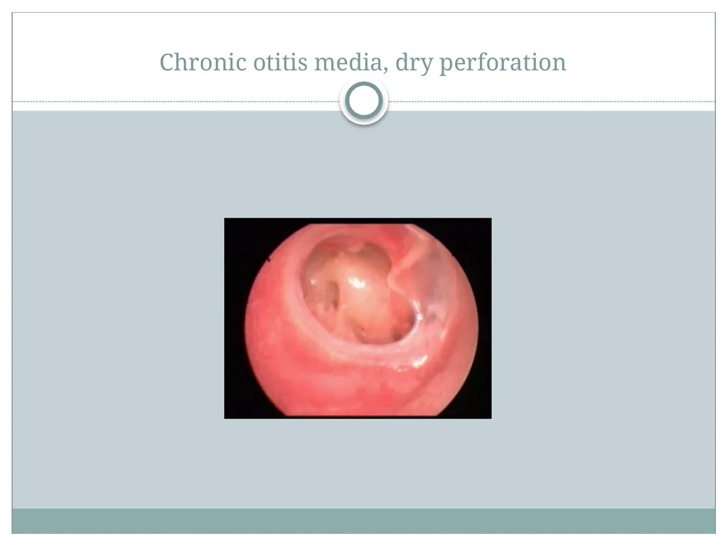
slide14. Chronic otitis media, dry perforation<br>
slide15. Chronic suppurative otitis media, “wet” or active stage<br>
slide16. Attic cholesteatoma<br>
slide17. Acute mastoiditis<br>
Tal Marom, MD and Sharon Ovnat Tamir, MD
Department of Otolaryngology-Head and Neck Surgery
Edith Wolfson Medical Center
Sackler Faculty of Medicine
Tel Aviv University
Holon, Israel Definitions of Otitis Media<br>
slide2. Acknowledgement This presentation is aimed for teaching purposes of students, residents and other allied healthcare workers
Please visit the International Society for Otitis Media website for more resources, www.otitismediasociety.org<br>
slide3. What are the Definitions of Otitis Media ?<br>
slide4. Definitions Otitis Media (OM): Refers to all forms of inflammation and infection of the middle ear. Active inflammation or infection is nearly always associated with a middle ear effusion (fluid in the middle ear space).
Acute Otitis Media (AOM): Presence of fluid behind the eardrum plus at least one of the following: bulging eardrum, red eardrum, recent discharge of pus from the external ear canal, fever, ear pain or irritability. A bulging eardrum, recent discharge of pus, and ear pain are the most reliable indicators of AOM.
Acute Otitis Media with Perforation (AOMwiP): Discharge of pus through a perforation (hole) in the eardrum within the last 6 weeks. The perforation is usually very small (a pinhole) when the eardrum first ruptures. And can heal and re-perforate after the initial onset of AOMwiP.
Acute Otitis Media without Perforation (AOMwoP): The presence of fluid behind the eardrum plus at least one of the following: bulging eardrum, red eardrum, fever, ear pain or irritability. There is no perforation of the eardrum.<br>
slide5. Definitions Chronic Suppurative Otitis Media (CSOM): Persistent ear discharge through a persistent perforation (hole) in the eardrum. Definition of CSOM varies in the duration of persistent ear discharge (from 2 weeks to 12 weeks). Importantly, the diagnosis of CSOM is only appropriate if the tympanic membrane perforation is seen and if it is large enough to allow the discharge to flow out of the middle ear space.
Cholesteatoma: This occurs when the normal lining skin of the eardrum accumulates in the middle ear or other part of the temporal bone. A diagnosis is made by the visualization of a white mass in the tympanic membrane or middle ear and surgical management is necessary for this condition.
Tympanostomy tube: A small tube surgically placed across the eardrum to re-establish ventilation of the middle ear. It is also called a ‘ventilation tube’, a ‘PE tube’ (pressure equalization tube), or a ‘grommet’.<br>
slide6. Definitions Mastoiditis: Infection of the mastoid air cells consisting part of the mastoid bone.
Tympanometry: An electro-acoustic measurement of the stiffness, mass and resistance of the middle ear (more simply described as mobility of the eardrum). This test can be used to describe normal or abnormal middle ear function or simply the presence or absence of fluid in the middle ear.
Otitis Media with Effusion (OME): Presence of fluid behind the eardrum without any acute symptoms. Other terms have also been used to describe OME (including ‘glue ear’, ‘serous otitis media’ and ‘secretory otitis media’, SOM). OME may be episodic or persistent. A type B tympanogram or reduced mobility of the eardrum on pneumatic otoscopy are the most reliable indicators of OME.<br>
slide7. Definitions Persistent (Chronic) Otitis Media with Effusion: Presence of fluid in the middle ear for more than 3 months without any symptoms or signs of inflammation.
Recurrent Acute Otitis Media (rAOM): The occurrence of 3 or more episodes of AOM in a 6 month period, or occurrence of 4 or more episodes in the last 12 months.
Myringoplasty: A surgical operation to repair a perforated of damaged eardrum.
Tympanoplasty: A surgical operation to correct damage to the middle ear and restore the integrity of the eardrum and ossicles of the middle ear.<br>
slide8. What is the difference between AOM and OME?<br>
slide9. Distinguishing AOM from OME Hoberman A. Clinical Pediatr 2002;41:373-390 )<br>
slide10. Acute otitis media with central perforation<br>
slide11. Otitis media with effusion Air bubbles
and fluid<br>
slide12. Ventilation tube (grommet)<br>
slide13. Otorrhea and granulations from a ventilation tube<br>
slide14. Chronic otitis media, dry perforation<br>
slide15. Chronic suppurative otitis media, “wet” or active stage<br>
slide16. Attic cholesteatoma<br>
slide17. Acute mastoiditis<br>