
Viral Pathogenesis “No virus is known to do good.




Description: Viral Pathogenesis No virus is known to do good. It has been well said that a virus is a piece of bad news wrapped up in protein. A few definitions Viral pathogenesis: process by which a virus causes disease Virulence: capacity of a
Related Topics
Download Presentation
"Viral Pathogenesis “No virus is known to do good." is the property of its rightful owner. Permission is granted to download and print the materials on this website for personal, non-commercial use only, and to display it on your personal computer provided you do not modify the materials and that you retain all copyright notices contained in the materials. By downloading content from our website, you accept the terms of this agreement.
Presentation Transcript
slide1. Viral Pathogenesis<br>
slide2. “No virus is known to do good. It has been well said that a virus is a piece of bad news wrapped up in protein.”<br>
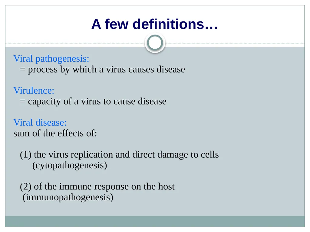
slide3. A few definitions… Viral pathogenesis:
= process by which a virus causes disease
Virulence:
= capacity of a virus to cause disease
Viral disease:
sum of the effects of:
(1) the virus replication and direct damage to cells
(cytopathogenesis)
(2) of the immune response on the host (immunopathogenesis)<br>
slide4. Viral pathogenesis is the process by which a viral infection leads to disease.
Viral pathogenesis is an abnormal situation of no value to the virus.
The majority of viral infections are subclinical. It is not in the interest of the virus to severely harm or kill the host.
The consequences of viral infections depend on the interplay between a number of viral and host factors.<br>
slide5. The ability of Viruses to cause disease is viewed at two distinct level:
Changes within the infected cell
Process within the infected patient<br>
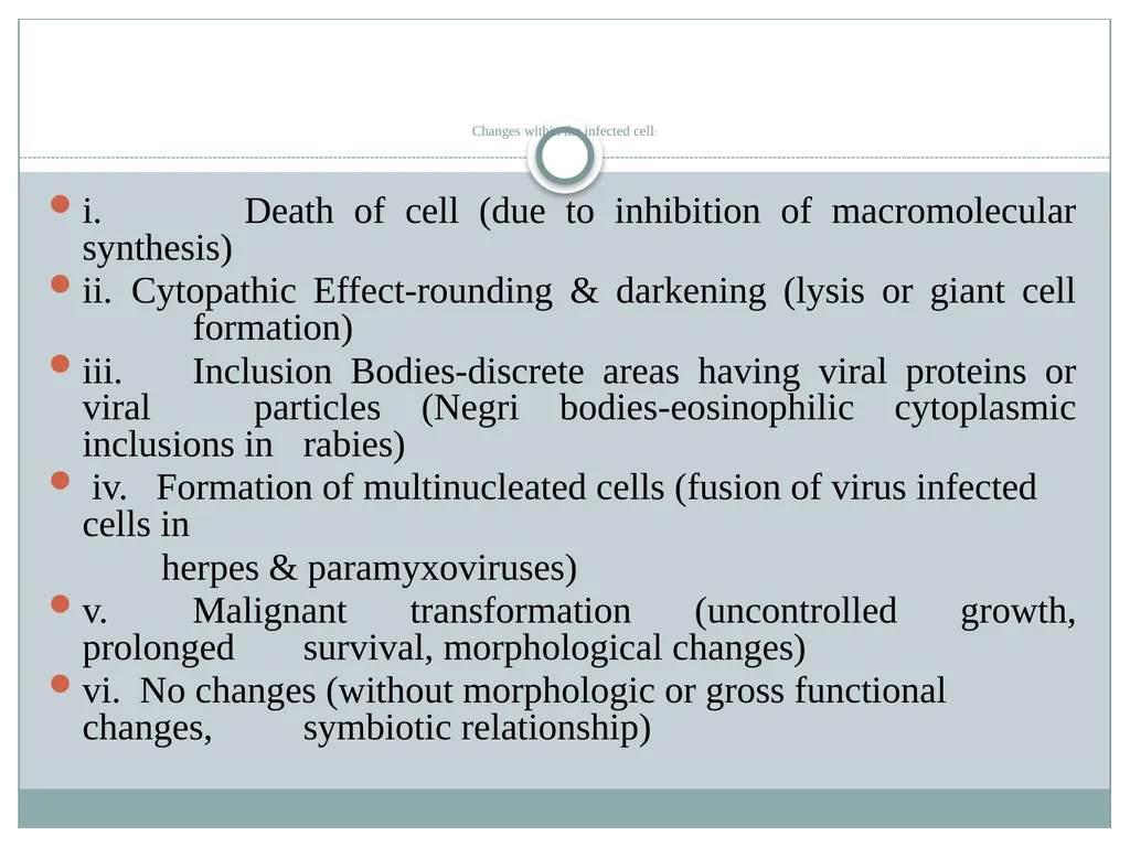
slide6. Changes within the infected cell: i. Death of cell (due to inhibition of macromolecular synthesis)
ii. Cytopathic Effect-rounding & darkening (lysis or giant cell formation)
iii. Inclusion Bodies-discrete areas having viral proteins or viral particles (Negri bodies-eosinophilic cytoplasmic inclusions in rabies)
iv. Formation of multinucleated cells (fusion of virus infected cells in
herpes & paramyxoviruses)
v. Malignant transformation (uncontrolled growth, prolonged survival, morphological changes)
vi. No changes (without morphologic or gross functional changes, symbiotic relationship)<br>
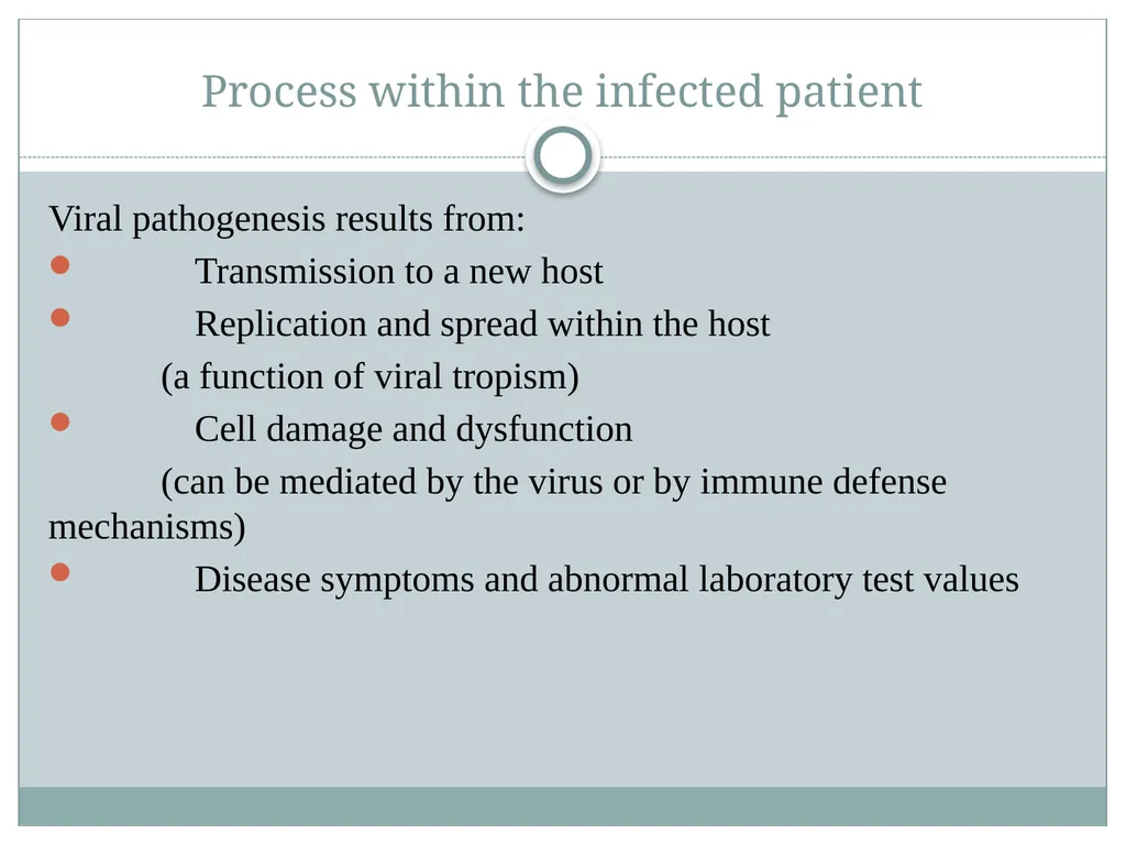
slide7. Process within the infected patient Viral pathogenesis results from:
Transmission to a new host
Replication and spread within the host
(a function of viral tropism)
Cell damage and dysfunction
(can be mediated by the virus or by immune defense mechanisms)
Disease symptoms and abnormal laboratory test values<br>
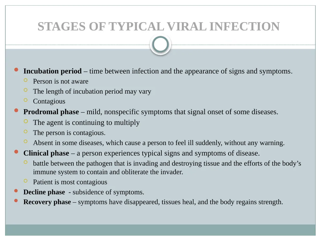
slide8. STAGES OF TYPICAL VIRAL INFECTION Incubation period – time between infection and the appearance of signs and symptoms.
Person is not aware
The length of incubation period may vary
Contagious
Prodromal phase – mild, nonspecific symptoms that signal onset of some diseases.
The agent is continuing to multiply
The person is contagious.
Absent in some diseases, which cause a person to feel ill suddenly, without any warning.
Clinical phase – a person experiences typical signs and symptoms of disease.
battle between the pathogen that is invading and destroying tissue and the efforts of the body’s immune system to contain and obliterate the invader.
Patient is most contagious
Decline phase - subsidence of symptoms.
Recovery phase – symptoms have disappeared, tissues heal, and the body regains strength.<br>
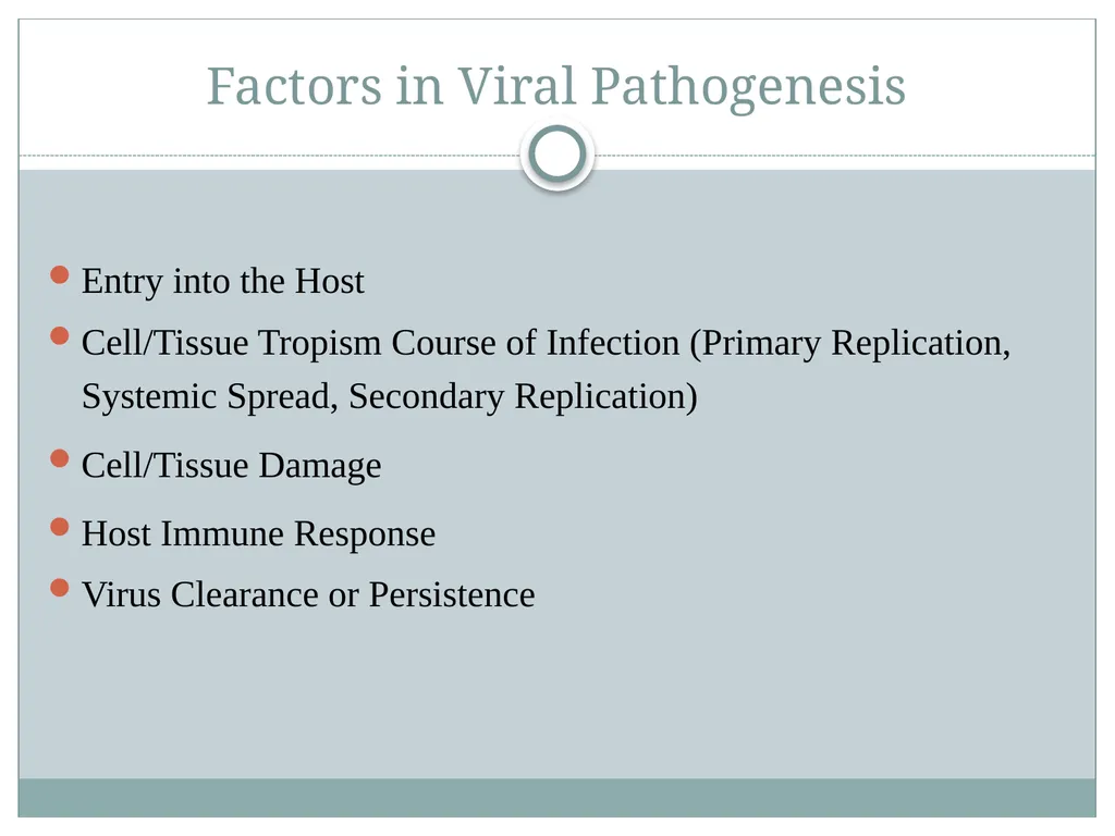
slide9. Factors in Viral Pathogenesis Entry into the Host
Cell/Tissue Tropism Course of Infection (Primary Replication, Systemic Spread, Secondary Replication)
Cell/Tissue Damage
Host Immune Response
Virus Clearance or Persistence<br>
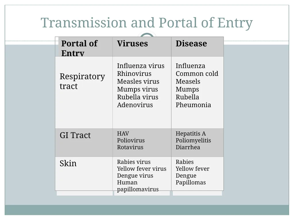
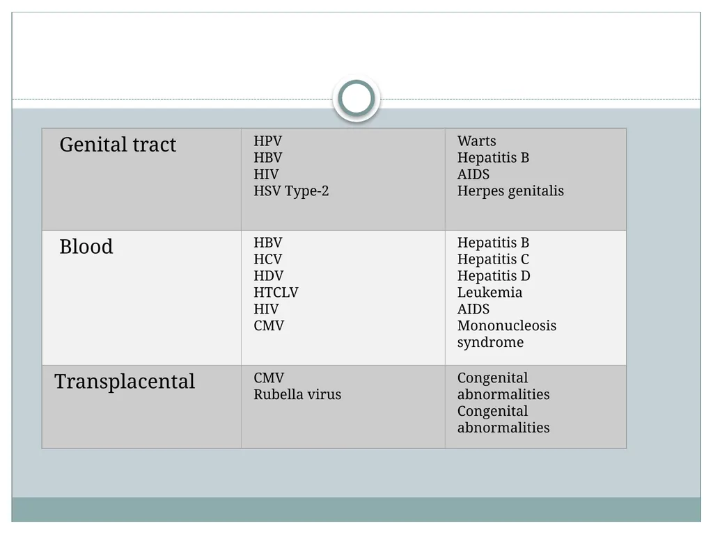
slide10. Transmission and Portal of Entry<br>
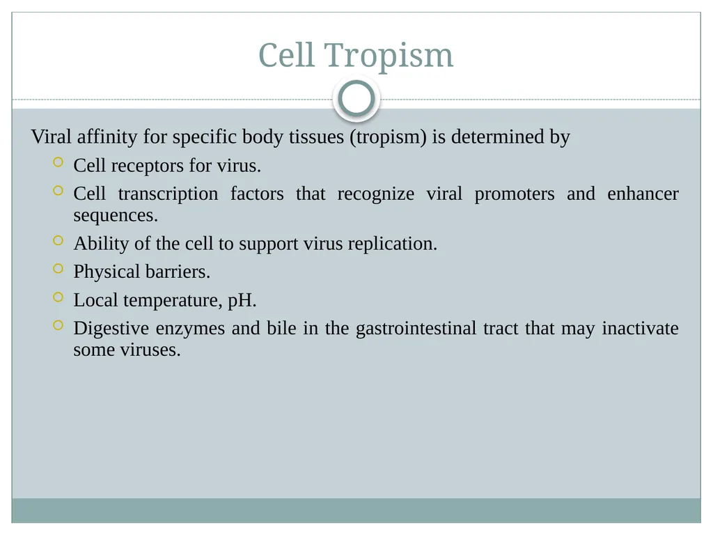
slide12. Cell Tropism Viral affinity for specific body tissues (tropism) is determined by
Cell receptors for virus.
Cell transcription factors that recognize viral promoters and enhancer sequences.
Ability of the cell to support virus replication.
Physical barriers.
Local temperature, pH.
Digestive enzymes and bile in the gastrointestinal tract that may inactivate some viruses.<br>
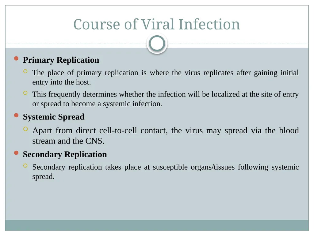
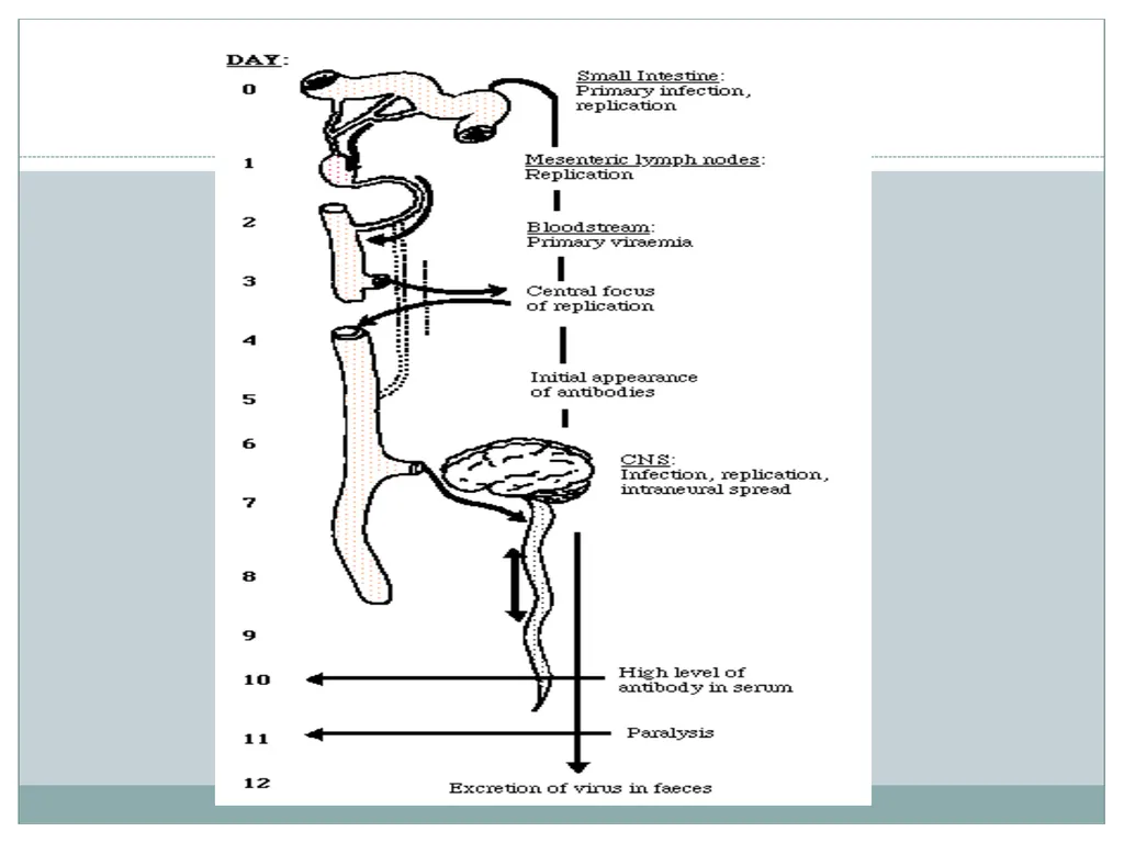
slide13. Course of Viral Infection Primary Replication
The place of primary replication is where the virus replicates after gaining initial entry into the host.
This frequently determines whether the infection will be localized at the site of entry or spread to become a systemic infection.
Systemic Spread
Apart from direct cell-to-cell contact, the virus may spread via the blood stream and the CNS.
Secondary Replication
Secondary replication takes place at susceptible organs/tissues following systemic spread.<br>
slide15. Cell Damage Viruses may replicate widely throughout the body without any disease symptoms if they do not cause significant cell damage or death.
Picornaviruses cause lysis and death of the cells in which they replicate, paralysis or death for Poliovirus.<br>
slide16. Immune Response The immune response to the virus probably has the greatest impact on the outcome of infection.
In the most cases, the virus is cleared completely from the body and results in complete recovery.
In other infections, the immune response is unable to clear the virus completely and the virus persists.
In a number of infections, the immune response plays a major pathological role in the disease.<br>
slide17. Viral Clearance or Persistence The majority of viral infections are cleared but certain viruses may cause persistent infections. There are 2 types of chronic persistent infections.
True Latency - the virus remains completely latent following primary infection e.g. HSV, VZV. Its genome may be integrated into the cellular genome or exists as episomes.
Persistence - the virus replicates continuously in the body at a very low level e.g. HIV, HBV, CMV, EBV.<br>
slide18. Mechanisms of Viral Persistence antigenic variation
immune tolerance, causing a reduced response to an antigen, may be due to genetic factors, pre-natal infection, molecular mimicry
restricted gene expression
down-regulation of MHC class I expression, resulting in lack of recognition of infected cells e.g. Adenoviruses
down-regulation of accessory molecules involved in immune recognition e.g. LFA-3 and ICAM-1 by EBV.<br>
slide19. Cytopathic effect Cytopathic effect or cytopathogenic effect (abbreviated CPE) refers to structural changes in host cells that are caused by viral invasion.
The infecting virus causes lysis of the host cell or when the cell dies without lysis due to an inability to reproduce.
Both of these effects occur due to CPEs. If a virus causes these morphological changes in the host cell, it is said to be cytopathogenic.
Common examples of CPE include rounding of the infected cell, fusion with adjacent cells to form syncytia, and the appearance of nuclear or cytoplasmic inclusion bodies<br>
slide20. CPEs and other changes in cell morphology are only a few of the many effects by cytocidal viruses.
When a cytocidal virus infects a permissive cell, the viruses kill the host cell through changes in cell morphology, in cell physiology, and the biosynthetic events that follow.
These changes are necessary for efficient virus replication but at the expense of the host cell.<br>
slide21. Common types Total destruction:
Total destruction of the host cell monolayer is the most severe type of CPE.
To observe this process, cells are seeded on a glass surface and a confluent monolayer of host cell is formed. Then, the viral infection is introduced.
All cells in the monolayer shrink rapidly, become dense in a process known as pyknosis, and detach from the glass within three days.
This form of CPE is typically seen with enteroviruses<br>
slide22. Subtotal destruction of the host cell monolayer is less severe than total destruction.
Similarly to total destruction, this CPE is observed by seeding a confluent monolayer of host cell on a glass surface then introducing a viral infection.
Subtotal destruction characteristically shows detachment of some but not all the cells in the monolayer.
It is commonly observed with some togaviruses (Chikungunya, rubella), some picornaviruses , and some types of paramyxoviruses.<br>
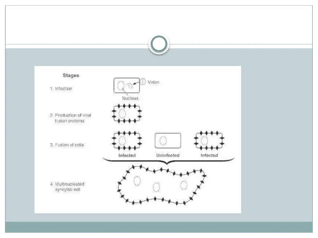
slide23. Syncytium
Syncytium is also known as cell fusion and polykaryon formation.
With this CPE, the plasma membranes of four or more host cells fuse and produce an enlarged cell with at least four nuclei.
Although large cell fusions are sometimes visible without staining, this type of CPE is typically detected after host cell fixation and staining.
Herpesviruses characteristically produce cell fusion as well as other forms of CPE.
Some paramyxoviruses may be identified through the formation of cell fusion as they exclusively produce this CPE.<br>
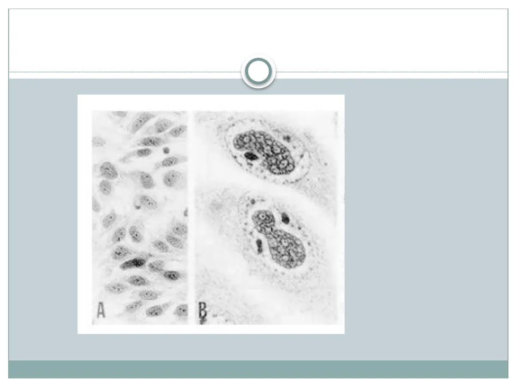
slide25. Inclusion bodies:
Inclusion bodies – insoluble abnormal structures within cell nuclei or cytoplasm – may only be seen with staining as they indicate areas of altered staining in the host cells.
Typically, they indicate the areas of the host cell where viral protein or nucleic acid is being synthesized or where virions are being assembled.
They may be single or multiple, small or large, and round or irregularly shaped. They may also be intra nuclear or intracytoplasmic<br>
slide2. “No virus is known to do good. It has been well said that a virus is a piece of bad news wrapped up in protein.”<br>
slide3. A few definitions… Viral pathogenesis:
= process by which a virus causes disease
Virulence:
= capacity of a virus to cause disease
Viral disease:
sum of the effects of:
(1) the virus replication and direct damage to cells
(cytopathogenesis)
(2) of the immune response on the host (immunopathogenesis)<br>
slide4. Viral pathogenesis is the process by which a viral infection leads to disease.
Viral pathogenesis is an abnormal situation of no value to the virus.
The majority of viral infections are subclinical. It is not in the interest of the virus to severely harm or kill the host.
The consequences of viral infections depend on the interplay between a number of viral and host factors.<br>
slide5. The ability of Viruses to cause disease is viewed at two distinct level:
Changes within the infected cell
Process within the infected patient<br>
slide6. Changes within the infected cell: i. Death of cell (due to inhibition of macromolecular synthesis)
ii. Cytopathic Effect-rounding & darkening (lysis or giant cell formation)
iii. Inclusion Bodies-discrete areas having viral proteins or viral particles (Negri bodies-eosinophilic cytoplasmic inclusions in rabies)
iv. Formation of multinucleated cells (fusion of virus infected cells in
herpes & paramyxoviruses)
v. Malignant transformation (uncontrolled growth, prolonged survival, morphological changes)
vi. No changes (without morphologic or gross functional changes, symbiotic relationship)<br>
slide7. Process within the infected patient Viral pathogenesis results from:
Transmission to a new host
Replication and spread within the host
(a function of viral tropism)
Cell damage and dysfunction
(can be mediated by the virus or by immune defense mechanisms)
Disease symptoms and abnormal laboratory test values<br>
slide8. STAGES OF TYPICAL VIRAL INFECTION Incubation period – time between infection and the appearance of signs and symptoms.
Person is not aware
The length of incubation period may vary
Contagious
Prodromal phase – mild, nonspecific symptoms that signal onset of some diseases.
The agent is continuing to multiply
The person is contagious.
Absent in some diseases, which cause a person to feel ill suddenly, without any warning.
Clinical phase – a person experiences typical signs and symptoms of disease.
battle between the pathogen that is invading and destroying tissue and the efforts of the body’s immune system to contain and obliterate the invader.
Patient is most contagious
Decline phase - subsidence of symptoms.
Recovery phase – symptoms have disappeared, tissues heal, and the body regains strength.<br>
slide9. Factors in Viral Pathogenesis Entry into the Host
Cell/Tissue Tropism Course of Infection (Primary Replication, Systemic Spread, Secondary Replication)
Cell/Tissue Damage
Host Immune Response
Virus Clearance or Persistence<br>
slide10. Transmission and Portal of Entry<br>
slide12. Cell Tropism Viral affinity for specific body tissues (tropism) is determined by
Cell receptors for virus.
Cell transcription factors that recognize viral promoters and enhancer sequences.
Ability of the cell to support virus replication.
Physical barriers.
Local temperature, pH.
Digestive enzymes and bile in the gastrointestinal tract that may inactivate some viruses.<br>
slide13. Course of Viral Infection Primary Replication
The place of primary replication is where the virus replicates after gaining initial entry into the host.
This frequently determines whether the infection will be localized at the site of entry or spread to become a systemic infection.
Systemic Spread
Apart from direct cell-to-cell contact, the virus may spread via the blood stream and the CNS.
Secondary Replication
Secondary replication takes place at susceptible organs/tissues following systemic spread.<br>
slide15. Cell Damage Viruses may replicate widely throughout the body without any disease symptoms if they do not cause significant cell damage or death.
Picornaviruses cause lysis and death of the cells in which they replicate, paralysis or death for Poliovirus.<br>
slide16. Immune Response The immune response to the virus probably has the greatest impact on the outcome of infection.
In the most cases, the virus is cleared completely from the body and results in complete recovery.
In other infections, the immune response is unable to clear the virus completely and the virus persists.
In a number of infections, the immune response plays a major pathological role in the disease.<br>
slide17. Viral Clearance or Persistence The majority of viral infections are cleared but certain viruses may cause persistent infections. There are 2 types of chronic persistent infections.
True Latency - the virus remains completely latent following primary infection e.g. HSV, VZV. Its genome may be integrated into the cellular genome or exists as episomes.
Persistence - the virus replicates continuously in the body at a very low level e.g. HIV, HBV, CMV, EBV.<br>
slide18. Mechanisms of Viral Persistence antigenic variation
immune tolerance, causing a reduced response to an antigen, may be due to genetic factors, pre-natal infection, molecular mimicry
restricted gene expression
down-regulation of MHC class I expression, resulting in lack of recognition of infected cells e.g. Adenoviruses
down-regulation of accessory molecules involved in immune recognition e.g. LFA-3 and ICAM-1 by EBV.<br>
slide19. Cytopathic effect Cytopathic effect or cytopathogenic effect (abbreviated CPE) refers to structural changes in host cells that are caused by viral invasion.
The infecting virus causes lysis of the host cell or when the cell dies without lysis due to an inability to reproduce.
Both of these effects occur due to CPEs. If a virus causes these morphological changes in the host cell, it is said to be cytopathogenic.
Common examples of CPE include rounding of the infected cell, fusion with adjacent cells to form syncytia, and the appearance of nuclear or cytoplasmic inclusion bodies<br>
slide20. CPEs and other changes in cell morphology are only a few of the many effects by cytocidal viruses.
When a cytocidal virus infects a permissive cell, the viruses kill the host cell through changes in cell morphology, in cell physiology, and the biosynthetic events that follow.
These changes are necessary for efficient virus replication but at the expense of the host cell.<br>
slide21. Common types Total destruction:
Total destruction of the host cell monolayer is the most severe type of CPE.
To observe this process, cells are seeded on a glass surface and a confluent monolayer of host cell is formed. Then, the viral infection is introduced.
All cells in the monolayer shrink rapidly, become dense in a process known as pyknosis, and detach from the glass within three days.
This form of CPE is typically seen with enteroviruses<br>
slide22. Subtotal destruction of the host cell monolayer is less severe than total destruction.
Similarly to total destruction, this CPE is observed by seeding a confluent monolayer of host cell on a glass surface then introducing a viral infection.
Subtotal destruction characteristically shows detachment of some but not all the cells in the monolayer.
It is commonly observed with some togaviruses (Chikungunya, rubella), some picornaviruses , and some types of paramyxoviruses.<br>
slide23. Syncytium
Syncytium is also known as cell fusion and polykaryon formation.
With this CPE, the plasma membranes of four or more host cells fuse and produce an enlarged cell with at least four nuclei.
Although large cell fusions are sometimes visible without staining, this type of CPE is typically detected after host cell fixation and staining.
Herpesviruses characteristically produce cell fusion as well as other forms of CPE.
Some paramyxoviruses may be identified through the formation of cell fusion as they exclusively produce this CPE.<br>
slide25. Inclusion bodies:
Inclusion bodies – insoluble abnormal structures within cell nuclei or cytoplasm – may only be seen with staining as they indicate areas of altered staining in the host cells.
Typically, they indicate the areas of the host cell where viral protein or nucleic acid is being synthesized or where virions are being assembled.
They may be single or multiple, small or large, and round or irregularly shaped. They may also be intra nuclear or intracytoplasmic<br>