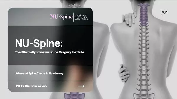
/
ADVANCES  IN  SPINE  IMAGING ON MRI ADVANCES  IN  SPINE  IMAGING ON MRI

ADVANCES IN SPINE IMAGING ON MRI - PowerPoint Presentation

williams
williams . @williams
Follow
27 views
Uploaded On 2024-02-09

ADVANCES IN SPINE IMAGING ON MRI - PPT Presentation

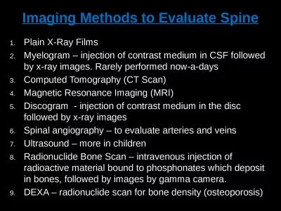
A noninvasive procedure to evaluate different types of tissue including the spinal cord intervertebral disks spaces between the vertebrae through which the nerves travel distinguish healthy tissue from diseased tissue ID: 1045359

dti diffusion nerve imaging diffusion dti imaging nerve images water spinal tissue matter mri white adc movement infiltration disc

Share:

Link:

Embed:

Download Presentation from below link

Download Presentation The PPT/PDF document "ADVANCES IN SPINE IMAGING ON MRI" is the property of its rightful owner. Permission is granted to download and print the materials on this web site for personal, non-commercial use only, and to display it on your personal computer provided you do not modify the materials and that you retain all copyright notices contained in the materials. By downloading content from our website, you accept the terms of this agreement.


Presentation Transcript

1. ADVANCES IN SPINE IMAGING ON MRI

2. A noninvasive procedure to evaluate different types of tissue, including the spinal cordintervertebral disks spaces between the vertebrae through which the nerves travel distinguish healthy tissue from diseased tissue.

3. Screen Shot 2019-02-11 at 12.44.17 PM

4. DIFFUSION ADC

5. To depict cellularityMalignant infiltration  dense compact tumor infiltration  inhibits free movement of water molecules, causing diffusion restriction The areas of restricted diffusion appear bright on DW-MRI and dark on ADC mapping

6.

7. T2* Imaging

8. T2* Imaging

9. utilize the physics of water molecule relaxation within a magnetic field to produce images  highlights the presence of proteoglycans, hydration levelConventional T2 weighted sagittal MRI sequences are used to create a subjective grading scale for disc health based on morphological features  lack specificityQuantitative T2* mapping may offer promise in more accurately determining the health of disc

10. Post Processed

11. Lumbar Plexus Imaging3D VIBEDESSSTIR 3D SPACE- T1-weighted images with high contrast and spatial resolution - relatively short acquisition timeDouble Echo Steady StateHigh SNRenables acquisition of high resolution 3D datasetscan obtain heavily T2W images as well as T1W or FLAIR images

12. Ganglions of lumbar plexus

13. Lateral femoral cutaneous nerveObturator nerve

14. Ilioinguinal nerve

15. Femoral nerve

16. Sciatic nerve

17. Diffusion Tensor Imaging(DTI)DTI is an advanced non-invasive technique of MR which enables visualization as well as qualitative and quantitative assessment of the integrity of the white matter tracts.DTI is used primarily for assessing white matter of the brain and spinal cord, because movement of the water particles in these structures is limited in directions perpendicular to the white matter fibres called anisotropic diffusion.

18. DTI

19.

20. Clinical applications of DTI in spinal cordTRAUMA

21. TUMOR

22. Degenerative myelopathyDemyelinating Infectious diseases

23. UTILITYIncreases diagnostic confidenceAdditional raw dataUltra structural detailing

24. THANK YOU