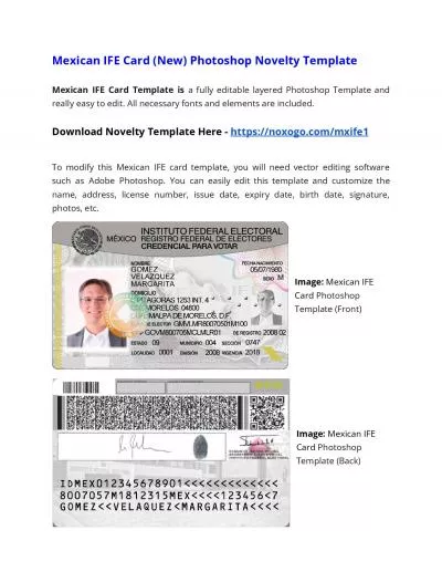
/
The Journal of Neuroscience August 1991 IfE 22992294 The Journal of Neuroscience August 1991 IfE 22992294

The Journal of Neuroscience August 1991 IfE 22992294 - PDF document

willow
willow . @willow
Follow
342 views
Uploaded On 2022-10-26

The Journal of Neuroscience August 1991 IfE 22992294 - PPT Presentation

Mineralocorticoid Hormones Suppress Serotonininduced Hyperpolarization of Rat Hippocampal CA 1 Neurons Marian Joil Wouter Heseni2 and E Ronald de Kloet 145Division of Molecular Biology ID: 960579

binding steroid application receptor steroid binding receptor application aldosterone membrane pyramidal responses neurons ca1 min response corticosterone 1989 kloet

Share:

Link:

Embed:

Download Presentation from below link

Download Pdf The PPT/PDF document "The Journal of Neuroscience August 1991 ..." is the property of its rightful owner. Permission is granted to download and print the materials on this web site for personal, non-commercial use only, and to display it on your personal computer provided you do not modify the materials and that you retain all copyright notices contained in the materials. By downloading content from our website, you accept the terms of this agreement.


Presentation Transcript

The Journal of Neuroscience, August 1991, If(E): 2299-2294 Mineralocorticoid Hormones Suppress Serotonin-induced Hyperpolarization of Rat Hippocampal CA 1 Neurons Marian Joi$l~,~~* Wouter Hesen,i*2 and E. Ronald de Kloet* ‘Division of Molecular Biology, Institute of Molecular Biology and Medical Biotechnology, and *Rudolf Magnus Institute, University of Utrecht, The Netherlands Pyramidal neurons in the rat CA1 hippocampal area contain intracellular mineralocorticoid receptors (Ml%) and gluco- corticoid receptors (GRs) to which the adrenal hormone cor- ticosterone can bind with differential affinity. The pyramidal neurons also have high amounts of SHT,, receptors, which mediate a membrane Corticosteroid hormones, which are secreted by the adrenal gland, readily cross the blood-brain barrier and bind to two types of intracellular receptors in the brain: mineralocorticoid receptors (MRs) and glucocorticoid receptors (GRs; Reul and Kloet, 1985; for reviews, see McEwen et al., 1986; Funder, 1986; de Kloet, 1991). Pyramidal neurons in the rat hippocampal CA1 area are particularly rich in both MRs and GRs (Gerlach and McEwen, 1972; Fuxe et al., 1985; Arriza et al., 1988; van Ee- kelen et al., 1988; Herman et al., 1989): Recent electrophysio- logical studies with in vitro, they specifically affect the spike frequency accommodation and af- terhyperpolarization (AHP) associated with a short depolarizing Received Oct. 16, 1990; revised Jan. 29, 1991; accepted Feb. 20, 1991. This work was supported by C and C Huygens Grant H88-I45 from The Neth- erlands oraanization for scientific research tNW0). We thank W. Sutanto for assistance, Roussel-Uclaf for the gift of RU 38486 RU 28362, Organon International for corticosterone, aldosterone, and 5-CT, Ciba-Geigy for baclofen, and Duphar for spiperone. Correspondence should he addressed to M. J&ls, Department of Experimental Zoology, University of Amsterdam, current

step (Joels and Kloet, 1989, Kerr et al., 1989). Thus, corticosterone induces, via MRs, a decrease in the spike frequency accommodation and AHP (Joels and Kloet, 1990) that is gradually overriden by a GR-mediated increase in the AHP amplitude (Joels and Kloet, 1989). In vitro autoradiographical studies have shown that the CA1 area of rat hippocampus displays also a high density of binding sites for 5-HT; in particular, ligands for the 5-HT,, receptor subtype bind strongly to the dorsal CA1 hippocampal region (Deshmukh et al., 1983; Marcinkiewicz et al., 1984; Pazos and Palacios, 1985). Electrophysiological data showed that 5-HT,, agonists induce a Materials and Methods All the experiments (72 different experiments) were performed in male Wistar rats (120-l 70 gm), adrenalectomized under ether anesthesia ap- proximately 1 week before the electrophysiological experiment, as de- scribed elsewhere (Ratka et al.. 1988: Joels and Kloet. cycle (lights on 8:00 A.M.) and received food and saline (after ADX) ad libitum. Electrophysiology. On the day of the experiment, the rat was placed in a clean cage and decapitated after 30-60 min; trunk blood was col- lected for measurement of plasma corticosterone levels. All adrenal- ectomized animals displayed plasma corticosterone levels well below 1 fig corticosterone per 100 ml plasma. The brain was removed from the skull and dipped in ice-cold artificial cerebrospinal fluid (ACSF) of the following composition: 124 mM NaCl, 3.5 mM KCl, 1.25 mM NaH,PO,, 1.5 mM MgS0,.7H,O, 2.0 mM CaCl,, 25 mM NaHCO,, and mM The Journal of Neuroscience, August 1991, 1 f(9) 2289 glucose. Slices (350 pm) were prepared from the dorsal hippocampus on McIlwain tissue chopper. The slices were placed in a perfusion system, submerged, and continuously superfused (2-3 ml/min) with warm (32°C) oxygenated (95% O,, 5% CO,) ACSF. Intracellular recording was performed with 4 M KAc-filled micropi- p

ettes (impedance, 80-150 MQ), which were placed in the CA1 pyra- midal cell layer. The signals were transferred to an Axoclamp 2A am- plifier ( 10 x amplification) and continuously displayed on Gould digital oscilloscope. The membrane potential and applied current were regis- tered on Gould 2200 chart recorder and, in some cases, on Vetter videocassette recorder for later analysis. From each neuron, we recorded the resting membrane potential, input resistance (from current-voltage relationship with 150-msec current pulses of -0.6 to 0.2 nA), sponta- neous activity, and spike accommodation and AHP evoked by a Membrane bindina assay. Hippocampal tissue slices (350 pm) were prepared at 4°C with; McIlwaintissue chopper. The slices were perfused for 1 hr ACSF equilibrated with 95% O,, 5% &M, pH 7.7) incubated for 10 min at 37°C to eliminate endogenous 5-HT, and centrifugated again at 50,000 x g for 10 min (4°C). Membrane pellets were stored overnight at -80°C and resuspended in 50 vol of incubation buffer containing Tris-HCl (50 mM, pH 7.7), CaCl, (4 mM), ascorbic acid (O.l%), and pargyline (10 PM). The suspension was homogenized and incubated at 37°C for 15 min. The membrane binding assay was performed as described by Gozlan et PM 5-HT. Membranes were collected by filtration through Whatman GF/B filters. The filters were washed with 2 x 5 ml of ice-cold Tris-HCl(50 mM, pH 7.7). placed in 2 ml of soluene overnight, and counted after addition of the appropriate scin- tillation fluid. Nonspecific binding under these conditions was less than 15% of the total binding. In the absence of membranes, less than 1% of total radioactivity was bound by the filter. All binding assays were performed in triplicate. In a total of six experiments, we compared 5-HT,, receptor binding characteristics after aldosterone treatment with binding in nontreated tissue. Results In total, we recorded from 126 CA 1 cells, ident

ified as pyramidal neurons as described by Schwartzkroin (1975, Average resting KM 5-HT. This con- centration range is in with previous reports on 5-HT in intact animals (Andrade and Nicoll, 1987; Joels et al., 1990). We observed that application of 30 nM corticosterone, par- ticularly with concomitant administration of 500 nM of the se- lective GR antagonist RU 38486 (Philibert, 1984; Gagne et al., 1985), markedly reduced the hyperpolarizing response to 10 PM 5-HT recorded 14 hr after steroid application (Fig. 1A). On average, application of corticosterone in the presence of the GR antagonist RU 38486, I I I I and 500 5 d or with 3 no and an and 2 but not A 5CT 1 J ADX I 10 - 5CT 1 rnin ADX 1 aldosterone B 5HT SHT 5CT Figure 3. Responses to a 5-HT,, agonist are diminished by aldoste- rone. A, The response of a CA1 pyramidal cell to the 5-HT,, agonist 5-CT (0.1 PM) is diminished approximately 90 min after a 20-min perfusion with 3 nM aldosterone (lower record) when compared with the response obtained before steroid treatment (upper record). Duration of the 5-CT application is horizontal bar. Downward deflections represent the voltage responses to constant current pulses (0.3 nA, 150 msec). In the upper record, the membrane potential was brought to the pretreatment level by injection of positive DC current just before 5-CT administration was terminated. As illustrated here, responses to 5-CT, in particular those recorded before steroid appli- cation, were not in all cases readily reversible. B, Membrane hyper- polarizations (left) and resistance decreases (right) obtained in the same set of CA1 pyramidal neurons (n = 5) by 10 PM 5-HT and 0.1 MM of the 5-HT,, ligand 5-CT (mean + SEM). Compared to the responses obtained before steroid perfusion (open bars), both 5-HT- and 5-CT- evoked responses (gray/black and hatched bars, respectively) were sig- nificantly reduced after a 20-mi

n perfusion with 3 nM aldosterone. The data were tested with a Student’s t test (significance, p 0.05). membrane hyperpolarization (2.0 -t 0.7 mV; 11 neurons) and resistance decrease (21.0 -t 2.0%) obtained after 3 nM aldo- sterone was still significantly (Student’s t test, p 0.05) smaller than in neurons recorded before steroid application (5.7 f 0.4 mV and 32.0 f 1.9% for changes in membrane potential and resistance, respectively; 24 neurons). Andrade et al. (1986) have suggested that the 5-HT,, receptor is linked directly through a G-protein to the same K+ conduc- 0 Figure 4. 5-HT,, receptor data point is the mean of a triplicate determination. Specific binding in this experiment is total binding minus the binding persisting in the presence of 10 PM 5-HT (nonspecific binding). Bound/Free, bound over free )H-80H-DPAT. Without steroid treatment: K, = 1.51 nM, B,,, = 1.67 pMr/mg protein (r,,, = 0.99; open diamonds); after aldosterone application: KD = 1.25 �nM &a. = 1.53 phl/mg protein (r,,, 1 .O; solid diamonds). sponses, because neither the depolarization nor the decrease in accommodation, which are both induced by 5-HT through a noncharacterized receptor (Andrade and Nicoll, 1987), was af- fected by the MR ligands. The fact that the steroid action has a slow onset and persists for many hours in vitro after termi- nation of the steroid application is compatible with the notion that it involves an MR-mediated genomic action rather than a direct membrane-associated event. In our attempt to establish the target for the steroid in the pathway from 5-HT receptor binding to opening of K+ channels, we observed that the 5-HT,, receptor-binding properties were only marginally affected by the steroid B,,, observed after aldosterone treatment can fully explain the rather large aldosterone-induced reduction of responses to 5-HT. The MR-linked effect on the 5-HT response is probab

ly also not due to receptor desensiti- zation, because (1) repeated application of 5-HT, either before or after steroid application, yielded very stable responses, and (2) 5-HT responses were significantly reduced even when the slice had not been exposed previously to 5-HT. Finally, it is unlikely that the steroid effect is only aimed at the K+ conduc- tance linked to the 5-HT,, receptor. Thus, it has been proposed that the 5-HT,, receptor is, through a G-protein, coupled to the same in vivo administration of estrogen enhances 5-HT responses in the hip- pocampal slice (Beck et al., 1989), probably by changing 5-HT- mediated inhibition of adenylate cyclase activity (Clarke and Maayani, 1990). It should be noted, though, that differences in The Journal of Neuroscience, August 1991, 1 f(9) 2293 A Al l!zz ADX A2 badofen I ADX aldostercne 110 mV - 1 tin Figure 5. Responses to baclofen are not diminished by aldosterone. A, Response to 3 PM baclofen obtained in a CA1 pyramidal neuron recorded before steroid application (Al) and after a brief application of 3 nM aldosterone (A2, upper fruce). Note that the baclofen response is not affected by aldosterone treatment, while the hyperpolarization induced by 10 PM 5-HT for the estrogen- and MR-mediated effects and the fact that estrogen enhances whereas mineralocorticoids suppress 5-HT responsiveness suggest that the two steroids may well have different mechanisms of action. Alternatively, the effect of min- eralocorticoids on the 5-HT response could also be caused by actions on synthesis or activation of regulatory proteins that are involved in processes between the 5-HT,, receptor and the K channels linked to the 5-HT,, receptor, such as G-proteinxou- pled phosphorylation (Saito et al., 1989). All our experiments were performed in hippocampal slices and employed in vitro administration of steroids, with the ad- vantage to study selectively steroid-transmitte

r in vivo corticosterone application showed that 5-HT receptor binding in the dorsal CA 1 area was reduced by the steroid (Biegon et al., 1985; de Kloet et al., 1986). The presently observed weak steroid effects on 5-HT binding may be partly explained by the fact that, in contrast to previous studies, our experiments were performed with in vitro appli- cation of the steroid to slices, so that (1) steroids cannot affect 5-HT synthesis in the midbrain, (2) 5-HT input fibers to the hippocampus are no longer intact, and (3) in vivo application of steroid hormones used in the previous studies and the presently used in vitro steroid administration may well have resulted in differences in time course for the MR-mediated action on the 5-HT system. In an earlier study, we have shown that corticosterone reduces the norepinephrine-evoked reduction of cell firing accommo- dation in CA1 pyramidal neurons (Joels and Kloet, 1989). Compounds selective for GRs could mimic the effect of corti- costerone on this response. Because accommodation in CA1 pyramidal Andrade R, Nicoll RA (1987) Pharmacologically distinct actions of serotonin on single pyramidal neurones of the rat hippocampus re- corded in vitro. J Physiol (Lond) 394:99-l 24. Andrade R, Malenka RC, Nicoll RA (1986) A G binding protein couples serotonin and GABA, receptors to the same channels in hippocampus. Science 243: 126 l-l 265. Arriza JL, Simerly RB, Swanson L (1988) Neuronal mineralocorticoid as a mediator of glucocorticoid response. Neuron 1:887-900. Azmitia EC, McEwen BS (1974) Adrenalcortical influence on rat brain tryptophan hydroxylase activity. Brain Res 78:29 l-302. Beck SG (1989) 5Carboxyamidotryptamine mimics only the 5-HT elicited hyperpolarization of hippocampal pyramidal cells via 5-HT,, receptor. Neurosci Lett 99:101-106. Beck SG, Clarke WP, Goldfarb J (1989) Chronic estrogen effects on 5-hydroxytryptamine mediated responses DD 95-165.