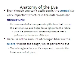
/
Light in Medicine    Even though man is now very efficient at making artificial Light in Medicine    Even though man is now very efficient at making artificial

Light in Medicine Even though man is now very efficient at making artificial - PowerPoint Presentation

winnie
winnie . @winnie
Follow
27 views
Uploaded On 2024-02-02

Light in Medicine Even though man is now very efficient at making artificial - PPT Presentation

light the sun is still the major source of light in the world The sun is both beneficial and hazardous to our health Light has some interesting properties many of which are used in ID: 1044064

laser light visible wavelengths light laser wavelengths visible medicine skin energy absorbed beam sun called retina heat tissue ultraviolet

Share:

Link:

Embed:

Download Presentation from below link

Download Presentation The PPT/PDF document "Light in Medicine Even though man is ..." is the property of its rightful owner. Permission is granted to download and print the materials on this web site for personal, non-commercial use only, and to display it on your personal computer provided you do not modify the materials and that you retain all copyright notices contained in the materials. By downloading content from our website, you accept the terms of this agreement.


Presentation Transcript

1. Light in Medicine

2. Even though man is now very efficient at making artificial light, the sun is still the major source of light in the world. The sun is both beneficial and hazardous to our health.Light has some interesting properties, many of which are used in medicine: -1. The speed of light changes when it goes from material into another. The ratio of the speed of light in a vacuum to its speed in a given material is called the index of refraction. If a light beam meets a new material at an angle other than perpendicular, it bends, or is refracted. This property permits light to be focused and is the reason we can read and see objects clearly.

3. 2. Light behaves both as a wave and as a particle. As a wave it produces interference and diffraction, which are of minor importance in medicine. As a particle it can be absorbed by a single molecule. When a light photon is absorbed its energy is used in various ways. It can cause a chemical change in the molecule that in turn can cause an electrical change. This is basically what happens when a light photon is absorbed in one of the sensitive cells of the retina (the light-sensitive part of the eye). The chemical change in a particular point of the retina triggers an electrical signal to the brain to inform it that a light photon has been absorbed at that point.

4. 3. When light is absorbed, its energy generally appears as heat. This property is the basic for the use in medicine of IR light to heat tissues. Also, the heat produced by laser beams is used to "weld" a detached retina to the back of the eyeball and to coagulate small blood vessels in the retina.

5. 4. Sometimes when photon is absorbed, a lower energy light photon is emitted. This property is known as fluorescence, it is the basis of the fluorescent lightbulb. Certain materials fluoresce in the presence of UV light, sometimes called "black light," and give off visible light. The amount of fluorescence and the color of the emitted light depend on the wavelength of the UV light and on the chemical composition of the material that is fluorescing. One way fluorescence is used in medicine is in the detection of porphyria, a condition in which the teeth fluoresce red when irradiated with UV light. Another important application is in fluorescent microscopes.

6. 5. Light is reflected to some extent from all surfaces. There are two types of reflection. Diffuse reflection occurs when rough surfaces scatter the light in many directions. Specular reflection is more useful type of reflection; it is obtained from very smooth shiny surfaces such as mirrors where the light is reflected at an angle that is equal to the angle at which it strikes the surface. Mirrors are used in many medical instruments.

7. Measurement of light and its units The three general categories of light-UV, visible, and IR-are defined in terms of their wavelengths. Wavelengths of light used to be measured in microns (1μ=10-6m) or in angstroms (1Å=10-10 m), but at present the recommended unit is the nanometer (1nm=10-9m). Ultraviolet light has wavelengths from about 100 to 400 nm; visible light extends from about 400 to 700 nm; and IR light extents from about 700 to over 104 nm.

8. Visible light is measured in photometric units that relate to how light is seen by the average human eye. In photometry the quantity of light striking a surface is called illuminance and the intensity of a light source is called its luminance. All light radiation, including UV and IR radiation, can be measured in radiometric units. In radiometry the quantity of light striking a surface is called irradiance and the intensity of a light source is its radiance. The wavelengths of light fit into the whole spectrum of electromagnetic radiation. Note that light has wavelengths much shorter than TV and radio waves but much longer than X-rays and gamma rays.

9. Applications of visible light in medicine An obvious use of visible light in medicine is to permit the physician to obtain visual information about the patient regarding, for example, the color of his skin and the presence of abnormal structures in or on his body. It is quite easy for a physician to examine the skin under normal lighting conditions, but when she wishes to look into a body opening she is faced with the practical problem of getting light into the opening without obstructing the view. Like a lot of tricks, this one is done with mirrors. The curved surface focuses the light at the region of interest. More sophisticated instruments, such as the ophthalmoscope for looking into the eyes and the otoscope for looking into the ears, use basically the same principle.

10. A number of instruments, called endoscopes, are used for viewing internal body cavities. Special purpose endoscopes are often given names indicating their purpose. For example, cystoscopes are used to examine the bladder, proctoscopes are used for examining the rectum, and bronchoscopes are used for examining the air passages into the lungs. Some endoscopes are rigid tubes with a light source to illuminate the area of interest. Many of them are equipped with optical attachments to magnify the tissues being studied.

11. The development of fiberoptic techniques permitted the construction of flexible endoscopes. Flexible endoscopes can be used to obtain information from regions of the body that cannot be examined with rigid endoscopes, such as the small intestine and much of the large intestine. Some flexible endoscopes are over a meter in length. The image obtained with a flexible endoscope is not as good as that obtained with a rigid endoscope, but often the only alternative to a flexible endoscopic examination is exploratory surgery. Flexible endoscopes usually have an opening or channel that permits the physician to take samples of the tissues (biopsies) for later microscopic examination.

12. Since light contains energy that largely appears as heat when it is absorbed, there is a limit on the amount of light that can be used in endoscopy. For endoscopy, the heating can be reduced by reducing the IR light from the source with special IR absorbing glass filters. In this cold-light endoscopy the light source contains very little IR radiation and the heating of tissues is minimized. Transillumination is the transmission of light through the tissues of the body. Most of us have at one time or another shone a flashlight through our fingers to see the red glow that is produced. The glow is primarily red because most of the other colors in the beam are absorbed by the red blood cells; the red light is the only important component that is transmitted.

13. Transillumination is used clinically in the detection of hydrocephalus (water-head) in infants. Since the skull of young infants is not fully calcified, light is able to penetrate to the inside of the skull; if there is an excess of relatively clear cerebrospinal fluid (CSF) in the skull, light is scattered to different parts of the skull producing patterns characteristic of hydrocephalus. The special transilluminating device uses a 150W projection bulb as the light source. The device has a two-position trigger switch. The infant is taken into a dark room for the study; after a few minutes of dark adaptation the physician pulls the trigger to its first position, which turns on a red light that permits him to find the patient. The physician then points the barrel at the part of the head to be studied and pulls the trigger to the second position, which turns off the red light and turns on the intense white light used for the study. Infrared absorbing glass in the beam removes almost all of the IR radiation so that the light striking the infant is primarily visible light.

14. The physician can then insert a needle attached to a syringe into the area of collapse to remove the air between the lung and chest wall, causing the lung to reinflate. Transillumination is also used to detect pneumothorax (collapsed lung) in infants. The bright light penetrates the thin front chest wall and reflects off the back chest wall to indicate the degree of pneumothorax. The sinuses, the gums, the breasts, and the testes have also been studied with transillumination.

15. Visible light has an important therapeutic use. Since light is a form of energy and is selectively absorbed in certain molecules, it should not be surprising that it can cause important physiological effects. Many premature infants have jaundice, a condition in which an excess of bilirubin is excreted by the liver into the blood. Relatively recently (1958) it was discovered that most premature infants recover from jaundice if their bodies are exposed to visible light (phototherapy).

16. Applications of ultraviolet and infrared light in medicine The wavelengths adjacent to the visible spectrum also have important uses in medicine. Ultraviolet photons have energies greater than visible photons, while IR photons have lower energies. Because of their higher energies, UV photons are more useful than IR photons. Ultraviolet light with wavelengths below about 290nm is germicidal-that is, it can kill germs-and it is sometimes used to sterilize medical instruments. Ultraviolet light also produces more reactions in the skin than visible light. Some of these reactions are beneficial, and some are harmful. One of the major beneficial effects of UV light from the sun is the conversion of molecular products in the skin into vitamin D. Dermatologists have also found that UV light improves certain skin conditions.

17. Ultraviolet light from the sun affects the melanin in the skin to cause tanning. However, UV light can produce sunburn as well as tan the skin. The wavelengths that produce sunburn are around 300nm, just at the edge of the solar spectrum. The amount of 300nm light in the sun's spectrum depends on the amount of atmosphere that the sunlight must pass through. Ordinary window glass permits some near UV to be transmitted but absorbs the sunburn component.

18. Solar UV light is also the major cause of skin cancer in humans. The high incidence of skin cancer among people who have been exposed to the sun a great deal, such as fishermen and agricultural workers, may be related to the fact that the UV wavelengths that produce sunburn are also very well absorbed by the DNA in the cells. Skin cancer usually appears on those portions of the body that have received the most sunlight, such as the tip of the nose, the tops of the ears, and the back of the neck.

19. You probably know that the sky is blue because light of short (blue) wavelengths is scattered more easily than light of long wavelengths. Ultraviolet light has even shorter wavelengths than blue light and is scattered even more easily. About half of the UV light hitting the skin on a summer day comes directly from the sun and the other half is scattered from the air in other parts of the sky. Thus you can get a sunburn even when you are sitting in the shade under a small tree. Even when the sky is completely covered with clouds about one half of the UV light gets through. Ultraviolet light cannot be seen by the eye because it is absorbed before it reaches the retina. The large percentage of near-UV light absorbed by the lens may be the cause of some cataracts (opacities of the lens).

20. About half of the energy from the sun is in the IR region. The warmth we feel from the sun is mainly due to the IR component. The IR rays are not usually hazardous even though they are focused by the cornea and lens of the eye onto the retina. However, looking at the sun through a filter (e.g., plastic sunglasses) that removes most of the visible light and allows most of the IR wavelengths through can cause a burn on the retina. Some people have damaged their eyes in this way by looking at the sun during a solar eclipse. Dark glasses absorb varying amounts of the IR and UV rays from the sun.

21. Heat lamps that produce a large percentage of IR light with wavelengths of 1000 to 2000 nm are often used for physical therapy purposes. Infrared light penetrates further into the tissues than visible light and thus is better able to heat deep tissues. Two types of IR photography are used in medicine: reflective IR photography and emissive IR photography. The latter, which uses the long IR heat waves emitted by the body that give an indication of the body temperature, is usually called thermography. Reflective IR photography, which uses wavelengths of 700 to 900 nm to show the patterns of veins just below the skin. Some of these veins are visible to the eye, but many more can be seen on a near-IR photograph of the skin. Since the temperature at the skin depends on the local blood flow, a thermogram with good resolution shows the venous pattern much like a near-IR photograph.

22. There is considerable variation in the venous patterns of normal individuals. Even in the same individual the venous patterns in the two breasts may be quite different. Cancer and other diseases can cause changes in the venous pattern, but these changes can be masked by the normal variations. Also, a layer of fat beneath the skin can reduce the appearance of the venous pattern. Infrared can also be used to photograph the pupil of the eye without stimulating the reflex that changes its size.

23. Lasers in medicine A laser is a unique light source that emits a narrow beam of light of a single wavelength (monochromatic light) in which each wave is in phase with the others near it (coherent light). Laser is an acronym for Light Amplification by Stimulated Emission of Radiation. While the basic theory for lasers was proposed by Albert Einstein in 1917, the first successful laser was not made until 1960, when T. H. Maiman produced a laser beam from a ruby crystal. Since 1960 scientists have made many types of lasers using gases and liquids as well as solids as the laser materials.

24. In a laser, energy that has been stored in the laser material (e.g., ruby) is released as a narrow beam of light-either as a steady beam continuous wave (CW) or as an intense pulse. The beam remains narrow over long distances and can be thought of as an ideal "spot" light. A laser beam can be focused to a spot only a few microns in diameter. When all of the energy of the laser is concentrated in such a small area, the power density (power per unit area) becomes very large. The total energy of a typical laser pulse used in medicine, which is measured in millijoules (mJ), can be delivered in less than a microsecond, and the resultant instantaneous power may be in megawatts. The output of a pulsed laser is usually measured by the heat produced in the detector (calorimetric method). The output of a low-power CW laser is often measured with a photodetector such as a silicon photocell (often called a solar cell).

25. Since in medicine lasers are used primarily to deliver energy to tissue, the laser wavelength used should be strongly absorbed by tissue. The short wavelengths (400 to 600 nm) are always absorbed better than the long wavelengths (~700nm). Laser energy directed at human tissue causes a rapid rise in temperature and can destroy the tissue. The amount of damage to living tissue depends on how long the tissue is at the increased temperature. For example, tissue can withstand 70oC for 1sec. However, not all laser damage is due to heat. It also produces noticeable damage due to photochemical effects.

26. The laser is routinely used in clinical medicine only in ophthalmology. Its effectiveness in treating certain types of cancer and its usefulness as a "bloodless knife" for surgery are under active investigation. Lasers are also being used in medical research for special three-dimensional imaging called holography. In ophthalmology lasers are primarily used for photocoagulation of the retina, that is, heating a blood vessel to the point where the blood coagulates and blocks the vessel.

27. The amount of laser energy needed for photocoagulation depends on the spot size used. In general, the proper dose is determined visually by the ophthalmologist at the time of the treatment. The minimum amount of laser energy that will do observable damage to the retina is called the Minimal Reactive Dose (MRD). For example, the MRD for 50µm spot in the eye is about 2.4mJ delivered in 0.25sec. Photocoagulation is useful for repairing retinal tears or holes that develop prior to retinal detachment. When the retina is completely detached, the laser is of no help. A complication of diabetes that affects the retina, called diabetic retinopathy, can also be treated with photocoagulation.

28. Protective glasses must be worn in medical laser areas to protect the eyes of the patient and the workers. Since the laser energy is concentrated in a narrow beam for long distances, even a reflected beam can be a hazard; thus the walls and other surfaces in a laser installation should have low reflectivity (e.g., flat black paint). The area should have adequate warning signs and a system that prevents outsiders from entering while lasers are in use.

29. Applications of microscopes in medicine There have been few breakthroughs in science that have had as great an impact as the invention of the microscope by Leeuwenhoek (~1670). The use of the microscope in the pathology laboratory is as common as the use of the thermometer in the clinic. The standard light microscope usually can be set at any of several magnifications by changing the power of the eyepiece or of the objective lens. The highest magnification that can be obtained is limited by the wavelength of visible light. Since the wavelengths of visible light range from 400 to 700 nm (0.4 to 0.7 μm), the smallest object that can be resolved is about 1μm in diameter. Since most cells are 5 to 50 μm in diameter, this type of microscope is adequate for resolving all but subcellular objects.

30. If you put a thin slice of tissue under a microscope you will not see much because most cells are transparent to all wavelengths of visible light-red blood cells are an exception. In order to distinguish different cells it is usually necessary to stain them with a chemical that strongly absorbs certain visible wavelengths. Other techniques in addition to staining are useful in microscopy. One technique takes advantage of the different indexes of refraction of different cell parts. Since light travels at different speeds in the various parts of a cell, the phase relationships of the light waves change in passing through a specimen. The phase-contrast microscope takes advantage of this phenomenon to allow cell structures to be seen without the use of stain. In this type of microscope, a light beam that passes through the tissue is combined with a reference beam directed through an optically uniform zone. The combined beams interfere, producing dark areas where there is destructive interference and light areas where there is constructive interference. The darkness depends on the degree of interference.