/
Dr Payal Garg et al GorlinGoltz Syndrome Dr Payal Garg et al GorlinGoltz Syndrome

Dr Payal Garg et al GorlinGoltz Syndrome - PDF document

ximena
ximena . @ximena
Follow
346 views
Uploaded On 2022-10-12

Dr Payal Garg et al GorlinGoltz Syndrome - PPT Presentation

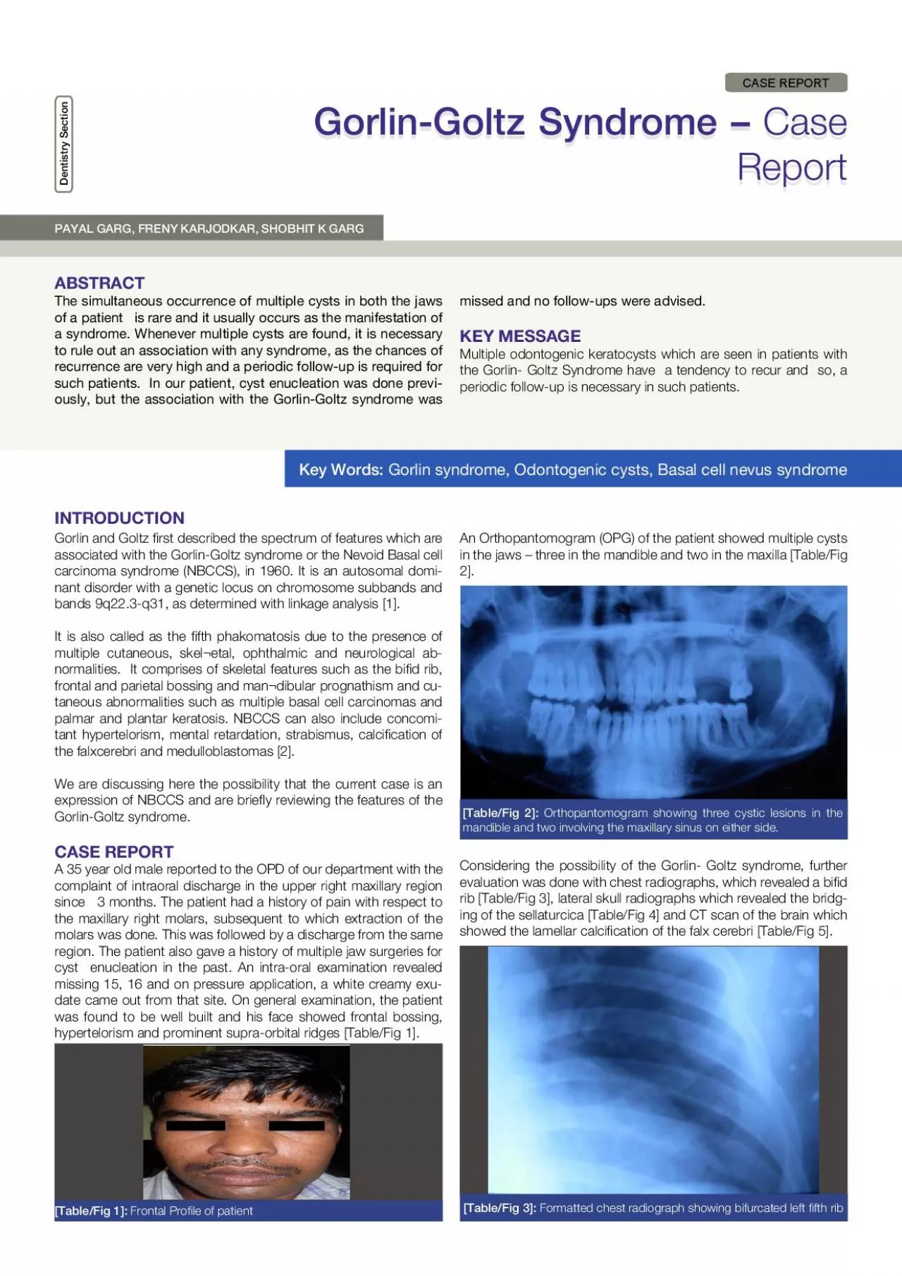
wwwjcdrnet Journal of Clinical and Diagnostic Research 2011 Apr Vol52393395 394 TableFig 4 Formatted lateral skull radiograph showing bridging of the sella turcica TableFig 6 Axial ID: 959041

gorlin syndrome goltz oral syndrome gorlin oral goltz patient fig table nbccs cell multiple basal cysts 150 follow cyst

Share:

Link:

Embed:

Download Presentation from below link

Download Pdf The PPT/PDF document "Dr Payal Garg et al GorlinGoltz Syndrome" is the property of its rightful owner. Permission is granted to download and print the materials on this web site for personal, non-commercial use only, and to display it on your personal computer provided you do not modify the materials and that you retain all copyright notices contained in the materials. By downloading content from our website, you accept the terms of this agreement.


Presentation Transcript

Dr. Payal Garg, et al, Gorlin-Goltz Syndrome www.jcdr.net Journal of Clinical and Diagnostic Research. 2011 Apr, Vol-5(2):393-395 394 [Table/Fig 4]: Formatted lateral skull radiograph showing bridging of the sella turcica. [Table/Fig 6]: Axial CT image showing expansile lytic lesions in maxilla bilaterally with cortical breech. [Table/Fig 7]: Hematoxylin& Eosin (H & E) stained section shows satellite cyst. [Table/Fig 5]: Axial CT brain image showing falx calcication. CT scan of the jaw of the patient revealed ve expansile osteolytic lesions, two in the maxilla and three in the mandible, with thinning and breech of the cortical plates. Maxillary lesions, one on either side, were seen involving the alveolus [Table/Fig 6] and were also seen to be extending into the maxillary sinus and the retrobulbar region of the orbits. An impacted tooth was seen in left maxillary lesion. On the right side, there was loss of the cortex in the maxillary tuberosity region. The mandibular lesion on the right side involved the ramus. On the left side, one lesion was seen in the periapical region with respect to the second premolar and another one was seen in the posterior part of the mandibular body, extending into the angle and the ramus. The lesions also involved the mandibular canal on the left side. Surgical enucleation of all the ve cysts was done, followed by curettage. The tissues were sent for histo-pathological examination and all the cysts were found to be odontogenic keratocysts. DISCUSSION The diagnosis of the Gorlin-Goltz syndrome is made clinically by using the criteria which are suggested by Evans and others [3]. Two major or one major and two minor criteria should be satised for a positive diagnosis. Our patient had three major features of NBCCS, namely bid rib, multiple odontogenic keratocysts in the jaw and lamellar calcica - tion of the falx and minor features such as the bridging of the sella turcica, frontal bossing, prominent supraorbital ridges and hyper - telorism, thus suggesting it to be a case of the Gorlin-Goltz syn - drome. Regarding the site predilection, OKCs which are associ - ated with NBCCS are more common in the mandible with 69% involvement, as compared to 31% in the maxilla. In the mandible, 43% OKCs occurs in the molar ramus region, followed by 18% in the incisor-¬canine area. In the maxilla, 14% OKCs were found to occur in the incisor-¬canine area, followed by molar tuberosity with 12%, 7% in the mandibular premolar region and 3% in the maxillary premolar region. Regarding the male to female ratio, it was 1:0.62 for OKCs which were not associated with NBCCS and 1:1.22 for OKCs in NBCCS. This shows that simple keratocysts are more common in males, but that more females seem to have NBCCS [4]. Based on histopathological studies, parakeratiniza¬tion, intramural epithelial remnants and satellite cysts were found to be more fre - quent among the OKCs which were associated with NBCCS than in the solitary keratocysts [5]. In our patient, the lining of the OKCs revealed the presence of parakeratinization and epithelial remnants in the connective tissue wall, thus indi¬cating the association with NBCCS. Multiple satellite and daughter cysts were also seen by histopathology in our patient [Table/Fig 7]. The term “multiple cysts” does not necessarily mean that the pa - tient must have more than one cyst at a given time; rather it refers to the occurrence of cysts over the life time of the patient [6]. Our patient also had a history of multiple cyst enucleations in the past. www.jcdr.net Dr. Payal Garg, et al, Gorlin-Goltz Syndrome Journal of Clinical and Diagnostic Research. 2011 Apr, Vol-5(2): 393-395 395 REFERENCES: [5] [6] [7] [4] [1] [2] [3] [8] [9] [10] [11] are at risk, medulloblastomas necessitate a neurological examina - tion every 6 months, and intermittent MRIs should be considered in children who are younger than 7 years of age. Odontogenic kera - tocysts require dental follow-up visits, including a periodic radio - graphical evaluation every 6 months, especially in childhood and early adolescence [11]. Finally, the whole family of the patients with the Gorlin-Goltz syn - drome should be examined and genetic counseling should be of - fered, as it is inherited as an autosomal domi

nant disorder. NAME, ADDRESS, TELEPHONE, E-MAIL ID OF THE CORRESPONDING AUTHOR: Dr. Payal Garg Address- H. No. 727, Sector 31, Gurgaon – 122001 E-mail – payalshobhit@rediffmail.com, garg.payal4@gmail.com Phone: 9899259434, 0124- 4117785 AUTHORS: 1. Dr. PAYAL GARG Dept of ODMR, SGT Dental College, Farukh Nagar Road, Vill.Budhera, Gurgaon, Haryana (India) Dept of ODMR, Nair Hospital and Dental College, Mumbai Central, Mumbai, Maharashtra (India) Department of Radiodiagnosis, MEDICITY – The Medanta, Gurgaon, Haryana (India) NAME OF DEPARTMENT(S) / INSTITUTION(S) TO WHICH THE WORK IS ATTRIBUTED: Date of Submission: Dec 06, 2010 Peer Review Completion: Jan 16, 2011 Date of Acceptance: Jan 21, 2011 Date of Final Publication: Apr 11, 2011 DECLARATION ON COMPETING INTERESTS: No competing Interests 2. Dr. FRENY KARJODKAR 3. Dr. SHOBHIT K GARG Gailani MR, Bale SJ, Leffell DJ, et al. Developmental defects in Gorlin syndrome related to a putative tumor suppressor gene on chromosome. Cell 1992;69(1):111-7. Lindeboom JA, Kroon FH, de Vires J, van den Akker HP. Multiple recur¬rent and de novo odontogenickeratocysts associated with oral-facial-digital syndrome. Oral Surg Oral Med Oral Pathol Oral RadiolEndod 2003; 95(4):458–62. Evans DG, Sims DG, Donnai R. Family implications of neonatal Gorlin s syndrome. Arch Dis Child 1991; 66:1162-3. Karthiga S Kannan, Sivapatha B Sundharam, RManikandan. Nevoid basal cell carcinoma syndrome. IJDR 2006; 17(1): 50-53. Dominguez FV, Keszler A. Comparative study of keratocysts, as - sociated and non-associated with nevoid basal cell carcinoma syn - drome. J Oral Pathol1988; 17(1):39–42. Woolgar JA, Rippin JW, Browne RM. The odontogenickeratocyst and its occurrence in the nevoid basal cell carcinoma syndrome. Oral Surg Oral Med Oral Pathol 1987; 64(6):727–30. Bakaeen G, Rajab LD, Sawair FA, Hamdan MA, Dallal ND. Nevoid basal cell carcinoma syndrome: a review of the literature and a re - port of a case. Int J Paediatr Dent 2004; 14(4):279–87. Myoung H, Hong SP, Hong SD, Lee JI, Lim CY, Choung PH, and others. Odontogenickeratocyst: review of 256 cases for recurrence and clinico¬pathologic parameters. Oral Surg Oral Med Oral Pathol Oral RadiolEndod 2001; 91(3):328–33. Stoelinga PJ. Excision of the overlying, attached mucosa, in con - junction with cyst enucleation and treatment of the bony defect with carnoy solution. Oral MaxillofacSurgClin N Am 2003; 15:407–14. Christina Kaloaeropoulou, PetrosZampakis, SantraKazantzi, Pan - telisKraniotis and Nicholas S Mastronikolis. Gorlin-Goltz Syndrome – incidental nding on routine CT scan following car accident. Cas - es Journal 2009; 2:9087.doi:10.1186/1757-1626-2-9087. Berg Daniel. Nevoid Basal Cell Carcinoma Syndrome: Follow up [Internet] 2010.[Updated 2009 March 12; cited 2010 Jan 20] Avail - able from http:// emedicine.medscape.com/article/1101196- fol - low-up There is no specic laboratory test to diagnose NBCCS, although the affected patients may have high levels of cyclic adenosine mo - nophosphate and impaired phosphate diuresis on parathormone challenge [7]. The treatment of the Gorlin Goltz syndrome is in accordance with the generally accepted rules for the treatment of basal cell car - cinomas and keratocysts in other patients. Radiation should be avoided, as it may trigger off the development of other tumors in the adjacent skin areas. Cystectomy, including the removal of the bony walls of the resulting cavity, is an adequate surgical treatment for the odontogenic keratocysts. In the treatment of the recurrent OKCs which are associated with NBCCS, the overlying surface epithelium should be excised along with the cystic lining to prevent recurrences from the residual epithelial islands and microcysts [8]. In addi¬tion, the use of Carnoy’s solution following cyst enucle - ation (applied only over the areas where the cyst is attached to the mucosa) and cryosurgery (because of the unique ability of liquid nitrogen to devitalize the bone in situ while leaving the inorganic framework untouched) is advocated to kill the epithelial remnants and the dental lamina within the osseous structures and to thus, prevent recurrences [9]. CONCLUSION Our case highlights the importance of the awareness of this rare syndrome, especia

lly in young patients without any skin lesions. It is useful to keep in mind the existence of this syndrome and to recognize the presence of some major criteria that are easily rec - ognizable in the CT scan of the head and neck, to thus establish the diagnosis, to offer the opportunity for frequent follow-ups and to therefore, increase the chances for better overall survival rates [10]. In this case, the patient had also undergone surgeries in the past for cyst enucleation, but the possibility of the Gorlin-Goltz syn - drome was not considered at that time and therefore, no frequent follow-ups were advised to the patient. It is important to follow up the patients with diagnosed syndromes for the rest of their lives, because they can produce new odontogenic cysts and new basal cell carcinomas almost continuously. Basal Cell Carcinomas require frequent follow-up care, 3-4 times a year (or more), to achieve an early diagnosis and to plan the treatment. In young children who Dentistry Section Gorlin-Goltz Syndrome – Case Report ABSTRACT INTRODUCTION The simultaneous occurrence of multiple cysts in both the jaws of a patient is rare and it usually occurs as the manifestation of a syndrome. Whenever multiple cysts are found, it is necessary to rule out an association with any syndrome, as the chances of recurrence are very high and a periodic follow-up is required for such patients. In our patient, cyst enucleation was done previ - ously, but the association with the Gorlin-Goltz syndrome was PAYAL GARG, FRENY KARJODKAR, SHOBHIT K GARG [Table/Fig 1]: Frontal Prole of patient [Table/Fig 2]: Orthopantomogram showing three cystic lesions in the mandible and two involving the maxillary sinus on either side. [Table/Fig 3]: Formatted chest radiograph showing bifurcated left fth rib Gorlin and Goltz rst described the spectrum of features which are associated with the Gorlin-Goltz syndrome or the Nevoid Basal cell carcinoma syndrome (NBCCS), in 1960. It is an autosomal domi - nant disorder with a genetic locus on chromosome subbands and bands 9q22.3-q31, as determined with linkage analysis [1]. It is also called as the fth phakomatosis due to the presence of multiple cutaneous, skel¬etal, ophthalmic and neurological ab - normalities. It comprises of skeletal features such as the bid rib, frontal and parietal bossing and man¬dibular prognathism and cu - taneous abnormalities such as multiple basal cell carcinomas and palmar and plantar keratosis. NBCCS can also include concomi - tant hypertelorism, mental retardation, strabismus, calcication of the falxcerebri and medulloblastomas [2]. We are discussing here the possibility that the current case is an expression of NBCCS and are briey reviewing the features of the Gorlin-Goltz syndrome. CASE REPORT A 35 year old male reported to the OPD of our department with the complaint of intraoral discharge in the upper right maxillary region since 3 months. The patient had a history of pain with respect to the maxillary right molars, subsequent to which extraction of the molars was done. This was followed by a discharge from the same region. The patient also gave a history of multiple jaw surgeries for cyst enucleation in the past. An intra-oral examination revealed missing 15, 16 and on pressure application, a white creamy exu - date came out from that site. On general examination, the patient was found to be well built and his face showed frontal bossing, hypertelorism and prominent supra-orbital ridges [Table/Fig 1]. An Orthopantomogram (OPG) of the patient showed multiple cysts in the jaws – three in the mandible and two in the maxilla [Table/Fig 2]. Considering the possibility of the Gorlin- Goltz syndrome, further evaluation was done with chest radiographs, which revealed a bid rib [Table/Fig 3], lateral skull radiographs which revealed the bridg - ing of the sellaturcica [Table/Fig 4] and CT scan of the brain which showed the lamellar calcication of the falx cerebri [Table/Fig 5]. missed and no follow-ups were advised. KEY MESSAGE Multiple odontogenic keratocysts which are seen in patients with the Gorlin- Goltz Syndrome have a tendency to recur and so, a periodic follow-up is necessary in such patients. Key Words: Gorlin syndrome, Odontogenic cysts, Basal cell nevus syndrome CASE REPOR