/
DIABETES  AND  LIPID DIABETES  AND  LIPID

DIABETES AND LIPID - PowerPoint Presentation

yoshiko-marsland
yoshiko-marsland . @yoshiko-marsland
Follow
426 views
Uploaded On 2016-05-24

DIABETES AND LIPID - PPT Presentation

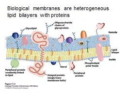
SKHALILZADEH Lipids are hydrophobic molecules that are insoluble in water They are in cell membranes as a major form of stored nutrients triglycerides as precursors of adrenal and gonadal ID: 332593

cholesterol ldl apo hdl ldl cholesterol hdl apo plasma levels risk triglyceride vldl triglycerides statins hepatic acids chd statin patients total fatty

Share:

Link:

Embed:

Download Presentation from below link

Download Presentation The PPT/PDF document "DIABETES AND LIPID" is the property of its rightful owner. Permission is granted to download and print the materials on this web site for personal, non-commercial use only, and to display it on your personal computer provided you do not modify the materials and that you retain all copyright notices contained in the materials. By downloading content from our website, you accept the terms of this agreement.


Presentation Transcript

Slide1

DIABETES AND LIPID

S-KHALILZADEHSlide2

Lipids are hydrophobic molecules that are insoluble in water. They are in cell membranes

as a major form of stored nutrients (triglycerides),

as precursors of adrenal and

gonadal

steroids and bile acids

as extracellular and intracellular messengers (e.g., prostaglandins,

phosphatidylinositol

).

Lipoproteins provide a vehicle for transporting the complex lipids in the blood as water-soluble complexes and deliver lipids to cellsSlide3

Fatty acids vary in length and in the number and position of double bonds

Saturated

fatty acids

lack

double

bonds

unsaturated fatty acids have one or more double bonds.

Monounsaturated fatty acids have one double bond, and polyunsaturated fatty acids (PUFAs) have two or more. Slide4

Cholesterol is a four-ring hydrocarbon with an eight-carbon side chain.

It is a major component of cell membranes and as a precursor of steroid hormones (adrenal and

gonadal

hormones) and bile acids

In the blood, about two thirds of the cholesterol is

esterified

Slide5
Slide6

Triglycerides consist of three fatty acid molecules

esterified

to a glycerol molecule Triglycerides store fatty acids and form large lipid droplets in adipose tissue. They are also transported as a component of certain lipoproteins. When triglycerides are hydrolyzed in

adipocytes

, free fatty acid (FFA) are released to be used as a source of energySlide7
Slide8

Chylomicrons

are the largest of the plasma lipoproteins (>1000 Å in diameter) ,float after ultracentrifugation of plasma.

They are composed of 98% to 99% lipid (85%-90% triglyceride) and 1% to 2% protein

Chylomicrons

are present in postprandial plasma (but are absent after an overnight fast) and contain apo-B48,

apo

-AI,

apo

-AIV,

apo

-E, and the C

apolipoproteins

Slide9

VLDLs are particles 300 to 700 Å

They are composed of 85% to 90% lipid (about 55% triglyceride, 20% cholesterol, and 15%

phospholipid

) and 10% to 15% protein. The distinctive

apolipoprotein

is apo-B100, the hepatic form of

apo

-B. VLDLs also contain

apo

-E and C

apolipoproteins

Slide10

IDLs present in low concentrations in the plasma and are intermediate in size and composition between VLDL and LDL

Their proteins are apo-B100 and apo-E.

The IDLs are precursors of LDLs and represent metabolic products of VLDL catabolism in the plasma by the action of lipases.

IDLs are often considered to be VLDL remnants and to be atherogenic.Slide11

LDLs are about 200 Å in diameter, are the major cholesterol-carrying lipoproteins in the plasma; about 70% of total plasma cholesterol is in LDL. LDLs are composed of approximately 75% lipid (about 35%

cholesteryl

ester, 10% free cholesterol, 10% triglyceride, and 20%

phospholipid

) and 25% protein. Apo-B100 is the principal protein in these particles, with trace amounts of

apo

-ESlide12

The clearance of LDL is mediated by apo-B100. The affinity of apo-B100 for the LDL receptor is lower than that of

apo

-E, and clearance of LDL is much slower (with a half-life of 2 to 3 days).

Compared with apo-B100–containing LDLs,

apo

-E–containing lipoproteins have 20-fold greater affinity for the LDL receptorSlide13

HDLs are small particles (70-120 Å in diameter)

HDLs contain about 50% lipid (25%

phospholipid

, 15%

cholesteryl

ester, 5% free cholesterol, and 5% triglyceride) and 50% protein Their major

apolipoproteins

are

apo

-AI (65%),

apo

-AII (25%), and smaller amounts of the C

apolipoproteins

and

apo

-E Slide14

Apo-E is a minor component of a subclass of HDL referred to as HDL

1

, but about 50% of total plasma

apo

-E is in this HDL fraction. The major classes of HDLs lack

apo

-E and do not interact with the LDL receptorSlide15

Apolipoproteins


 Understanding the major functions of the different

apolipoproteins

is important clinically, because defects in

apolipoprotein

metabolism lead to abnormalities in lipid handlingSlide16
Slide17

Apolipoproteins

A-I — Structural protein for HDL; activator of lecithin-cholesterol

acyltransferase

(LCAT).

A-II — Structural protein for HDL; activator of hepatic lipase.

A-IV — Activator of lipoprotein lipase (LPL) and LCAT.

B-100 — Structural protein for VLDL, IDL, LDL, and

Lp

(a);

ligand

for the LDL receptor; required for assembly and secretion of VLDL.

B-48 — Contains 48 percent of B-100; required for assembly and secretion of

chylomicrons

; does not bind to LDL

receptor. Slide18

C-I — Activator of LCAT.

C-II — Essential cofactor for LPL.

C-III — Interferes with

apo

-E mediated clearance of triglyceride-enriched lipoproteins by cellular receptors ; inhibits triglyceride hydrolysis by lipoprotein lipase and hepatic lipase ,interferes with normal endothelial function . Slide19

D — May be a cofactor for

cholesteryl

ester transfer protein (CETP).

E —

Ligand

for hepatic

chylomicron

and VLDL remnant receptor, leading to clearance of these lipoproteins from the circulation;

ligand

for LDL receptor

. Slide20

Human LPL is synthesized by

adipocytes

, by

myocytes

in skeletal and cardiac muscle, and by macrophages but is not produced by

hepatocytes

.

LPL is transported to the capillary endothelial cells where it interacts with

chylomicrons

and VLDL in the circulation and mediates the hydrolysis of their triglycerides to FFAs.

The fatty acids are stored as triglyceride in

adipocytes

and in the formation of hepatic VLDL.Slide21

Hepatic lipase is primarily a

phospholipase

but also possesses triglyceride

hydrolase

activity

It is synthesized by

hepatocytes

Hepatic lipase is transported from the liver to the capillary endothelium of the adrenals, ovaries, and testes, where it functions in the release of lipids from lipoproteins for use in these organs.

Its activity is increased by androgens and reduced by estrogensSlide22

EXOGENOUS PATHWAY OF LIPID METABOLISM 

starts with the intestinal absorption of dietary cholesterol and fatty acids The mechanisms regulating the amount of dietary cholesterol that is absorbed are unknown.

Sitosterolemia

is a rare

autosomal

recessive disorder associated with

hyperabsorption

of cholesterol and plant sterols from the intestine . Slide23

Within the intestinal cell, free fatty acids combine with glycerol to form triglycerides, and cholesterol is

esterified

by

acyl

-coenzyme A:cholesterol

acyltransferase

(ACAT) to form cholesterol esters

Triglycerides and cholesterol are assembled

intracellularly

as

chylomicrons

.

The main

apolipoprotein

is B-48, but C-II and E are acquired as the

chylomicrons

enter the circulation. Apo B-48 permits lipid binding to the

chylomicron

but not to LDL receptor.Slide24

Apo C-II is a cofactor for LPL which makes the

chylomicrons

smaller by hydrolyzing the core triglycerides and releasing free fatty acids. The free fatty acids are then used as an energy source, converted to triglyceride, or stored in adipose tissue. The end-products of

chylomicron

are

chylomicron

remnants that are cleared from the circulation by hepatic

chylomicron

remnant receptors for which

apo

E is a high-affinity

ligand

. Slide25
Slide26

ENDOGENOUS PATHWAY OF LIPID METABOLISM 

begins with the synthesis of VLDL by the liver

Microsomal

triglyceride transfer protein is essential for the transfer of the bulk of triglycerides

into the

endoplasmic reticulum for VLDL assembly

They include

apo

C-II which acts as a cofactor for LPL,

apo

C-III which inhibits this enzyme, and

apo

B-100 and E which serve as

ligands

for LDL receptor Slide27

The triglyceride core of VLDL particles is hydrolyzed by lipoprotein lipase. During

lipolysis

, the core of the VLDL particle is reduced, generating VLDL remnant particles (also called IDL) that are depleted of triglycerides via a process similar to the generation of

chylomicron

remnants

. Slide28

Some of the excess surface components in the remnant particle, including

phospholipid

,

unesterified

cholesterol, and

apolipoproteins

A, C and E, are transferred to HDL

VLDL remnants can either be cleared from the circulation by the

apo

B/E (LDL) or the remnant receptors or remodeled by hepatic lipase to form LDL particles

. Slide29


                                                                  Slide30

LDL can be internalized by hepatic and

nonhepatic

tissues. Hepatic LDL cholesterol can be converted to bile acids and secreted into the intestinal lumen.

LDL cholesterol internalized by

nonhepatic

tissues can be used for hormone production, cell membrane synthesis, or stored in the

esterified

formSlide31

Circulating LDL can also enter macrophages and some other tissues through the unregulated scavenger receptor. This pathway can result in excess accumulation of intracellular cholesterol and the formation of foam cells which contribute to the formation of

atheromatous

plaquesSlide32

These cholesterol-enriched cells can rupture, releasing oxidized LDL, intracellular enzymes, and oxygen free radicals that can further damage the vessel wall. Oxidized LDL induces apoptosis of vascular smooth muscle and human endothelial cells via activation of a protease which suggests a mechanism for the response to injury hypothesis of atherosclerosisSlide33
Slide34

anti-atherogenic effect of HDL

Apolipoprotein

A-I on the surface of HDL serves as a signal to mobilize cholesterol esters from intracellular pools. After diffusion of cholesterol onto HDL, the cholesterol is

esterified

to cholesterol esters by LCAT, a plasma enzyme that is activated primarily by

apolipoprotein

A-I.

HDL can act as an acceptor for cholesterol released during

lipolysis

of triglyceride-containing lipoproteinsSlide35
Slide36
Slide37
Slide38

Diabetes mellitus

insulin deficiency and poor

glycemic

control lead to increases in the plasma levels of triglycerides and

apo

-B–containing lipoproteins insulin deficiency results in impaired LPL activity and diminished clearance of triglyceride-rich particles.

Insulin deficiency also enhances

lipolysis

, which increases FFA flux to the liver, increased FFA flux drives triglyceride and VLDL synthesis and secretion

. Slide39

Plasma levels of LDL are increased in some but not all subjects. the

hyperlipidemia

in type 2 diabetes is often characterized by an increase in small, dense LDLs which are particularly

atherogenic

a portion of the plasma LDL undergoes

glycosylation

, which can increase binding to arterial wall

proteoglycans

and susceptibility to oxidation.Slide40

(CHD) are common in industrialized societies

There is a direct relation between the plasma levels of total and LDL cholesterol and the risk of CHD and mortality

LDL cholesterol lowering in moderate to high-risk patients leads to a reduction in cardiovascular events

Abnormalities of plasma lipids (

dyslipidemia

) other than LDL cholesterol are common in patients with early onset CHD

HDL cholesterol levels are related to absolute CHD event rates in treated

hypercholesterolemic

subjects with and without baseline clinical CHD

Screening tests for

dyslipidemia

are widely available Slide41

Guidelines developed by the NCEP in 2001 recommend that a complete plasma lipid profile (total cholesterol, LDL-C, HDL-C, and triglycerides) be measured in all adults 20 years of age and older at least once every 5 yearsSlide42

The ATP III recommendations for the treatment of hypercholesterolemia are based on the LDL-cholesterol (LDL-C)and are influenced by the coexistence of CHD and the number of cardiac risk factors.

There are five major steps to determining an individual's risk category, which serves as the basis for the treatment

guidelines Slide43

Step 1 — The first step in determining patient risk is to obtain a fasting lipid profileSlide44

Step 2 — CHD equivalents, that is, risk factors that place the patient at similar risk for CHD events as a history of CHD itself, are identified :

Diabetes mellitus

Symptomatic carotid artery disease

Peripheral arterial disease

Abdominal aortic aneurysm

Multiple risk factors that confer a 10-year risk of CHD >20 percent Slide45

Step 3 — Major CHD factors other than LDL are identified:

Cigarette smoking

Hypertension (BP ≥140/90 or antihypertensive medication)

Low HDL-C (<40 mg/

dL

)

Family history of premature CHD (in male first degree relatives <55 years, in female first degree relative <65 years)

Age (men ≥45 years, women ≥55 years)

HDL-C ≥60 mg/

dL

counts as a "negative" risk factor; its presence removes one risk factor from the total countSlide46

STEP-4

If two or more risk factors other than LDL (as defined in step 3) are present in a patient without CHD or a CHD equivalent (as defined in step 2), the 10-year risk of CHD is assessed using the ATP III modification of the Framingham risk tablesSlide47

Step 5 — The last step in risk assessment is to determine the risk category that establishes the LDL goal, when to initiate therapeutic lifestyle changes, and when to consider drug therapy Slide48

Total-to-HDL-cholesterol ratio 

Among men, a ratio of 6.4 or more identified a group at 2 to 14 percent greater risk than predicted from serum total or LDL-C

Among women, a ratio of 5.6 or more identified a group at 25 to 45 percent greater risk than predicted from serum total or LDL-C Slide49

Non-HDL-cholesterol 

Non-HDL-C is defined as the difference between the total cholesterol and HDL-C. Non-HDL-C includes all cholesterol present in lipoprotein particles that is considered

atherogenic

, including

LDL,lp

(a),IDL and VLDL .

It has been suggested that the non-HDL-C fraction may be a better tool for risk assessment than LDL-CSlide50

ATP III identifies the

non-HDL-C concentration as a secondary target of therapy in people who have high triglycerides ≥200 mg/dl.

The goal for non-HDL-C in this circumstance is a concentration that is 30 mg/

dL

(0.78

mmol

/L) higher than that

for LDL-CSlide51
Slide52

 A standard serum lipid profile consists of total cholesterol, triglycerides, and HDL-cholesterol. Lipoprotein analysis should be performed after 9 to 12 hours of fasting to minimize the influence of postprandial

hyperlipidemia

. Either a plasma or serum specimen can be used; the serum cholesterol is approximately 3 percent lower than the plasma valueSlide53
Slide54
Slide55
Slide56

HMG-CoA Reductase Inhibitors

Inhibition of cholesterol biosynthesis up-regulates cellular LDL receptors and enhances clearance of LDL from the plasma into cells.Slide57

Statins

Competitive inhibitors of 3-hydroxy-3-methylglutaryl coenzyme

A (HMG-

CoA

)

reductase

, which catalyzes an early,

ratelimiting

step in cholesterol biosynthesisSlide58

Chemistry

The

statins

possess a side group that is structurally similar to HMG-

CoA

Mevastatin, lovastatin, simvastatin, and pravaslatin

are fungal metabolites

fluvastatin

,

atorvastatin

, and

rosuvastatin

are entirely synthetic

Lovastatin

and

simvastatin

are

lactone

prodrugs

that are modified in the liver to active

hydroxy

acid forms

Since they are lactones, they are less soluble in water than are the other

statinsSlide59

Mechanism of Action

Statins

exert their major

effectreduction

of LDL levels-through a

mevalonic

acid-like moiety that competitively inhibits HMG-

CoA

reductase

.

By reducing the conversion of HMG-

CoA

to

mevalonate

,

statins

inhibit an early and rate-limiting step in cholesterol biosynthesisSlide60
Slide61

Inhibition of hepatic cholesterol synthesis, results in increased expression of the LDL receptor gene

Degradation of LDL receptors also is reduced

The greater number of LDL receptors on the surface of

hepatocytes

results in increased removal of LDL from the blood,

statins

also can reduce LDL levels by enhancing the removal of LDL precursors (VLDL and IDL) and by decreasing hepatic VLDL production Slide62

Triglyceride Reduction by Statins.

Triglyceride levels >250

mgldl

are reduced substantially by

statins

,

If baseline triglyceride levels are below 250 mg/dl. Reductions in triglycerides do not exceed 25% irrespective of the dose or

statin

used

simvastatin

and

atorvastatin

, 80,mg/day;

rosuvastatin

, 40 mg/day experience a 35% to 45% reduction in fasting triglyceride levelsSlide63

Effect of Statins on HDL-C Levels

patients with elevated LDL-C levels and gender- appropriate HDL-C levels (40 to 50

mgldl

for men; 50 to 60 mg/dl for women). an increase in HDL-C of 5% to 10% was observed, irrespective of the dose or

statin

employed

In patients with reduced HDL-C levels (35 mg/dl)

statins

may differ in their effects on HDL-C levels (

Simvastatin

>

Atorvastatin

)Slide64

Effects of Slatins on LDL-C Levels

Statins

lower LDL-C by 20% to 55% depending on the dose and

statin

used

Maximal effects on plasma cholesterol levels are achieved within 7 to 10 days.

The

statins

are effective in almost all patients with high LDL-C levels.

The exception is patients with homozygous familial hypercholesterolemia,

the partial response in these patients is due to a reduction in hepatic VLDL synthesis associated with the inhibition of HMG-

CoA

reductase

-mediated cholesterol synthesisSlide65
Slide66
Slide67

potency

rosuvastatin > atorvastatin > simvastatin > pravastatin = lovastatin > fluvastatinSlide68

Nonlipid roles of statins

Endothelial Function

Plaque Stability

Inflammation

Lipoprotein Oxidation

CoagulationSlide69

Statins

and Endothelial Function

Statin

therapy enhances endothelial production of the vasodilator nitric oxide, leading to improved endothelial function after a month of therapySlide70

Statins and Plaque Stability.

They reportedly inhibit

monocyte

infiltration into the artery wall in a rabbit model

Inhibit macrophage secretion of matrix

metalloproteinases

in vitro

modulate the

cellularity

of the artery wall by inhibiting proliferation of smooth muscle cells and enhancing apoptotic cell deathSlide71

Statins and Inflammation

Statins

decreased the risk of CHD and levels of C-reactive protein (CRP, an independent marker for inflammation and high CHD risk) independently of cholesterol loweringSlide72

Coagulation

Statins

reduce platelet aggregation

reduce the deposition of platelet thrombi in the porcine aorta model

the different

statins

have variable effects on fibrinogen levelsSlide73

SECONDARY BENEFITS

Bone metabolism

Hypertension

Heart failure 

Dementia 

Cancer prevention

Renal function 

Sepsis and infections Slide74

Hepatotoxicity

Elevation in hepatic

transaminase

to values greater than three times the upper limit of normal

Incidence as great as 1%

The incidence appeared to be dose related

liver failure one case per million person-years of use

measure

alanine

aminotransferase

(ALT) at baseline and thereafter when clinically indicated.Slide75

Patients taking 80-mg doses (or 40 mg of

rosuvastatin

) should have their ALT checked after 3 months. If the ALT values are normal, it is not necessary to repeal the ALT test unless clinically indicatedSlide76

Myopathy

myopathy

as any muscle disease

myalgia

as muscle ache or weakness without increased serum CK levels

myositis

as muscle symptoms with elevated CK levels

rhabdomyolysis

as muscle symptoms with marked CK elevations (>10 times upper limit of normal [ULN]) plus an elevated serum

creatinine

.

(FDA) defines

rhabdomyolysis

as organ damage, typically renal insufficiency, with a CK level greater than 10,000 IU/LSlide77

Myopathy

major adverse effect

The incidence of

myopathy

is quite low (~0.0 1%). but the risk of

myopathy

and

rhabdomyolysis

increases in proportion to plasma

statin

concentrations

Factors inhibiting

statin

catabolism are associated with increased

myopathy

risk, including advanced age (especially >80 years of age), hepatic or renal dysfunction.

perioperative

periods. multisystem disease (especially in association with diabetes mellitus), small body size, and untreated hypothyroidismSlide78

RISK FACTORS FOR STATIN MYOPATHY

conditions that increase

statin

serum and muscle concentration

factors that increase muscle susceptibility to injurySlide79

Asymptomatic patients

Routine surveillance of CK levels is not required except in high-risk patients

If CK measured

CK < 5× ULN: reassurance

CK ≥5 - <10× ULN: monitor for symptoms and periodical CK determination

CK ≥10× ULN: stop

statin

and reconsider risks and benefits of

statin

treatmentSlide80

Factors related to an increase in statin serum level

Statin

dose

Small body frame

Decreased

statin

metabolism and excretion

Drug–drug interactions

Grapefruit juice (possibly also pomegranate &

starfruit

)

Hypothyroidism and diabetes mellitus

Advanced age

Liver disease

Renal diseaseSlide81

Factors related to muscle predisposition

Alcohol consumption

Drug abuse (cocaine, amphetamines, heroin)

Heavy exercise

Baseline muscular disease:

Multisystemic

diseases: diabetes mellitus, hypothyroidism

Inflammatory or inherited metabolic muscle defectsSlide82

Bile acid sequestrants

The bound bile acids are excreted in the

feces.The

increased excretion of bile acids causes increased oxidation of cholesterol to form bile acids in

hepatocytes

, and the resultant up-regulation of hepatic LDL receptors in turn lowers plasma LDL concentrations.

side effects are limited to local effects in the gastrointestinal system (e.g., bloating, gas, constipation)Slide83

these agents can lower plasma cholesterol levels by 15% to 25%.

they can increase plasma triglyceride levels and must be used with caution in patients predisposed to

hypertriglyceridemia

. Slide84

because they bind negatively charged molecules in the intestine, these agents can interfere with the absorption of other medications, including

levothyroxine

,

digoxin

,

warfarin

, and

thiazide

diuretics. Therefore, resins are given at least 4 hours before or 1 hour after other medications.

Colesevelam

does not bind other drugs to the same extent as the resins

cholestyramine

and

colestipol

.Slide85

niacin

typically 2.0 to 6.0 g/day lower both total and LDL cholesterol by 15% to 30%, lower triglyceride levels by 30% to 40%, and raise HDL-C levels by 15% to 25%. Maximum HDL increases usually occur with therapeutic doses of 1.5 to 2.0 g/day. Niacin also lowers plasma

Lp

(a) concentrations by up to 40%.

The mechanism whereby niacin affects plasma lipids is unclear, but it seems to be associated with decreased hepatic VLDL production.Slide86

The most troublesome side effect of niacin is a flushing syndrome that occurs shortly after taking the medicine. Flushing can be minimized by initiating therapy with small doses (e.g., 100 mg) and gradually increasing the dosage to the therapeutic range over weeks to months.

taking an aspirin about 1 hour before the niacin can diminish the flushing, possibly by inhibiting prostaglandin-mediated side effects.Slide87

The most serious complication of niacin

therapy is

hepatotoxicity

, and therapy should be accompanied by monitoring of serum liver function tests

therapy should be discontinued if

transaminases

reach >3 times normal. Because

hepatotoxicity

appears to be more common with sustained-release preparations of niacin, the immediate-release form is preferred.

Other side effects of niacin therapy include impairment or worsening of glucose tolerance and

hyperuricemia

.Slide88

The fibric acid derivatives

clofibrate

,

gemfibrozil

, and

fenofibrate

—lower plasma triglycerides by about 40% and increase HDL-C levels by about 10% but have only minor effects on LDL-C. These agents act by activating the (PPAR) a, a nuclear hormone receptor that is expressed in the liver and other tissues. This results in increased fatty acid oxidation, increased LPL synthesis, and reduced expression of

apo

-CIII, all of which contribute to lowering plasma triglycerides. Slide89

Side effects include gastrointestinal discomfort and possibly an increased incidence of cholesterol gallstones (documented for

clofibrate

).

Fibric

acid derivatives should be used with great caution in the setting of renal insufficiency

Fenofibrate

, which does not interfere with

statin

metabolism and has a much lower risk of causing

myopathy

, is the preferred

fibrate

to use in combination with a

statin

.Slide90

Omacor

is prepared in capsule form containing a gram of oil, which includes a total of 840 mg of EPA plus DHA. At the recommended dosage of four capsules daily given to patients who have triglycerides of 500-2000 mg/

dL

,

Omacor

lowers triglycerides by about 50%, raises HDL-C by about 10%, lowers VLDL-C by about 40%, and raises LDL-C by about 50%. Overall the total cholesterol-to-HDL-C ratio is reduced by about 20% and the non-HDL-C is lowered by about 10%Slide91
Slide92

Chylomicronemia Syndrome

Patients with the

chylomicronemia

syndrome often present with acute pancreatitis and severe

hypertriglyceridemia

(triglycerides >22.6 mm/L [2000 mg/

dL

]).

These patients should be treated with total fat restriction until the triglyceride level falls to a safe range

The goal is to maintain the triglyceride level at less than 11.3 mm/L (1000 mg/

dL

) and preferably less than 4.5 mm/L (400 mg/

dL

).

such patients often require a triglyceride-lowering drug, such as a

fibrate

or niacin, to maintain the plasma triglyceride level in a range that prevents subsequent episodes of pancreatitisSlide93