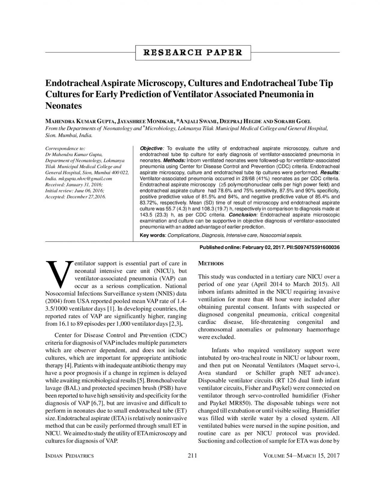
PDF-211V 54 15 2017Endotracheal Aspirate Microscopy Cultures and Endotra

Author : yvonne | Published Date : 2022-08-16

Objective RR 212V 54 15 2017UPTAENTILATORASSOCIATEDEONATESTESsuction was required for the presence of secretionsSuctioning was performed by using sterile feeding

Presentation Embed Code

Download Presentation

Download Presentation The PPT/PDF document "211V 54 15 2017Endotracheal Aspirate Mic..." is the property of its rightful owner. Permission is granted to download and print the materials on this website for personal, non-commercial use only, and to display it on your personal computer provided you do not modify the materials and that you retain all copyright notices contained in the materials. By downloading content from our website, you accept the terms of this agreement.

211V 54 15 2017Endotracheal Aspirate Microscopy Cultures and Endotra: Transcript


Objective RR 212V 54 15 2017UPTAENTILATORASSOCIATEDEONATESTESsuction was required for the presence of secretionsSuctioning was performed by using sterile feeding tube orsuction catheter and syringe. Fire Protection Laboratory Methods Day. June 25, 2014. Paul M. Anderson. Graduate Research Assistant. University of Maryland. Department of Fire Protection Engineering. Transmission Electron Microscopy. Laboratory 1. 2/4/14. Examination of the Peripheral Smear. Intro to Bone Marrow Morphology. Neutrophil. Eosinophil. Lymphocyte. Band (young neutrophil). Monocyte. Neutrophil. Basophil. Eosinophil. Large granular lymphocyte (LGL). Fire Protection Laboratory Methods Day. June 25, 2014. Paul M. Anderson. Graduate Research Assistant. University of Maryland. Department of Fire Protection Engineering. Transmission Electron Microscopy. Jenny . Malmstrom. AFM invented by Binning and co-workers in 1986. . Belongs to the Scanning Probe Microscopy family. Binning et al., Physics Review Letters 1986. AFM invented by Binning and co-workers in 1986. . Lecture . 07: . Confocal Microscopy. Adding the Third Dimension. Andres Collazo, Director Biological Imaging Facility. Wan-. Rong. (Sandy) Wong, Graduate Student, TA. Lecture . 7: Confocal Microscopy. Microscopy is the technical field using microscopes to view samples and objects that can not be seen without unaided eye (objects that are not within the resolution range of the normal eye). There are three well-known branches of microscopy: optical, electron and scanning probe microscopy.. . Histiocytic. proliferation in bone marrow post T-ALL remission. Initial Diagnostic Bone Marrow with T-ALL. 400X H&E. 400x H&E. Phenotype of T-ALL by . Flow . Cytometry. Subsequent, consecutive Bone Marrow Biopsies post T-ALL Remission with . for students attending the NU Bound Program These courses are for the Spring 2022 semesterfourof the following courses with the NU Bound England Program Calculus for Business MATH 1231 Calculus for KAARTHIK J. INTRODUCTION. A fluorescence microscope is a optical microscope that uses fluorescence and phosphorescence instead of , or I addition to reflection and absorption to study properties of organic or inorganic substances.. Outline. Introduction. Optical Microscopy. Types of Optical Microscopes. Confocal Microscopy. Laser Scanning Confocal Microscopy. Examples. Optical Microscopy. Optical microscopy is the oldest type of microscope and uses visible light and a system of lenses to create an image of the sample. AIM : T THEORY: Microscope is the most commonly used piece of apparatus in the laboratory. It produces greatly enlarged images of minute objects. LIGHT MICROSCOPE A Light Microscope can be simple or (SEM) Electron Microscopy (SEM) and TEM Scanning electron microscopy is used for inspecting topographies of specimens at very high magnifications using a piece of equipment called the scanning electr DO NOT . . perform blood cultures for “surveillance” in the absence of signs and symptoms of sepsis. BOTTLE PREPARATION. : . Disinfect . the rubber stopper of each bottle with 70% . alcohol. Do . ELECTRON. . MICROSC. O. PY. . . . Electron. . micros. c. opy. . (EM). . is a technique. . u. s. ed. . for. . obtaining. . high re. s. olu. t. ion. . images. . of. . biological and non-biologi.

Download Document

Here is the link to download the presentation.
"211V 54 15 2017Endotracheal Aspirate Microscopy Cultures and Endotra"The content belongs to its owner. You may download and print it for personal use, without modification, and keep all copyright notices. By downloading, you agree to these terms.

Related Documents