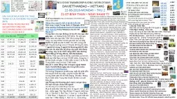
University of Pittsburgh Medical Center 72yearold male with fever of unknown origin Patient complains of fluctuating fevers for the last 3 weeks He has diplopia due to CN IV palsy but no ID: 933359
Download Presentation The PPT/PDF document "Lananh Nguyen, M.D. Division of Neuropa..." is the property of its rightful owner. Permission is granted to download and print the materials on this web site for personal, non-commercial use only, and to display it on your personal computer provided you do not modify the materials and that you retain all copyright notices contained in the materials. By downloading content from our website, you accept the terms of this agreement.
Slide1
Lananh Nguyen, M.D.Division of NeuropathologyUniversity of Pittsburgh Medical Center
72-year-old male with fever of unknown origin
Slide2Patient complains of fluctuating fevers for the last 3 weeks.He has diplopia due to CN IV palsy but no surgical or family history.
Clinical history
2
Slide3What is the differential diagnosis for fever of unknown origin (FUO)?3
Slide4What is the differential diagnosis for fever of unknown origin (FUO)?4
Slide5Patient has myalgias, joint pain, headache and jaw claudication.Scalp was tender to palpation.Imaging was negative for mass lesions.Serologies and cultures were negative.Prophylactic antibiotics were initiated without significant improvement.
Additional history and workup results
5
Slide6A temporal artery biopsy was ordered with a suspicion for an inflammatory disorder.The biopsy specimen was sent to pathology for evaluation.What did the clinician do next and why?
6
Slide7Gross description:5 x 0.2 cm tissue labeled “left temporal artery”
Serial sections of the entire vessel
Slide8Why is it important to evaluate the entire length of the vessels?8
Slide9Inflammatory lesions show patchy distribution hence, you need to evaluate for skip lesions.Skip lesions are just that, non contiguous lesions along the length of the vessels. Why is it important to evaluate the entire length of the vessels?9
Slide10This is one section on the slide. Name the histologic layers indicated by ***. What is your interpretation?
***
***
***
Slide11This is a normal section. This is an artery as evident by the thick muscular wall.
Media - muscular wall
Adventitia
Intima
Slide12This is another section on the slide. What vessel layer are we in and what do you see?
Slide13There is inflammation in the
vasa
vasorum
, branches of the temporal artery
Slide14This is another section. What do you see?
Slide15This section shows significant inflammation around and infiltrating the vessel wall.
Slide16Higher magnification shows inflammatory cells in the adventitia and infiltrating the media.
Slide17This is another section where severe inflammation can be appreciated even at low power.
Slide18Higher magnification shows inflammatory cell infiltration with disruption of the internal elastic lamina, the structure which separates the intima from the media.
Internal elastic lamina, note the squiggly pink line
Slide19Another section shows loss of vessel integrity. No granulomas or giant cells were identified.
Slide20Final diagnosisARTERY, LEFT TEMPORAL, BIOPSY: POSITIVE FOR ARTERITIS.
Slide21What is the treatment?21
Slide22What is the treatment?Glucocorticoids, even prior to confirmatory biopsy. This is key to preventing anterior ischemic optic neuropathy (otherwise known as permanent visual loss). Preferably, biopsy should be perform soon after administration.22
Slide23Discussion – Take home points23Giant cell arteritis (GCA) is a large vessel vasculitis (aorta and primary branches with a predilection for extracranial arteries)Pathologically, giant cells may not be present and
skip lesions
can occur. Therefore, evaluate multiple levels and evaluate the vasa vasorum (branches of the temporal artery) for evidence of inflammatory cells.
Mean age 70;
female:male 2:1
SIGNS AND SYMPTOMS
: new-onset headache, fever, visual symptoms (blindness,
amaurosis
fugax, diplopia
),
TA/scalp tenderness, jaw claudication, weight loss
LABS
: elevated ESR/CRP but also anemia, hypoalbuminemia, transaminitis. SIADH Is associated with GCA
40% with GCA have polymyalgia rheumatica (PMR) but only 10% with PMR have GCA on temporal artery biopsy. It’s a spectrum.
American College of Rheumatology criteria
for diagnosis: 3 out of the following 5
Age
>
50
New headache
Temporal artery abnormality
ESR
>
50
Abnormal temporal artery biopsy