/
Lecture 13 :Protein synthesis Lecture 13 :Protein synthesis

Lecture 13 :Protein synthesis - PowerPoint Presentation

alida-meadow
alida-meadow . @alida-meadow
Follow
576 views
Uploaded On 2017-10-25

Lecture 13 :Protein synthesis - PPT Presentation

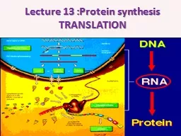
TRANSLATION Proteins involve in many body structures According to central dogma the genetic information flow from DNA the RNA via transcription process then it will translate later to functional protein ID: 599231

codon site structure rna site codon rna structure protein bond chain step triplet amino bind initiation group peptide types

Share:

Link:

Embed:

Download Presentation from below link

Download Presentation The PPT/PDF document "Lecture 13 :Protein synthesis" is the property of its rightful owner. Permission is granted to download and print the materials on this web site for personal, non-commercial use only, and to display it on your personal computer provided you do not modify the materials and that you retain all copyright notices contained in the materials. By downloading content from our website, you accept the terms of this agreement.


Presentation Transcript

Slide1

Lecture 13 :Protein synthesis TRANSLATION Slide2

Proteins involve in many body structures Slide3

According to central dogma , the genetic information flow from DNA the RNA via transcription process then it will translate later to functional protein. Slide4
Slide5

Basic structure of protein: the structural unit of protein is amino acid (A.A).The main structure of A.A is 1- central C atom called α- carbon. .2-Amino group gives the positive charge

3- carboxylic acid group gives the negative charge

4- R side chain which differ from one amino acid to another, the simplest R side group is H only to form

glycin

Slide6

The bond which bind the two adjacent A.A is called peptide bond with releasing of water molecules . Slide7

Usually the protein chain start with amino group (NH3) and end with Carboxyl group (COOH).binding 2-10 A.A give a rise to oligopeptide while binding more than that will form the polypeptide chain . This linear chain represent 1- the primary structure to the protein . Slide8

According to structure and configuration ,proteins can be divided to 4 types Slide9

2- the secondary structure formed due to folding of the primary structure via hydrogen bonds . Two types are well studied as secondary structur 1- α helix 2- β

pleated sheet Slide10

3- Tertiary structure is more complicated and we could find α helix and β pleated sheet in the same structure ,other types of bonds exist here like disulfide bond between cystine

A.A ,ionic bond ,hydrophobic bondSlide11
Slide12

4- Quaternary structure :more complicated result from folding the other structures and from more than subunits Slide13

TRANSLATIONIn this process the three types of RNA are involved each one of them play important role to complete protein synthesis . The Translation also involve 1-Initiation 2- Elongation 3- Termination step Slide14

Step one :initiation , it requires 1- m RNA (processed in Eukaryotic cell) 2- t RNA which will come charged (bind to A.A) and the first A.A is methionin cose the first codon is AUG (usually become as n formayl methionin ) .the AUG first codon in m RNA will face the triplet

codon in anti codon

arm in t RNA .3- small 30S ribosomal sub units thus the m RNA will bind to 16s r RNA via its ribosomal binding site .Slide15
Slide16

The initiation step requires initiation factors then the large sub unit bind the complex forming 70s initiation complex . The large subunit contain P site (peptidyl sit ) and A site (Amino -acyl site ).Slide17

The complete binding sites in the ribosomeSlide18

The first occupied site by the charged tRNA is P site followed by introducing A site and the triplet codon will determine the next incorporated A,A Slide19
Slide20

The 4 nitrogen base will form 64 possibilities and we have 20 A.A thus 2-3 triplet codon will code to the same A,A. beside we have one start codon (AUG) and 3 stop codon (UAA, UAG ,UGA) that they will not translated to any A.A. Slide21

2- Elongation step :here the peptide chain will elongate with the help of elongation factors .the A.A in the p site will bind with the A.A in A site via peptide bond with the add of peptidyl transferase .t RNA in p site now is empty ,convert to uncharged and leave the ribosome from Exit site .then the tRNA translocat from A to P site with 2 A.A . Now the A site empty and the complex will move for another triplet

codon Slide22
Slide23

The figure shows ribosome movement (from 5→3) as each 3 nitrogen base form one triplet codon and will translate to one A.A. the growing peptide chain will continue movning between P and A site along them RNA till reaching the stop codon. Slide24

Termination step: when the ribosome reached stop codon (UAG, UAA,UGA) and there is no anticodon in

tRNA so A site will remain empty causes releasing of free polypeptide and dissociation of the 2ribosomal subunits.

note: mRNA will degraded once it translated thus its concretion is low

while

m RNA in Eukaryotic cell more stable cos it contain cap structure at 5 end and poly A tail in 3 end . Slide25