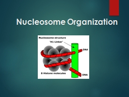
PPT-Nucleosome Organization Nucleosomes were first observed as particles in the electron microscope

Author : byrne | Published Date : 2023-07-22

Don and Ada Olins in 1974 and their existence and structure as histone octamers surrounded by approximately 200 base pairs of DNA were proposed by Roger Kornberg

Presentation Embed Code

Download Presentation

Download Presentation The PPT/PDF document "Nucleosome Organization Nucleosomes were..." is the property of its rightful owner. Permission is granted to download and print the materials on this website for personal, non-commercial use only, and to display it on your personal computer provided you do not modify the materials and that you retain all copyright notices contained in the materials. By downloading content from our website, you accept the terms of this agreement.

Nucleosome Organization Nucleosomes were first observed as particles in the electron microscope: Transcript


Don and Ada Olins in 1974 and their existence and structure as histone octamers surrounded by approximately 200 base pairs of DNA were proposed by Roger Kornberg The role of the nucleosome as a . This model comes with color corrected infinity optical system (CSIS), a movable large working distance condenser, 45 degree inclined trinocular head, high eye-point Plan eyepieces, infinity long working distance PLAN achromatic objectives and infinity long working distance PLAN phase contrast objectives. This microscope offers nine brightfield magnification powers, 40X, 60X, 100X, 150X, 200X, 300X, 400X, 600X & 900X, and six phase contrast settings, 100X, 150X, 200X, 300X, 400X & 600X. Its 30 watt adopt Kohler illumination system was designed for 90-240V wide voltage power supplies. : what, why and where?. Rob Brewster. Outline. What is a . nucleosome. ?. - how is DNA packaged/organized in Eukaryotes?. Why do . nucleosomes. form?. . - DNA is stiff, how do ~100 . bp. loops form so readily?. Jim Atherton. Development of Light Microscope. 1590 Hans Zacharias Janssen. 1660 Robert Hook. Onserved. . cells (cork). 1. Klein, Aaron E. The Electron Microscope: A Tool of Discovery. New . Y. ork,McGRaw. Tutor: Peter Harris. Click on the buttons to move between slides. .. (Make sure you are in “Slide Show” mode). The aims of this course are:. ● . to explain how X-rays are generated in the electron microscope. Compound Light Microscope. The compound light microscope is a common tool in a life science laboratory. A . compound light microscope. is an instrument that magnifies small objects so that they can be seen easily. . The Microscope. An understanding of cells and the ultrastructure of cells had to wait until objects so small could be visualized by magnification.. Origin of the Microscope. The first record of the invention of a microscope was by Hans and Zacharias Janssen (Netherlands) in 1590?. See Us at M&M 2020 in Milwaukee instrument that produces . enlarged . image of . object. Used to study organisms, cells, and cell parts. Increase image . of object . and . S. how object’s details. MAGNIFICATION. : . increase . of . Care. Parts. Focusing. Always carry with 2 hands. Do not force knobs. Always store covered. Keep objects clear of desk and cords. Only use lens paper for cleaning. Microscope Care. Eyepiece. Body. . What Is Microscope?. It is an instrument which deals with too small organisms that they cannot be seen distinctly with the naked eye. Microscopes provide the observer with enhanced . resolution. (ability to observe two nearby objects as distinct objects), . . Electron microscopy (EM) . is an electron beam which is focused into a small probe across the surface of a specimen . . The first electromagnetic lens was developed in 1926 by . Hans Busch. .. . An electron microscope is a microscope that uses a beam of accelerated electrons as a source o f illumination. As the wavelength of an electron can be up to 100,000 times shorter than that of visibl Rob Brewster. Outline. What is a . nucleosome. ?. - how is DNA packaged/organized in Eukaryotes?. Why do . nucleosomes. form?. . - DNA is stiff, how do ~100 . bp. loops form so readily?. Where do . To explain the advantages of using an Electron Microscope . To be able to carry out Simple Magnification Calculations. .

Download Document

Here is the link to download the presentation.
"Nucleosome Organization Nucleosomes were first observed as particles in the electron microscope"The content belongs to its owner. You may download and print it for personal use, without modification, and keep all copyright notices. By downloading, you agree to these terms.

Related Documents