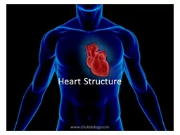
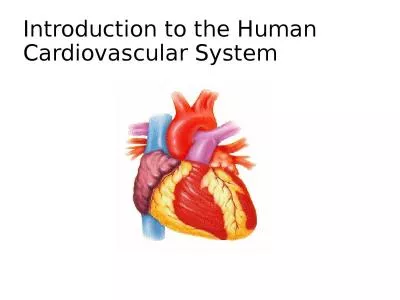
/
Heart  & Neck Vessels Assessment Heart  & Neck Vessels Assessment

Heart & Neck Vessels Assessment - PowerPoint Presentation

cecilia
cecilia . @cecilia
Follow
67 views
Uploaded On 2023-06-10

Heart & Neck Vessels Assessment - PPT Presentation

Prof Dr Salma Khadim Jehad Dr Ali Faris MSc Hassanain Mohammed Kadhim Lecture 7 Objectives At the end of this lab the students will be able to ID: 1000525

client amp left neck amp client neck left carotid jugular side palpate note chest artery heart spaces apex intercostal

Share:

Link:

Embed:

Download Presentation from below link

Download Presentation The PPT/PDF document "Heart & Neck Vessels Assessment" is the property of its rightful owner. Permission is granted to download and print the materials on this web site for personal, non-commercial use only, and to display it on your personal computer provided you do not modify the materials and that you retain all copyright notices contained in the materials. By downloading content from our website, you accept the terms of this agreement.


Presentation Transcript

1. Heart & Neck Vessels AssessmentProf Dr. Salma Khadim Jehad Dr. Ali Faris M.Sc Hassanain Mohammed KadhimLecture -7-

2. Objectives:At the end of this lab, the students will be able to:1. Demonstrate the ability to safely & accurately complete heart & neck vessels assessment.2.Demonstrate the ability to accurately document heart & neck vessels assessment data in organized manner.

3. Equipment NeededI . Marking Pen2. Small centimeter3. Stethoscope with diaphragm & bell 4. Alcohol swab 5- Watch with second hand

4. Preparation1. To evaluate the carotid arteries, the client can be sitting.2. To assess the jugular veins & the pericardium, the person should be supine with the head & chest slightly elevated.3. Stand on the client's right side.4. The room should be warm.5. Ensure the female's privacy by keeping her breasts draped.6. Gently displace the breast upward, or ask the client to hold it out of the way.

5. Subjective data: 1. Cough 2. Nocturia3. Dyspnea 4. Fatigue 5. Past cardiac history6. Orothopnea 7. Cyanosis or pallor 8. Family cardiac history9. Edema 10.Chest pain 1 1. Personal habits

6. The Neck VesselsPalpate the carotid artery- Palpate each carotid artery medial to the sternomastoid muscle in the neck.- Avoid excessive pressure on the carotid sinus area.- Palpate gently.- Palpate only one carotid artery at atime. - Feel the contour & amplitude of the pulse.- Compromise finding to the other side.

7. Normal finding : Contour is smooth with rapid upstroke & slower down stroke. Strength is 2+ or moderate.Findings should be same bilaterally

8. Auscultation of the carotid artery- Keep the neck in a neutral position.- Lightly apply the bell of thestethoscope over the carotid artery atthree levels: o Angle of jaw. o Mid – line area. o Base of the neck. - Ask the client to take a breath. Exhale & hold it briefly while you listen. Normally no sound present

9. Inspect the Jugular Venous Pulse- Put the client in supine position anywhere from 30-40 degree angle. - Remove the pillow to avoid flexing of the neck.- Turn the client's head slightly away from the examined side.- Direct strong light onto the neck.- Note the external jugular veins overlying the stemomastoid muscle.- Look for pulsation of internal jugular veins in the area of suprasternal notch Internal jugular vein pulsations 3cm above sternal angel

10. Jugular vein

11. Measurment

12. Assessment

13. Assessment of Odema

14. Palpate the Apical impulse: - localize the apical impulse using one finger pad. - Ask the client to "exhale & then hold". - Role the client midway to the left. Note the following: Location Size Amplitude Apical impulse occupy only on intercostals space, the 5*, & be at or medial to the MCL. *2cm.Note: apical impulse is not palpable inobese or in clients with thick chest wall.

15. Palpation

16. Palpate across the precordium- Using the palm aspect of your four fingers, gently palpate the apex. - Search for any pulsation : Normally no pulsation Percussion- place your stationary finger in the client's 5th ICS over on left side of chest near the anterior axillary line. - Slide your hand toward yourself, percussing as you go. - Note the change of sound. .

17. The left border of cardiac dullness is at the midclavicular line in the 5' interspace, & slopes toward the sternum progress upward, so that by the 2"d interspace the border of dullness coincides with the left sternal border.

18. Auscultation- clean the end pieces with alcohol swab. - After you place the stethoscope, try closing your eyes briefly to get out any distraction.- Begin with the diaphragm end piece & note the following: Rate & rhythm Identify S1 & S2 Listen for murmurs

19. Normal finding: Rate range from 60-IOO bpm, & the rhythm is regular.Sl is louder than S2 at the apex,& S2 is louder than S1 at the base.Should not be heard.

20. S1 : produced by click of the ateroventracular valve S2 : produced by click of the semiluner valveAfter auscultating in supine position, role the client toward his \ her left side.- Listen with the bell at the apex. - Ask the client to sit up, lean forward slightly, & exhale.- Listen with the diaphragm firmly pressed at the base, right, & left side. - Check for the soft high-pitchedsound.

21. Where to place your stethoscopeAs with palpation of the heart, auscultation should proceed in a logical manner over 5 general areas on the anterior chest, beginning with the patient in the supine position. Examined with diaphragm, including: 1- Aortic region at the 2nd intercostal spaces at the right 2- Pulmonic region at the 3rd intercostal spaces at the left

22. 3- Erb’s point at the 4th intercostal spaces 4- Tricuspid region at the 5th intercostal spaces at the left border of sternum 5- Mitral region (near the apex of the heard between the 5th intercostal spaces in the mid- clavicular line) (apex of the heart).

23. Auscultation