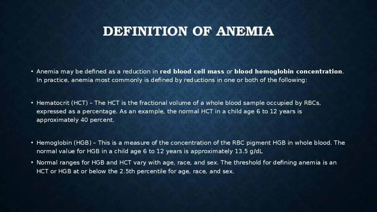
/
DEFINITION OF ANEMIA Anemia may be defined as a reduction in DEFINITION OF ANEMIA Anemia may be defined as a reduction in

DEFINITION OF ANEMIA Anemia may be defined as a reduction in - PowerPoint Presentation

clara
clara . @clara
Follow
356 views
Uploaded On 2022-02-14

DEFINITION OF ANEMIA Anemia may be defined as a reduction in - PPT Presentation

red blood cell mass or blood hemoglobin concentration In practice anemia most commonly is defined by reductions in one or both of the following Hematocrit HCT The HCT is the fractional volume of a whole blood sample occupied by RBCs expressed as a percentage As an example the norm ID: 908793

iron anemia blood deficiency anemia iron deficiency blood cell hemolysis due disease thalassemia hemoglobin age increase history hemolytic rbc

Share:

Link:

Embed:

Download Presentation from below link

Download Presentation The PPT/PDF document "DEFINITION OF ANEMIA Anemia may be defin..." is the property of its rightful owner. Permission is granted to download and print the materials on this web site for personal, non-commercial use only, and to display it on your personal computer provided you do not modify the materials and that you retain all copyright notices contained in the materials. By downloading content from our website, you accept the terms of this agreement.


Presentation Transcript

Slide1

DEFINITION OF ANEMIA

Anemia may be defined as a reduction in

red blood cell mass

or

blood hemoglobin concentration

. In practice, anemia most commonly is defined by reductions in one or both of the following:

Hematocrit (HCT) – The HCT is the fractional volume of a whole blood sample occupied by RBCs, expressed as a percentage. As an example, the normal HCT in a child age 6 to 12 years is approximately 40 percent.

Hemoglobin (HGB) – This is a measure of the concentration of the RBC pigment HGB in whole blood. The normal value for HGB in a child age 6 to 12 years is approximately 13.5 g/dL

Normal ranges for HGB and HCT vary with age, race, and sex. The threshold for defining anemia is an HCT or HGB at or below the 2.5th percentile for age, race, and sex.

Slide2

Slide3

Pathophysiology

The function of the RBC is to deliver oxygen from the lungs to the tissues and carbon dioxide from the tissues to the lungs. This is accomplished by using hemoglobin . In anemia, a decrease in the number of RBCs transporting oxygen and carbon dioxide impairs the body’s ability for gas exchange

The physiologic response to anemia varies according to acuity and the type of insult. Gradual onset may allow for compensatory mechanisms to take place. With anemia due to acute blood loss, a reduction in oxygen-carrying capacity occurs along with a decrease in intravascular volume, with resultant hypoxia and hypovolemia. Hypovolemia leads to hypotension, which is detected by stretch receptors in the carotid bulb, aortic arch, heart, and lungs. These receptors transmit impulses to the medulla, cerebral cortex, and pituitary gland.

In the medulla, sympathetic outflow is enhanced, while parasympathetic activity is diminished. Increased sympathetic outflow leads to norepinephrine release from sympathetic nerve endings and discharge of epinephrine and norepinephrine from the adrenal medulla. Sympathetic connection to the hypothalamic nuclei increases antidiuretic hormone (ADH) secretion from the pituitary gland.

 [3] 

ADH increases free water reabsorption in the distal collecting tubules.

Slide4

Pathophysiology

In response to decreased renal perfusion, juxtaglomerular cells in the afferent arterioles release renin into the renal circulation, leading to increased angiotensin I, which is converted by angiotensin-converting enzyme (ACE) to angiotensin II.

Angiotensin II has a potent pressor effect on arteriolar smooth muscle. Angiotensin II also stimulates the zona glomerulosa of the adrenal cortex to produce aldosterone. Aldosterone increases sodium reabsorption from the proximal tubules of the kidney, thus increasing intravascular volume. The primary effect of the sympathetic nervous system is to maintain perfusion to the tissues by increasing systemic vascular resistance (SVR). The augmented venous tone increases the preload and, hence, the end-diastolic volume, which increases stroke volume. Therefore, stroke volume, heart rate, and SVR all are maximized by the sympathetic nervous system. Oxygen delivery is enhanced by the increased blood flow

Slide5

Pathophysiology

To Increase Oxygen Delivery:

1.) Increase in blood flow

2.) Increase in red cell mass

3.) Increase oxygen unloading

    1.) Increase in blood flow

A.) Increased Cardiac Output Clinical Findings:

HR, Pulse pressure, murmurs, bruits, hyper-dynamic precordium

B.) Changes in Tissue Perfusion

Clinical Findings: Pallor

Slide6

Pathophysiology

 2.) Increase in red cell mass

EPO (kidney)

Reticulocytosis

, immature RBCs

Clinically: Bony pain with expansion of the marrow

3.) Increase oxygen unloading

2,3-diphosphoglycerate (2,3-DPG) in red blood cells increases in response to anemia/hypoxia and 

causes a shift of the oxygen dissociation curve

, allowing a more effective oxygen delivery

Slide7

symptoms:

Slide8

Slide9

How to approach anemia?

Slide10

EVALUATION OF ANEMIC PATIENT:

Physical examination

Detailed History

Essential laboratory tests

Slide11

Detailed History

Age of patient


— The age of the patient is important to consider because normal values of hematocrit (HCT) and hemoglobin (HGB) vary greatly with age and because different causes of anemia present at different ages

Birth to three months


– The most common cause of anemia in young infants is "

physiological anemia

," which occurs at approximately six to nine weeks of age. Common causes of

pathological anemia

in newborns include blood loss, immune hemolytic disease

Infants three to six months


– Anemia detected at three to six months of age suggests a hemoglobinopathy.

Slide12

Sex

 — Some inherited causes of anemia are X-linked (

eg

, G6PD deficiency and X-linked sideroblastic anemia) and occur most commonly in males.

In postmenarchal girls, excessive menstrual bleeding is an important cause of anemia.

Slide13

Race and ethnicity

 — Race and ethnic background are helpful in guiding the work-up for hemoglobinopathies and enzymopathies (

eg

, G6PD deficiency). HGB S and C are most commonly seen in Black and Hispanic populations; thalassemia syndromes are more common in individuals of Mediterranean and Southeast Asian descent; G6PD deficiency is more common among Jews, Greeks and Black populations

Slide14

Evaluation :

History

 — The evaluation of a child with anemia begins thorough history.

The degree of symptoms

,

past medical history

,

family history

,

dietary history

,

and developmental history

may provide important clues to the cause of anemia

Slide15

Symptoms


– Characterizing the symptoms helps elucidate the severity and chronicity of anemia and may identify patients with blood loss or hemolytic etiologies:

1-Symptoms attributable to anemia

 *

– Common symptoms of anemia include lethargy, tachycardia, and pallor.

Infants may present with irritability and poor oral intake.

Slide16

2-Symptoms of hemolysis

 – Changes in

urine color

,

scleral icterus

, splenomegaly or

jaundice

may indicate the presence of a hemolytic disorder. gallstones (

bilirubinate

) are a common complication of chronic hemolysis

Slide17

3-Bleeding symptoms

 – Specific questions related to bleeding from the

gastrointestinal tract

, including changes in stool color, blood in stools, and history of bowel symptoms, should be reviewed. Severe or chronic epistaxis also may result in anemia from blood loss and iron deficiency. In adolescent girls,

menstrual history

should be obtained, including duration and amount of bleeding. Severe

epistaxis

and/or heavy menstrual bleeding should raise suspicion for an underlying bleeding disorder .In patients who have symptoms of GI bleeding, history of additional GI symptoms should be elicited.

Slide18

4-Pica

 – The presence of pica, the intense craving for nonfood items, should be assessed given its strong association with iron deficiency. In young children, pica may manifest as craving dirt, rocks, and paper. In adolescents, craving for ice may be more common.

Slide19

Birth history

– The birth and neonatal history should include gestational age, duration of birth hospitalization, and history of jaundice and/or anemia in the newborn period. Results of newborn screening (which typically includes screening for sickle cell disease) should be reviewed.

History of anemia

– Previous complete blood counts (CBCs) should be reviewed, and, if prior anemic episodes occurred, they should be characterized (including duration, etiology, therapy, and resolution).

Slide20

Underlying medical conditions

– Past medical history and review of symptoms should be obtained to elucidate chronic underlying infectious or inflammatory conditions that may result in anemia.

Travel

to/from areas of endemic infection (

eg

, malaria, hepatitis, tuberculosis) should be noted 

Slide21

Drug and toxin exposure history

 Current and past medications should be reviewed with particular attention to oxidant drugs that can cause hemolysis, particularly in patients with underlying G6PD deficiency.

Possible environmental toxin exposure should be explored, including lead exposure and nitrates in well water.

Immune-mediated hemolysis (e.g., penicillin) Bone marrow suppression (e.g., chemotherapy) Phenytoin, increasing folate requirement all should be

reviewd

Slide22

Family history


  Family history of anemia should be reviewed. Family members with jaundice, gallstones, or splenomegaly should be identified. Asking if family members have undergone cholecystectomy or splenectomy may aid in the identification of additional individuals with inherited hemolytic anemias.

*It is also important to determine whether there is a family history of inflammatory bowel disease, intestinal polyps, colorectal cancer, hereditary hemorrhagic telangiectasia, von Willebrand disease, platelet disorders, or hemophilia.

Slide23

Dietary history


Dietary history

 – The dietary history is focused on assessing iron intake and, to a lesser degree, folate and vitamin b12 content. The type of diet, type of formula (if iron fortified), and age of infant at the time of discontinuation of formula or breast milk should be documented. In addition, the amount and type of milk the patient is drinking should be determined.

Infants and children who are exclusively fed goat's milk can develop anemia due to folate deficiency.

Exclusively breastfed infants who do not receive sufficient iron supplementation may be anemic at the time of the initial screening at age 9 to 12 months, whereas infants receiving iron-fortified formula until age 12 months are unlikely to be anemic at this time, though they may be at risk for iron deficiency during the second year of life after transitioning to cow's milk.

Strict vegetarian: vitamin B12 deficiency

Slide24

Developmental history 

Parents should be asked questions to determine if the child has reached age-appropriate developmental milestones. Developmental delay can be associated with iron deficiency, lead toxicity, 

vitamin B12

/

folic acid

 deficiency, and Fanconi anemia

Slide25

Physical examination

Slide26

1- General Appearance:

Pale , tired , conscious , oriented , shortness of breath

.

2- Vital signs:

Blood pressure and pulse

should be measured to be sure that hypovolemic shock isn’t present. If anemia or volume loss is mild to moderate, tachycardia may be present, but normal blood pressure is preserved.

3- Growth parameters:

Hc

,

Ht

, Wt.

4- Face/head: features

of extramedullary hematopoiesis : frontal bossing, prominence of malar eminence, depressed nasal bridge, exposed upper central teeth, dysmorphic features (like

fanconi

anemia).

5- Eyes

:

Scleral icterus suggests shortened red cell survival with hemolysis. Conjunctival pallor , Kayser-Fleischer ring suggest Wilson disease, Blue sclera suggests Iron deficiency or osteopetrosis

Slide27

6- Mouth:

Glossitis suggests Vitamin B12 deficiency, iron deficiency , angular stomatitis suggests iron deficiency

Telangiectasia Osler-Weber-

Rendu

syndrome

7- Neck:

Lymph nodes for malignancy.

8- Chest:

lung for infiltration due to malignancy, cardiac for murmurs.

Shield chest or widespread nipples for Diamond-

Blackfan

syndrome

9- Abdomen:

hepatosplenomegaly, scars (splenectomy), Nephromegaly or absent kidney

fanconi

anemia.

10- Hand:

absent thumb (Fanconi), koilonychia (iron deficiency).

Slide28

Slide29

11- Legs

: Edema "chronic renal failure”, rash.

12- Skin:

Café au lit spots (Fanconi), petechial, purpuric rash, bruises (bleeding), Vitiligo Vitamin B12 deficiency

Erythematous rash may suggest Parvovirus or Epstein-Barr virus infection, Butterfly rash may suggest SLE

13- Nerves

Irritable, apathy suggests Iron deficiency, Peripheral neuropathy for Deficiency of vitamins B12.

Slide30

Essential laboratory tests

Complete blood count

 — The CBC provides information about the RBCs and other cell lines (

ie

, white blood cells [WBCs] and platelets).

All cell lines should obtained to determine whether anemia is the result of a process limited to the erythroid line or a process that affects other marrow elements

Reticulocyte count.

To check problems in the production of RBCs

Blood smear

 — A review of the peripheral smear is an essential part of any anemia evaluation. Even if the patient's RBC are normal, review of the blood smear may reveal abnormal cells that can help identify the cause of anemia.

Slide31

Classifications of Anemias

# According to MCV :

1- Microcytic anemia

2- Macrocytic anemia

3- Normocytic anemia

Slide32

Microcytic anemia

#Causes :

1-

Iron deficiency

2-Thalassemia

3-Chronic inflammatory disease

4-Copper deficiency

5-Sideroblastic anemia

6-Aluminum, (?) lead intoxication

7-Hereditary pyropoikilocytoses

8-Hemoglobin CC

Slide33

Iron Deficiency Anemia

Iron deficiency is the most common nutritional deficiency in children. 

the most common cause of

anemia in the world, is approximately 9% in toddlers, 9-11% in adolescent females, and less than 1% in adolescent males.

#

Risk factors :

Infants fed cow’s milk when younger than 1 year of Age.

toddlers fed large volumes of cow’s milk.

Menstruating teenage girls who are not receiving supplemental iron.

bottle-fed toddlers who are receiving large volumes of cow’s milk and eat minimal amounts of food high in iron content.

children with chronic inflammatory diseases, even without chronic blood loss.

6) Blood loss particularly in older children and adolescents(lesion of the gastrointestinal (GI) tract, such as peptic ulcer, Meckel diverticulum, polyp, hemangioma, inflammatory bowel Disease ,or Infection with hookworm, and Helicobacter pylori .

7) Gastrointestinal disease — Dietary iron is absorbed primarily within the duodenum. Malabsorption of iron may occur in diseases affecting the duodenum, including celiac disease; Crohn disease, giardiasis; or any surgical resection of the proximal small intestine.

Slide34

7) Several conditions in the perinatal period can increase the risk for iron deficiency anemia (IDA) during the first three to six months of life : Maternal iron deficiency , Fetal-maternal hemorrhage (FMH) , Twin-twin transfusion syndrome (TTTS) , Other perinatal hemorrhagic events

8)

Premature infants due to less maternal-fetal iron transfer, smaller total blood volume at birth, blood loss through phlebotomy, and poor gastrointestinal absorption 

9) Exclusive

breastfeeding without initiation of adequate iron supplementation by six months of age (Breast-fed infants are less likely to have iron deficiency than bottle-fed infants because, although there less iron in breast milk, this iron is more efficiently absorbed. However, infants who continue to be exclusively breast fed in the second half of the first year of life are at risk for iron deficiency.)

10)

Inefficient absorption due to dietary sources of iron with low bioavailability :

Types of iron in diet :

Heme dietary sources (fish, poultry, and meat) have a higher bioavailability of iron than do

nonheme

(vegetable) sources 

Factor increase absorption :

. Ascorbic acid (vitamin C) enhances absorption of

nonheme

iron by converting fe+3 into fe+2 (absorbable )

Factors decrease absorption :


tannates

(teas), bran foods rich in phosphates, and

phytates

(plant fiber, especially in seeds and grains) .

Slide35

Iron Deficiency Anemia

#

Clinical Manifestations:

1-Cardiovascular manifestations of anemia.

2-central nervous system (CNS) abnormalities (apathy, irritability, poor concentration) from alterations of iron-containing enzymes

(monoamine oxidase) and cytochromes .

3-Poor muscle endurance.

4-Gastrointestinal dysfunction.

5-Immune :  increase the risk for bacterial infection because the iron-binding proteins transferrin and

lactoferrin

have bacteriostatic effects, Similarly, there is some evidence that iron supplementation results in increased susceptibility to or reactivation of dormant infections, such as malaria or tuberculosis .

6-Neurodevelopmental — In young children, IDA is associated with impaired neurocognitive development, including slower visual and auditory processing

7-Brittle nails.

8-koilonychia (spoon-like nail deformity).

9-hair loss.

10-Pica.

11-Angular cheilitis: inflammation and fissuring of the corners of the mouth/ Atrophic glossitis: erythematous, edematous, painful tongue with loss of tongue papillae (smooth, bald appearance)

12-IDA can be associated with Plummer-Vinson syndrome (PVS):Triad of iron deficiency anemia, post-cricoid dysphagia, and upper esophageal webs.

Slide36

Iron Deficiency Anemia

#

Lab investigations:

CBC : low MCV , low MCH , MCHC , high RDW

iron study :

Serum iron (Low).

Total iron-binding capacity (High).

Serum ferritin (Low).

C) Others :

Free erythrocyte protoporphyrin (High).

Hemoglobin A2 or F (Normal).

Reticulocytes (Low).

Blood film: microcytic ( small RBC ) , hypochromic ( increase central pallor ) ,

poikilocytosis

(variation in shape) and

anisocytosis

(variation in size).

Slide37

Slide38

Iron Deficiency Anemia

# Management:

In an otherwise healthy child, a

therapeutic trial of oral iron

is the best diagnostic study for iron deficiency as long as the

child is re-examined and a response is documented.

The response to oral iron includes:

rapid subjective improvement, especially in neurologic function (within 24to 48 hours)

reticulocytosis (48 to 72 hours).

increase in hemoglobin levels (4 to 30 days) (

An appropriate response is indicated by a rise in hemoglobin of >1 g/

dL

within four weeks for children with mild anemia (hemoglobin 9 to 11 g/

dL

) or within two weeks for those with moderate or severe anemia (hemoglobin <9 g/

dL

).

repletion of iron stores (in 1 to 3 months).

## The usual therapeutic dose of 4 to 6 mg/day of elemental iron induces an increase in hemoglobin of 0.25 to

0.4 g/dL/day (a 1%/day increase in hematocrit).

Iron may be given alone or with water, juice, or acidic fruits (eg, citrus, strawberries, or applesauce), with care to ensure that the entire dose is taken. Milk and/or dairy products should be avoided for approximately one hour before and two hours after each dose. ##If the hemoglobin level fails to increase within 2 weeks after iron treatment; then  careful re-evaluation for ongoing blood loss, development of infection, poor compliance, or other causes ofmicrocytic anemia.

Slide39

Prevention

The introduction of iron-enriched solid foods at 6 months of age, followed by a transition to a limited amount of cow’s milk and increased solid foods at 1 year, can help prevent iron deficiency anemia.

Avoid unmodified (nonformula) cow's milk until age 12 months; after 12 months of age, limit milk intake to no more than 20

oz

(600 mL) daily.

Bottle-fed infants should receive an iron-containing formula until 12 months of age.

Exclusively breast-fed infants older than 6 months of age should receive an iron supplement.

Adolescent females who are menstruating should have a diet enriched with iron-containing foods.

Slide40

Thalassemia

The

thalassemias

are a group of disorders in which the normal ratio of alpha globin to beta globin production is disrupted due to a disease-causing variant in one or more of the globin genes. This abnormal alpha- to beta-chain ratio causes the unpaired chains to precipitate and causes destruction of red blood cell precursors in the bone marrow (ineffective erythropoiesis) and circulation (hemolysis).

#

Types:

Beta Thalassemia

:

due to point mutation in chromosome 11 which leads to decrease in B chain production .

Beta thalassemia minor (trait): one defective allele ( heterozygote ) : clinical : usually asymptomatic . HB electrophoresis : increase HB A2

<

3.5 %

Beta thalassemia major (Cooley anemia): two defective alleles ( homozygote ) : clinical ( it become symptomatic after 6 months of age when HBF start to decline :1) Severe hemolytic anemia that often requires transfusions 2) secondary iron overload due to hemolysis, transfusion, or both → secondary hemochromatosis 3) Growth retardation 4) extra medullary hematopoiesis : hepatosplenomegaly , Skeletal deformities chipmunk facies: (high forehead, prominent zygomatic bones, and maxilla) 5) marrow expansion (crew cut appearance in skull X ray ), 6) Transient aplastic crisis (secondary to infection with parvovirus B19).

Hb

electrophoresis : increase HBA2 & HBF

Sickle cell beta thalassemia: a combination of one defective

β-

globin allele and one defective

HbS

allele. Clinical ; mild to moderate sickle cell anemia depend on the amount of

β-globin synthesis : if no synthesis ---< increase sickling . Beta thalassemia is present in Africa with an estimated rate of heterozygosity of approximately 1 percent .

Slide41

Alpha Thalassemia:

due to deletion in alpha gene on CH 16 .

Silent carrier (minima form): one defective allele (-

α/αα)

. Clinical : no anemia ( silent carrier )

Alpha thalassemia trait (minor form) Two defective alleles: 1- trans (-

α/-α

) which is more common in African population 2- cis ( --/

αα)

more common in Asian . Clinical : mild hemolytic anemia, Jaundice, anemia at birth . cis mutation may worse outcome to the offspring .

3) Hemoglobin H disease (intermedia form):three defective alleles (--/-

α) →

results in excessive production of pathologically altered

HbH

(4 beta chains ) , clinical : Chronic hemolytic anemia that may require transfusions → secondary iron overload due to hemolysis, transfusion, or both → secondary hemochromatosis, Hepatosplenomegaly, Skeletal deformities (less common) Compared to thalassemia beta, symptoms in adults are generally less severe.

4) Hemoglobin Bart disease (major form): four defective alleles (--/-‑) → results in excessive production of pathologically altered

Hb

Bart (consists of four

γ-

chains (

γ-

tetramers).Clincal : Intrauterine ascites and hydrops fetalis, severe hepatosplenomegaly, and often cardiac and skeletal anomalies Incompatible with life (death in utero or shortly after birth).

Slide42

Thalassemia

*** Lab investigations:

Initial investigations:

CBC Characteristic findings:

microcytic hypochromic anemia (i.e., MCV < 80, MCH < 27)/ Hb levels: variable depending on the subtype/ Other red cell indices: Normal RDW, Higher RBC count more than iron deficiency anemia.

Hemolysis evaluation:

Coombs test: negative/ ↓ Haptoglobin/ ↑ LDH/low reticulocytes/ Liver chemistries: hyperbilirubinemia (indirect)/ Iron studies (particularly ferritin) : expected to be normal in thalassemia.

Peripheral blood smear findings include:

HbH

inclusion bodies/ Target cells/ Teardrop cells/

Anisopoikilocytosis

Erythroblasts.

4

)

Mentzer

index

:The RBC count is usually elevated. As a result, if the mean corpuscular volume (MCV) divided by the RBC count is less than 12.5, the diagnosis is suggestive of thalassemia trait.

confirmatory diagnostic studies:

Hb-electrophoresis (qualitative analysis):

Genetic studies (PCR-based): to determine specific diagnosis and mutations.

Slide43

Other causes of target cells :

HBC disease

Liver disease

Asplenia

Slide44

Thalassemia

*** Management:

Transfusion therapy:

*No treatment is required for children with

thalassemia minor

.

*This is the mainstay of management for thalassemia major , intermedia and hemoglobin H disease (

β4

tetramers). Transfusion dependency: can fluctuate for individual patients depending on the subtype, severity, and external factors.

*Non-transfusion-dependent patients: only require either occasional or short-term regular blood transfusions for acute needs. *Transfusion-dependent patients: require lifelong regular transfusions (e.g., every 2–5 weeks).

Additional therapies:

Folic acid: should be considered in patients with: Thalassemia major or intermedia/ episodic supplementation for Thalassemia minor during periods of acute physiological stress (e.g., infections).

Fetal hemoglobin induction: hydroxyurea may help induce fetal hemoglobin, reducing symptoms and the need for transfusions.

Slide45

Thalassemia

Sp

lenectomy:

Splenectomy may be appropriate for individuals with thalassemia (typically beta thalassemia) who have one or more of the following findings [

9

]:

●Severe anemia due to thalassemia (

eg

, persistent symptomatic anemia not due to iron deficiency or other non-thalassemia conditions)

●A dramatic increase in transfusion requirement (

eg

, doubling of transfusion requirement over the course of one year)

●Growth retardation

Hypersplenism

leading to other

cytopenias

(leukopenia [

eg

, absolute neutrophil count below 1000/

microL

], thrombocytopenia with a platelet count <10,000/microL)●Symptomatic splenomegaly (eg, abdominal discomfort, early satiety)●Splenic infarction or splenic vein thrombosis● Uncontrollable iron overload disease. **Avoid splenectomy in patients < 5 years old due to the risk of overwhelming postsplenectomy sepsis.**All patients receiving transfusion therapy should be periodically evaluated for iron overload disease and subsequent organ damage.

Slide46

Thalassemia

*** Complications:

● Osteoporosis – 23 percent , due to widening of the bone marrow spaces 

Extramedullary

hematopoiesis (radiologic evidence) – 21 percent

●Hypogonadism – 17 percent , from pituitary iron deposition

Cholelithiasis

(by ultrasound) – 17 percent , feature of chronic hemolytic anemia

●Thrombosis – 14 percent ,  Thalassemia major and intermedia are considered to be

hypercoagulable

states (venous more than arteries )

●Pulmonary hypertension – 11 percent

●Abnormal liver function – 10 percent

●Leg ulcers – 8 percent , due to  iron overload, and reduced tissue oxygenation .

●Hypothyroidism – 6 percent, iron deposition 

●Diabetes mellitus – 2 percent , iron deposition 

Bony masses

 – In the most severely symptomatic children, erythroid bone marrow may invade the bony cortex and break through bone, setting up masses of ectopic erythroid cell colonies in the thoracic or pelvic cavities or sinuses

● Heart failure(4 percent) and arrhythmias -  Cardiovascular disease is the major cause of death, due to heart failure and/or fatal arrhythmias

Slide47

Growth impairment

 :

This applies to individuals with beta thalassemia major and some with thalassemia intermedia phenotypes. Contributing factors include

●Chronic anemia

●A

hypermetabolic

state due to ineffective

●Nutrient deficiencies related to

hypermetabolic

state and/or use of chelating agents, including folate, zinc, and vitamin E

●Toxicities of excess iron stores and/or iron chelation therapy ●

Endocrinopathies

(

eg

, hypogonadism with delayed puberty), typically due to excess iron deposition

Slide48

Normocytic anemia

Slide49

Hemolytic normocytic Intrinsic causes :

1)

Hemoglobinopathy

• Hemoglobin SS, S-C, S- thalassemia

2) Enzymopathy

• G6PD deficiency

• Pyruvate kinase deficiency

3)

Membranopathy

• Hereditary spherocytosis

Elliptocytosis

Ovalocytosis

Paroxysmal nocturnal

hemglobiurea

4) Extrinsic factors

1) MAHA

• DIC, HUS, TTP

2) Macroangiopathic HA

3) Immune hemolytic anemia• Autoimmune• Isoimmune• Drug-induced4) Others :• Abetalipoproteinemia• Burns• Wilson disease• Vitamin E deficiency

Slide50

Types of hemolysis :

Intra vascular hemolysis

:

hemolysis occur inside blood vessels .

Characteristics : increase LDH ( found inside RBC for glycolysis )/ decrease heptoglobin ( a plasma protein bind free HB ) / hemoglobinuria , hemosiderinuria , uroblinogen in urine . May see increase in unconjugated bilirubin .

Causes : MAHA , PNH , Macroangiopathic .

Extravascular hemolysis

:

rbc

hemolysis occur in spleen by macrophages .

Characteristics : increase LDH , uroblinogen in urine , increase in unconjugated bilirubin , small decrease in heptoglobin , splenomegaly . ( no hemoglobinuria , hemosiderinuria)

Causes ; PKD ,HBC ,Hereditary spherocytosis

Note :

some types of hemolytic anemia

exheibit

both intra & extravascular hemolysis ex ; G6PD , SCA

Slide51

HEREDITARY SPHEROCYTOSIS

***Etiology:

Congenital RBC membrane protein defect , in proteins interacting with RBC cytoskeleton ex of Frequently affected 

proteins

:

Spectrin

//

Ankyrin

//Band3 // Protein 4.2 ,

which result in small , round RBC membrane with less surface area ( increase MCHC ) and no central pallor . Extra vascular hemolysis .

Inheritance pattern Autosomal dominant (∼ 75% of cases) Autosomal recessive (∼ 25% of cases).

***Clinical manifestations:

1)Presentation is variable:

A) Mild HS: often asymptomatic.

B)Moderate HS: onset of symptoms in infancy or childhood.

C)Severe HS: onset of symptoms in newborns or even in utero (hydrops fetalis)

Pallor.

Jaundice (due to ↑ unconjugated bilirubin).

Splenomegaly with left upper quadrant pain.

Black pigment gallstones (made of calcium

bilirubinate

) may lead to cholecystitis.Aplastic crisis if parvo b 19 infection .

Slide52

HEREDITARY SPHEROCYTOSIS

***Lab investigations:

1) CBC:

Normocytic anemia: mean cell volume (MCV) within normal range (80-100

fL

) or slightly decreased.

↑ mean corpuscular hemoglobin concentration (MCHC).

↑ RDW.

2) ↑ Reticulocytes.

3) Findings of hemolytic anemia:

↑ Unconjugated bilirubin.

↓ Haptoglobin.

↑ LDH.

4)Coombs test: (negative)

to exclude autoimmune hemolytic anemia (positive Coombs test).

5)Osmotic fragility test: Positive.

6) EMA (eosin-5-maleimide) binding test : is an eosin-based fluorescent dye that binds to RBC membrane proteins, especially band 3 , The reduction in EMA binding is observed in RBCs when HS is due to band 3 deficiency or to deficiencies in other proteins such as

ankyrin

or

spectrin

6)Blood smear: Characteristic spherocytes (small round cells without central pallor).

Slide53

Slide54

HEREDITARY SPHEROCYTOSIS

#

Management

: As with most inherited hemolytic anemias, treatment is directed at preventing or minimizing complications of chronic hemolysis and anemia. There are no specific treatments directed at the underlying red blood cell (RBC) membrane defect.

Non-surgical treatments: Phototherapy/ exchange transfusions in cases of aplastic or hemolytic crisis or neonatal jaundice/ Folic acid supplementation to maintain erythropoiesis / Erythropoietin / blood transfusion .

Splenectomy: the Sole definitive treatment.

3)  Allogeneic hematopoietic cell transplantation (HCT) is not used in HS due to an unfavorable risk-benefit ratio,

***Complications:

Hemolytic crisis: esp. as a result of viral infection.

Aplastic crisis: following infection with parvovirus B19 (erythema

infectiosum

).

Megaloblastic anemia: folate and vitamin B12 deficiency may develop due to chronic hemolysis and high RBC turnover. Megaloblastic crisis: due to folate deficiency.

Bilirubinate

gallstone formation, possibly leading to cholecystitis, cholangitis, and pancreatitis.

Growth retardation and skeletal abnormalities due to bone marrow expansion.

Slide55

G6PD DEFICIENCY (AR)

Glucose-6-phosphate dehydrogenase (G6PD) deficiency is an inherited disorder caused by a genetic defect in the red blood cell (RBC) enzyme G6PD, which generates NADPH and protects RBCs from oxidative injury. G6PD deficiency is the most common enzymatic disorder of RBCs.

Defect in G6PD lead to RBC Susceptibility to damage by oxidant stress which lead to intra & extravascular hemolysis .

##Causes of oxidant stress

:

Fava beans.

Drugs: antimalarial drugs (e.g., chloroquine, primaquine), sulfa drugs (e.g., trimethoprim-sulfamethoxazole), nitrofurantoin, isoniazid, dapsone, NSAIDs, ciprofloxacin, chloramphenicol

Bacterial and viral infections (most common cause): Severe enzymatic deficiency can inhibit respiratory burst activity due to reduced NADPH production in phagocytes.

Inflammation: During an inflammatory reaction free radicals are produced and can diffuse into RBCs.

Metabolic acidosis

.

Slide56

***Clinical manifestations:

1) Most patients are asymptomatic.

2) Recurring hemolytic crises may occur, especially following triggers

Arise within 2–3 days after increased oxidative stress

, causing:

A)Sudden onset of back or abdominal pain.

B)Jaundice

C)Dark urine(hemoglobinuria ) .

D)Transient splenomegaly.

3) Recurrent severe infections causing symptoms of chronic granulomatous disease (Rarely, individuals with severe G6PD deficiency (

eg

, <20 percent activity at baseline) may have neutrophil dysfunction due to an impaired respiratory burst, with impaired bactericidal activity and recurrent infections with catalase-positive organisms)

Slide57

G6PD DEFICIENCY (AR)

*** Lab investigations:

1) Blood smear:

Heinz bodies: Oxidative stress causes hemoglobin denaturation. Hemoglobin then precipitates as small inclusions within the erythrocytes.

Bite cells: due to macrophages selectively removing the denatured hemoglobin inclusions of RBCs.

2) Signs of intravascular hemolysis:

A)Normocytic anemia

B)↑ Reticulocyte count

C)↑ Unconjugated bilirubin.

D)↑ LDH.

E)↓ Haptoglobin Hemoglobinuria.

Screening test: fluorescent spot test : Add glucose-6-phosphate and NADP → measure NADPH fluorescence Semi-quantitative 4) Confirmatory test: quantitative G6PD enzyme assays

## note : both the screening and confirmatory tests should be performed during remission.

***Management:

Avoid triggers. 

Blood transfusions should be performed only in rare, severe cases.

Slide58

Slide59

PYRUVATE

KINASE DEFIECIENCY(AR)

#

Pathophysiology:

Glucose is the only energy source in RBCs.

*Pyruvate kinase catalyzes the last step of glycolysis (i.e., irreversibly converts phosphoenolpyruvate into pyruvate) → Absence of pyruvate kinase → ATP deficiency in RBC → ATP deficiency disrupts the cation gradient along the RBC membrane → rigid RBCs → ↑ hemolysis (extravascular) → Accumulation of 2,3-bisphosphoglcyerate → ↑ release of O2 from Hb → masks symptoms of anemia.

***Clinical manifestations:

Newborn jaundice due to hemolysis and history of exchange transfusions Splenomegaly.

Pallor.

Fatigue.

weakness.

In rare cases: hydrops fetalis.

Slide60

PYRUVATE KINASE DEFIECIENCY(AR)

#

Diagnosis

:

↓ Pyruvate kinase enzyme activity.

PKLR gene mutation.

#Management:

Phototherapy and/or exchange transfusions.

Folic acid

In the case of severe anemia or excessively enlarged spleen: splenectomy.

***Complications:

 higher lifetime rates of osteoporosis, liver cirrhosis, and pulmonary hypertension, splenectomy and cholecystectomy rates.

Slide61

SICKLE CELL DISEASE(AR)

#

Pathophysiology:

Point mutation in the

β-

globin gene (chromosome 11) → glutamic acid replaced with valine (single amino acid substitution) (hydrophilic to hydrophobic ) → 2

α-

globin and 2 mutated

β-

globin subunits create pathological hemoglobin S (

HbS

).

Sickling is precipitated by low O2 , acidosis or dehydration which leads to hemolytic anemia ( intra & extra ) and

vaso-oclusive

disease.

Types :

Heterozygotes (

HbSA

): carry one sickle allele and one other (usually normal) → sickle cell trait.

Homozygotes (

HbSS

): carry two sickle alleles → sickle cell anemia.Glutamic acid can also be replaced with a lysine, creating hemoglobin CHemoglobin SC diseaseResults in a phenotype more severe than sickle cell trait but not as severe as sickle cell disease (fewer acute sickling events).

Slide62

Clinical presentation :

Sickle cell trait:

Often asymptomatic.

Painless gross hematuria due to renal papillary necrosis.

Recurrent urinary tract infections.

Renal medullary carcinoma.

Very rarely, symptoms of sickle cell disease may occur as a result of severe oxygen deficiency.

Slide63

Sickle cell disease: clinical and complication

1-

Chronic compensated hemolytic anemia

 — Sickled cells undergo hemolysis, with a typical red blood cell (RBC) lifespan of approximately 17 days (one-seventh that of normal RBCs), leading to chronic hemolytic anemia .

2-

sever anemia

: occur due to :

A) Aplastic crisis : Most cases in children follow infection with human parvovirus B19, which specifically invades proliferating erythroid progenitors .

b) Splenic sequestration crisis — Splenic sequestration crisis is a potentially life-threatening complication of SCD characterized by an acute drop in hemoglobin level, typically two g/

dL

below baseline. This occurs when RBCs are captured and pool within the spleen. A large percentage of the total blood volume can become sequestered in the spleen, leading to hypovolemic shock and death. present with a rapidly enlarging spleen and a marked decrease in hemoglobin level despite persistent reticulocytosis 

c)

Hyperhemolytic

crisis — 

Hyperhemolytic

crisis refers to the sudden exacerbation of hemolysis with worsening anemia despite ongoing reticulocyte production# causes : 1-  multiply-transfused patients, consistent with a delayed hemolytic transfusion reaction 2- Infections and/or drug exposure may be responsible for increased hemolysis in some cases.  3- associated with acute

vaso

-occlusive events including acute chest syndrome and acute painful episodes.

Slide64

3-

INFECTION

:

Infection is a major cause of morbidity and mortality in patients with SCD. Mechanisms include functional

hyposplenism

or

asplenism

, reduced tissue perfusion, presence of an indwelling catheter (

eg

, for chronic transfusion), splinting, and hypoventilation.

Bacteremia

 – Encapsulated organisms, especially 

Streptococcus

pneumoniae

 and 

Haemophilus

influenzae

. Other common organisms include 

Escherichia coli

, Staphylococcus aureus

, and Salmonella species●Meningitis – Encapsulated organisms, especially S. pneumoniae. H. influenzae ●Pneumonia – Mycoplasma pneumoniae, Chlamydia pneumoniae (which together account for about 20 percent of cases), and Legionella. Respiratory viruses are also common causes of pulmonary infection, while S. pneumoniae and H. influenza type b are uncommon. ●osteomyelitis : by salmolella

Slide65

4-Vaso-occlusive phenomena•Acute

vaso

-occlusive pain (

m.c

) ;  previously called "sickle cell crises“

Pain episodes can begin as early as six months of age and typically last throughout life. In a series of children diagnosed with SCD at birth, one-third had experienced pain by the age of one year, two-thirds by the age of two years.

The sites of pain can include the back, chest, extremities, and abdomen

•Stroke

•Acute chest syndrome : a syndrome of fever, chest pain, hypoxemia, wheezing, cough, or respiratory distress in the setting of a new pulmonary infiltrate. ACS occurs in as many as half of patients with SCD and is one of the major reasons for hospitalization and a major cause of mortality.

Management of ACS in the acute setting includes analgesia, oxygen, incentive spirometry, bronchodilators, antibiotics, and transfusion.

•Renal infarction

Dactylitis

or bone infarction :  is

vaso

-occlusive pain in the small bones of the hands and feet that typically occurs in infants with SCD and children with SCD up to approximately four years of age. Pain may be severe with selling

•Myocardial infarction

•Priapism

•Venous thromboembolism –

Slide66

Chronic complications : Neurologic deficits or seizure disorderPulmonary conditions – (Asthma ,Pulmonary hypertension  and Sleep disordered breathing and nocturnal hypoxemia)

●Renal impairment and hypertension

●Osteoporosis and complications of bone infarction (Osteoporosis and Avascular necrosis and osteomyelitis )

●Cardiomyopathy with diastolic dysfunction

●Hepatotoxicity (transfusion iron overload or medications) and pigmented gallstones (chronic hemolysis)

●Delayed puberty and reduced growth

●Chronic leg ulcers

●Proliferative retinopathy

Auto splenectomy

Slide67

Complications

Slide68

Diagnosis Cbc

: normocytic

normochroic

mild to moderate anemia 

Increase LDH / decrease heptoglobin / increase UCB

HB electrophoresis : HBSS , The

HbF

level is usually slightly to moderately elevated and

HbA

is absent .

BLOOD film : crescent shape RBC ,

polychromasia

indicative of reticulocytosis, and Howell-Jolly bodies reflecting

hyposplenia

.

Slide69

Slide70

SICKLE CELL DISEASE(AR)

***Management:

Long term management:

Pneumococcal vaccines.

Meningococcal vaccines.

Daily penicillin prophylaxis (at least until the age of 5 years).

Hydroxyurea

Indications

: Frequent, acute painful episodes or other

vaso

-occlusive events/ Severe symptomatic anemia. If the response to hydroxyurea alone is not adequate

Combine with

erythropoietin+Blood

transfusions.

L-glutamine

Indications

: recurrent, acute complications of sickle cell disease.

Folic acid supplementation.

Splenectomy or partial splenectomy to prevent recurrent splenic sequestration.

Slide71

***Management:

Management of acute sickle cell crisis:

1) Prompt and adequate supportive treatment:

A)Hydration.

B)Pain management with nonsteroidal anti-inflammatory agents.

C)Thromboembolic prophylaxis.

D)Nasal oxygen.

E)Bed rest.

Blood

transfusion

Indications

: Acute, severely symptomatic anemia (e.g., aplastic crisis)/ Secondary prophylaxis of acute

vaso

-occlusive crisis/ Surgery (preoperative transfusions).

Exchange transfusions (

erythrocytapheresis

)

Indications: acute

vaso

-occlusive crisis/ splenic sequestration crisis.

Curative therapy:Allogeneic bone marrow transplantation

Indications: homozygotes, children < 16 years with severe disease.

Slide72

Autoimmune hemolytic anemia

Autoimmune hemolytic anemia (AIHA) is a collection of disorders characterized by the presence of autoantibodies that bind to the patient's own erythrocytes, leading to premature red cell destruction (hemolysis) and, when the rate of hemolysis exceeds the ability of the bone marrow to replace the destroyed red cells, to anemia and its attendant signs and symptoms.

Classification:

Warm-reactive AIHA – The most common form of primary AIHA in children, accounting for 60 to 90 percent of cases, involves warm-reactive autoantibodies, usually immunoglobulin (Ig)G, that bind preferentially to the red cells at 37°C, leading to extravascular hemolysis mainly in the spleen, with resulting anemia, jaundice and, occasionally, splenomegaly. In some cases, IgG is present in sufficient quantity and proximity to fix complement, resulting in features of concomitant intravascular hemolysis.

Slide73

Cold agglutinin disease – Cold agglutinin disease is relatively uncommon in children, amounting to approximately 10 percent of cases, most commonly occurring after Mycoplasma pneumoniae or Epstein-Barr virus (EBV) infection. In this disorder, IgM autoantibodies bind erythrocyte I/

i

or rarely anti-PR antigens at colder temperatures and fix complement, which leads to anemia, either due to complement-mediated intravascular hemolysis or immune-mediated extravascular clearance, mainly by hepatic macrophages.

The initial laboratory evaluation of the child with AIHA includes all of the following:

●Complete blood count with red blood cell (RBC) indices and platelet count

●Reticulocyte count

●Review of the peripheral blood smear

●Direct antiglobulin test (DAT, formerly called the direct Coombs test)

●Urinalysis, both dipstick and microscopic

●Serum markers of hemolysis, including indirect bilirubin, lactate dehydrogenase (LDH), aspartate aminotransferase (AST), alanine amino transferase (ALT), and either serum haptoglobin (for patients ≥18 months old) or plasma free hemoglobin

●Blood urea nitrogen (BUN) and creatinine

Slide74

Autoimmune hemolytic anemia

Slide75

Macrocytic anemia

Megaloblastic anemia :

Vitamin B12 and Folate deficiencies.

Non-megaloblastic anemia :

Fanconi anemia

Aplastic anemia

Pearson syndrome

Diamond-

blackfan

anemia

Hypothyroidism

Chronic liver disease

Down syndrome

Slide76

Megaloblastic anemia

Megaloblasts in bone marrow

High MCV

Peripheral blood shows

hypersegmented

neutrophils

Causes: Folic acid and vitamin B12 deficiency

Both vitamins are r

equired for DNA synthesis and red cell maturation

Slide77

Vitamin B12 deficiency anemia

Causes:

Malabsorption:

↓ Intrinsic factor (IF): Gastrectomy, Autoimmune gastritis

Reduced uptake of IF-vitamin B12 complex in terminal ileum due to:

Alcohol use disorder, Crohn disease, celiac disease, Surgical resection of the ileum.

Malnutrition: Strict vegan diets

Increased demand: during pregnancy, breastfeeding, fish tapeworm infection, and leukemia.

Slide78

Folic acid deficiency anemia

Causes:

low dietary intake, malabsorption, or vitamin-drug interactions.

Deficiency can develop within a few weeks of birth because infants require 10 times as much folate as adults.

Heat-sterilizing home-prepared formula can decrease the folate content by half. Evaporated milk and goat’s milk are low in folate.

Patients with chronic hemolysis (sickle cell anemia, thalassemia) may require extra folate.

Other conditions with risk of deficiency include: pregnancy, alcoholism, and treatment

with anticonvulsants (phenytoin) or antimetabolites (methotrexate).

Slide79

Megaloblastic anemia

Clinical: weakness, fatigue, failure to thrive, irritability, pallor, glossitis, diarrhea,

vomiting, jaundice, many neurologic symptoms (

peripheral neuropathy, degeneration of the spinal cord, or demyelination of white matter of brain).

Treatment: diet modification, supplements.

Slide80

Aplastic anemia

Etiology:

In a child with aplastic anemia, pancytopenia evolves as the hematopoietic elements of the bone marrow disappear and the marrow is replaced by fat. The disorder may be induced by drugs such as chloramphenicol and felbamate or by toxins such as benzene. Aplastic anemia also may follow infections, particularly hepatitis and infectious mononucleosis.

Laboratory Studies:

A bone marrow biopsy is crucial to determine cellularity or the extent of depletion of the hematopoietic elements.

Treatment:

For children with severe aplastic anemia—defined by an absolute reticulocyte count less than 50,000/

μL

, absolute neutrophil count less than 500/mm3, platelet count less than 20,000/mm3, and bone marrow cellularity on biopsy specimen less than 25% of normal—the treatment of choice is hematopoietic stem cell transplantation (HSCT)

Slide81

Fanconi anemia

Etiology:

Fanconi anemia is a constitutional form of aplastic anemia, a group of genetic defects

in proteins involved in DNA repair have been identified in Fanconi anemia, which is inherited in an autosomal recessive manner. Acute leukemia develops in10% of cases.

Clinical Manifestations:

microcephaly, absent thumbs, café-au-lait spots, cutaneous hyperpigmentation, short stature, high MCV and hemoglobin F, horseshoe or absent kidney, leukemic transformation.

Diagnosis: positive chromosome-breaking effect to

diepoxybutane

(DEB) or mitomycin C (MMC) tests.

Slide82

Fanconi anemia

Treatment:

Corticosteroids and androgens

Bone marrow transplant definitive

Slide83

Diamond-

Blackfan

anemia

Congenital erythroid aplasia that classically presents in infancy. It is characterized by a progressive normochromic, usually macrocytic, anemia; congenital malformations (in approximately 50 percent of patients); and predisposition to cancer.

Physical features

:

Craniofacial abnormalities

●Neck anomalies

●Thumb anomalies

●Urogenital anomalies

●Cardiac anomalies (atrial and ventricular septal defects)

●Hypogonadism

●Intellectual disability

●Other skeletal abnormalities

Slide84

Laboratory findings:

Anemia

Reticulocytopenia.

White blood cell count is typically normal

Evidence of stress erythropoiesis – Patients with DBA usually have an elevated fetal Hgb

Normal bone marrow cellularity – Bone marrow examination shows normal overall cellularity with decreased or absent erythroid precursors

Elevated erythrocyte adenosine deaminase (

eADA

) activity

Treatment:

The mainstays of therapy for DBA are glucocorticoids and blood transfusion [49]. Hematopoietic cell transplantation (HCT) has been employed with success in steroid-refractory patients.

Slide85

Pearson syndrome

Etioligy

:

Pearson syndrome (sideroblastic anemia and pancreatic dysfunction) is a congenital multisystem disorder characterized by severe anemia, ring

sideroblasts

in the bone marrow, neutropenia, thrombocytopenia, and exocrine pancreatic insufficiency. It is caused by mitochondrial DNA deletions .

Symptoms:

frequent diarrhea, stomach

pain,difficulty

gaining weight, diabetes.

No treatment other than supportive care. Death usually occur due to

sepsis,liver

or renal failure.