/
Concept 3.4: Lipids are a diverse group of hydrophobic mole Concept 3.4: Lipids are a diverse group of hydrophobic mole

Concept 3.4: Lipids are a diverse group of hydrophobic mole - PowerPoint Presentation

debby-jeon
debby-jeon . @debby-jeon
Follow
444 views
Uploaded On 2016-05-08

Concept 3.4: Lipids are a diverse group of hydrophobic mole - PPT Presentation

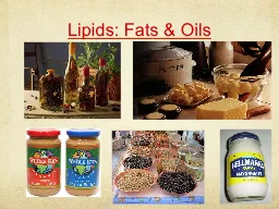
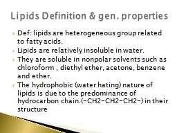
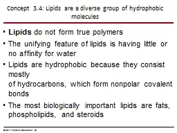
Lipids do not form true polymers The unifying feature of lipids is having little or no affinity for water Lipids are hydrophobic because they consist mostly of hydrocarbons which form nonpolar covalent bonds ID: 310632

figure 2014 acids education 2014 figure education acids pearson amino protein structure dna proteins acid molecules function fatty rna

Share:

Link:

Embed:

Download Presentation from below link

Download Presentation The PPT/PDF document "Concept 3.4: Lipids are a diverse group ..." is the property of its rightful owner. Permission is granted to download and print the materials on this web site for personal, non-commercial use only, and to display it on your personal computer provided you do not modify the materials and that you retain all copyright notices contained in the materials. By downloading content from our website, you accept the terms of this agreement.


Presentation Transcript

Slide1

Concept 3.4: Lipids are a diverse group of hydrophobic molecules

Lipids do not form true polymersThe unifying feature of lipids is having little or no affinity for waterLipids are hydrophobic because they consist mostly of hydrocarbons, which form nonpolar covalent bondsThe most biologically important lipids are fats, phospholipids, and steroids

© 2014 Pearson Education, Inc.Slide2

Fats

Fats are constructed from two types of smaller molecules: glycerol and fatty acidsGlycerol is a three-carbon alcohol with a hydroxyl group attached to each carbonA fatty acid consists of a carboxyl group attached to a long carbon skeleton

© 2014 Pearson Education, Inc.Slide3

Figure 3.12

(b) Fat molecule (triacylglycerol)

Glycerol

(a) One of three dehydration reactions in the synthesis of a fat

Ester linkage

Fatty acid

(in this case, palmitic acid)Slide4

Fats separate from water because water molecules hydrogen-bond to each other and exclude the fats

In a fat, three fatty acids are joined to glycerol by an ester linkage, creating a triacylglycerol, or triglyceride

© 2014 Pearson Education, Inc.Slide5

Fatty acids vary in length (number of carbons) and in the number and locations of double bonds

Saturated fatty acids have the maximum number of hydrogen atoms possible and no double bondsUnsaturated fatty acids have one or more double bonds

© 2014 Pearson Education, Inc.Slide6

Figure 3.13

(a) Saturated fat

(b) Unsaturated fat

Structural

formula of a

saturated fat

molecule

Structural

formula

of an

unsaturated

fat molecule

Space-filling

model of

stearic acid,

a saturated

fatty acid

Space-filling

model of oleic

acid, an

unsaturated

fatty acid

Double bond

causes bending.Slide7

Fats made from saturated fatty acids are called saturated fats and are solid at room temperature

Most animal fats are saturatedFats made from unsaturated fatty acids, called unsaturated fats or oils, are liquid at room temperaturePlant fats and fish fats are usually unsaturated

© 2014 Pearson Education, Inc.Slide8

The major function of fats is energy storage

Fat is a compact way for animals to carry their energy stores with them

© 2014 Pearson Education, Inc.Slide9

Phospholipids

In a phospholipid, two fatty acids and a phosphate group are attached to glycerol The two fatty acid tails are hydrophobic, but the phosphate group and its attachments form a hydrophilic headPhospholipids are major constituents of cell membranes

© 2014 Pearson Education, Inc.Slide10

Figure 3.14

(a) Structural formula

(b) Space-filling model

Hydrophilic

head

(d) Phospholipid

bilayer

(c) Phospholipid

symbol

Hydrophobic

tails

Choline

Phosphate

Glycerol

Fatty acids

Hydrophilic head

Hydrophobic tailsSlide11

When phospholipids are added to water, they self-assemble into a bilayer, with the hydrophobic tails pointing toward the interior

This feature of phospholipids results in the bilayer arrangement found in cell membranes

© 2014 Pearson Education, Inc.Slide12

Concept 3.5: Proteins include a diversity of structures, resulting in a wide range of functions

Proteins account for more than 50% of the dry mass of most cellsProtein functions include defense, storage, transport, cellular communication, movement, and structural support

© 2014 Pearson Education, Inc.Slide13

Figure 3.16a

Enzymatic proteins

Storage proteins

Defensive proteins

Transport proteins

Enzyme

Function: Selective acceleration of

chemical reactions

Function: Storage of amino acids

Example: Digestive enzymes catalyze the

hydrolysis of bonds in food molecules.

Ovalbumin

Amino acids

for embryo

Examples: Casein, the protein of milk, is

the major source of amino acids for baby

mammals. Plants have storage proteins

in their seeds. Ovalbumin is the protein

of egg white, used as an amino acid

source for the developing embryo.

Examples: Hemoglobin, the iron-containing

protein of vertebrate blood, transports

oxygen from the lungs to other parts of the

body. Other proteins transport molecules

across cell membranes.

Function: Transport of substances

Transport

protein

Cell membrane

Antibodies

Bacterium

Virus

Function: Protection against disease

Example: Antibodies inactivate and help

destroy viruses and bacteria.Slide14

Figure 3.16b

Hormonal proteins

Contractile and motor proteins

Receptor proteins

Structural proteins

Example: Insulin, a hormone secreted by

the pancreas, causes other tissues to

take up glucose, thus regulating blood

sugar concentration.

Function: Coordination of an organism’s

activities

Normal

blood sugar

High

blood sugar

Insulin

secreted

Examples: Motor proteins are responsible

for the undulations of cilia and flagella.

Actin and myosin proteins are

responsible for the contraction of

muscles.

Function: Movement

Muscle tissue

Actin

Myosin

30

m

Connective tissue

60

m

Collagen

Examples: Keratin is the protein of hair,

horns, feathers, and other skin appendages.

Insects and spiders use silk fibers to make

their cocoons and webs, respectively.

Collagen and elastin proteins provide a

fibrous framework in animal connective

tissues.

Function: Support

Signaling molecules

Receptor

protein

Example: Receptors built into the

membrane of a nerve cell detect signaling

molecules released by other nerve cells.

Function: Response of cell to chemical

stimuliSlide15

Polypeptides

are unbranched polymers built from the same set of 20 amino acidsA protein is a biologically functional molecule that consists of one or more polypeptides

© 2014 Pearson Education, Inc.Slide16

Amino Acids

Amino acids are organic molecules with carboxyl and amino groupsAmino acids differ in their properties due to differing side chains, called R groups

© 2014 Pearson Education, Inc.Slide17

Figure 3.UN04

Side chain (R group)

Carboxyl

group

Amino

group

carbonSlide18

Figure 3.17

Nonpolar side chains; hydrophobic

Side chain

(R group)

Glycine

(Gly or G)

Alanine

(Ala or A)

Methionine

(Met or M)

Phenylalanine

(Phe or F)

Polar side chains; hydrophilic

Leucine

(Leu or L)

Isoleucine

(

le or

)

Tryptophan

(Trp or W)

Proline

(Pro or P)

Valine

(Val or V)

Serine

(Ser or S)

Threonine

(Thr or T)

Tyrosine

(Tyr or Y)

Asparagine

(

Asn

or

N

)

Cysteine

(Cys or C)

Glutamine

(

Gln

or

Q

)

Aspartic acid

(Asp or D)

Glutamic acid

(Glu or E)

Arginine

(Arg or R)

Lysine

(

Lys

or

K

)

Histidine

(His or H)

Electrically charged side chains; hydrophilic

Acidic (negatively charged)

Basic (positively charged)Slide19

Polypeptides

Amino acids are linked by peptide bondsA polypeptide is a polymer of amino acidsPolypeptides range in length from a few to more than a thousand monomers Each polypeptide has a unique linear sequence of amino acids, with a carboxyl end (C-terminus) and an amino end (N-terminus)

© 2014 Pearson Education, Inc.Slide20

Figure 3.18

New peptide

bond forming

Peptide bond

Side

chains

Back-

bone

Amino end

(N-terminus)

Carboxyl end

(C-terminus)

Peptide

bondSlide21

Protein Structure and Function

A functional protein consists of one or more polypeptides precisely twisted, folded, and coiled into a unique shape

© 2014 Pearson Education, Inc.Slide22

Figure 3.19

(a) A ribbon model

(b) A space-filling model

Groove

GrooveSlide23

The sequence of amino acids, determined

genetically, leads to a protein’s three-dimensional structureA protein’s structure determines its function

© 2014 Pearson Education, Inc.Slide24

Figure 3.20

Antibody protein

Protein from flu virusSlide25

Four Levels of Protein Structure

Proteins are very diverse, but share three superimposed levels of structure called primary, secondary, and tertiary structureA fourth level, quaternary structure, arises when a protein consists of more than one polypeptide chain

© 2014 Pearson Education, Inc.Slide26

The primary structure of a protein is its unique sequence of amino acids

Secondary structure, found in most proteins, consists of coils and folds in the polypeptide chainTertiary structure is determined by interactions among various side chains (R groups)Quaternary structure results from interactions between multiple polypeptide chains

© 2014 Pearson Education, Inc.Slide27

Figure 3.21a

Primary structure

Amino end

Carboxyl end

Primary structure of transthyretin

125

95

90

100

105

110

120

115

80

70

60

85

75

65

55

50

45

40

25

30

35

20

15

10

5

1

Amino

acidsSlide28

Figure 3.21aa

Primary structure

Amino end

25

30

20

15

10

5

1

Amino

acidsSlide29

Figure 3.21b

Secondary

structure

Tertiary

structure

Quaternary

structure

Transthyretin

polypeptide

Transthyretin

protein

pleated sheet

helixSlide30

Figure 3.21d

Hydrogen

bond

Disulfide

bridge

Polypeptide

backbone

Hydrophobic

interactions and

van der Waals

interactions

Ionic bondSlide31

Figure 3.21e

Collagen Slide32

Figure 3.21f

Heme

Iron

subunit

subunit

subunit

subunit

HemoglobinSlide33

Sickle-Cell Disease: A Change in Primary Structure

Primary structure is the sequence of amino acids on the polypeptide chainA slight change in primary structure can affect a protein’s structure and ability to function Sickle-cell disease, an inherited blood disorder, results from a single amino acid substitution in the protein hemoglobin

© 2014 Pearson Education, Inc.Slide34

Figure 3.22

subunit

subunit

Function

Red Blood Cell

Shape

Quaternary

Structure

Secondary

and Tertiary

Structures

Primary

Structure

Normal

hemoglobin

Sickle-cell

hemoglobin

Exposed hydro-

phobic region

Molecules crystallized

into a fiber; capacity to

carry oxygen is reduced.

Molecules do not

associate with one

another; each carries

oxygen.

1

2

3

4

5

6

7

1

2

3

4

5

6

7

Normal

Sickle-cell

5 m

5 mSlide35

What Determines Protein Structure?

In addition to primary structure, physical and chemical conditions can affect structureAlterations in pH, salt concentration, temperature, or other environmental factors can cause a protein to unravelThis loss of a protein’s native structure is called denaturation

A denatured protein is biologically inactive

© 2014 Pearson Education, Inc.Slide36

Figure 3.23-3

Denatured protein

Normal proteinSlide37

Protein Folding in the Cell

It is hard to predict a protein’s structure from its primary structureMost proteins probably go through several intermediate structures on their way to their final, stable shapeScientists use X-ray crystallography to determine

3-D protein structure based on diffractions of an

X-ray beam by atoms of the crystalized molecule

© 2014 Pearson Education, Inc.Slide38

Figure 3.24

Digital detector

Crystal

Experiment

Results

X-ray

source

X-ray

beam

X-ray diffraction

pattern

Diffracted

X-rays

DNA

RNA

RNA

polymerase Slide39

Concept 3.6: Nucleic acids store, transmit, and help express hereditary information

The amino acid sequence of a polypeptide is programmed by a unit of inheritance called a geneGenes are made of DNA, a nucleic acid made of monomers called nucleotides

© 2014 Pearson Education, Inc.Slide40

The Roles of Nucleic Acids

There are two types of nucleic acidsDeoxyribonucleic acid (DNA)Ribonucleic acid (RNA)DNA provides directions for its own replicationDNA directs synthesis of messenger RNA (mRNA) and, through mRNA, controls protein synthesis

© 2014 Pearson Education, Inc.Slide41

Figure 3.25-3

CYTOPLASM

Ribosome

Amino

acids

mRNA

Polypeptide

Synthesis

of protein

Movement of

mRNA into

cytoplasm

NUCLEUS

Synthesis

of mRNA

mRNA

DNA

3

2

1Slide42

The Components of Nucleic Acids

Nucleic acids are polymers called polynucleotidesEach polynucleotide is made of monomers called nucleotidesEach nucleotide consists of a nitrogenous base, a pentose sugar, and one or more phosphate groupsThe portion of a nucleotide without the phosphate group is called a nucleoside

© 2014 Pearson Education, Inc.Slide43

Animation

: DNA and RNA Structure

Right click slide / Select playSlide44

Figure 3.26

Sugar-phosphate backbone

(on blue background)

(a) Polynucleotide, or nucleic acid

(b) Nucleotide

(c) Nucleoside components

5 end

3 end

5C

5C

3C

3C

Phosphate

group

Sugar

(pentose)

Nitrogenous

base

Nucleoside

Nitrogenous bases

Pyrimidines

Cytosine (C)

Thymine

(T, in DNA)

Uracil

(U, in RNA)

Purines

Adenine (A)

Guanine (G)

Sugars

Deoxyribose (in DNA)

Ribose (in RNA)Slide45
Slide46

Each nitrogenous base has one or two rings that include nitrogen atoms

The nitrogenous bases in nucleic acids are called cytosine (C), thymine (T), uracil (U), adenine (A), and guanine (G)Thymine is found only in DNA, and uracil only in RNA; the rest are found in both DNA and RNA

© 2014 Pearson Education, Inc.Slide47

The Structures of DNA and RNA Molecules

RNA molecules usually exist as single polypeptide chains DNA molecules have two polynucleotides spiraling around an imaginary axis, forming a double helixIn the DNA double helix, the two backbones run in opposite 5→ 3

 directions from each other, an arrangement referred to as

antiparallelOne DNA molecule includes many genes

© 2014 Pearson Education, Inc.Slide48

Animation

: DNA Double Helix

Right click slide / Select playSlide49

Video

: DNA Stick ModelSlide50

Figure 3.27

(a) DNA

Sugar-phosphate

backbones

5

5

3

3

(b) Transfer RNA

Base pair joined

by hydrogen bonding

Hydrogen bonds

Base pair joined

by hydrogen

bondingSlide51

DNA and Proteins as Tape Measures of Evolution

The linear sequences of nucleotides in DNA molecules are passed from parents to offspringTwo closely related species are more similar in DNA than are more distantly related speciesMolecular biology can be used to assess evolutionary kinship

© 2014 Pearson Education, Inc.Slide52

Figure 3.UN06Slide53

Figure 3.UN06aSlide54

Figure 3.UN06b