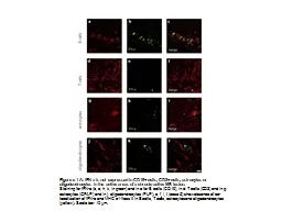
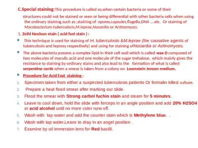
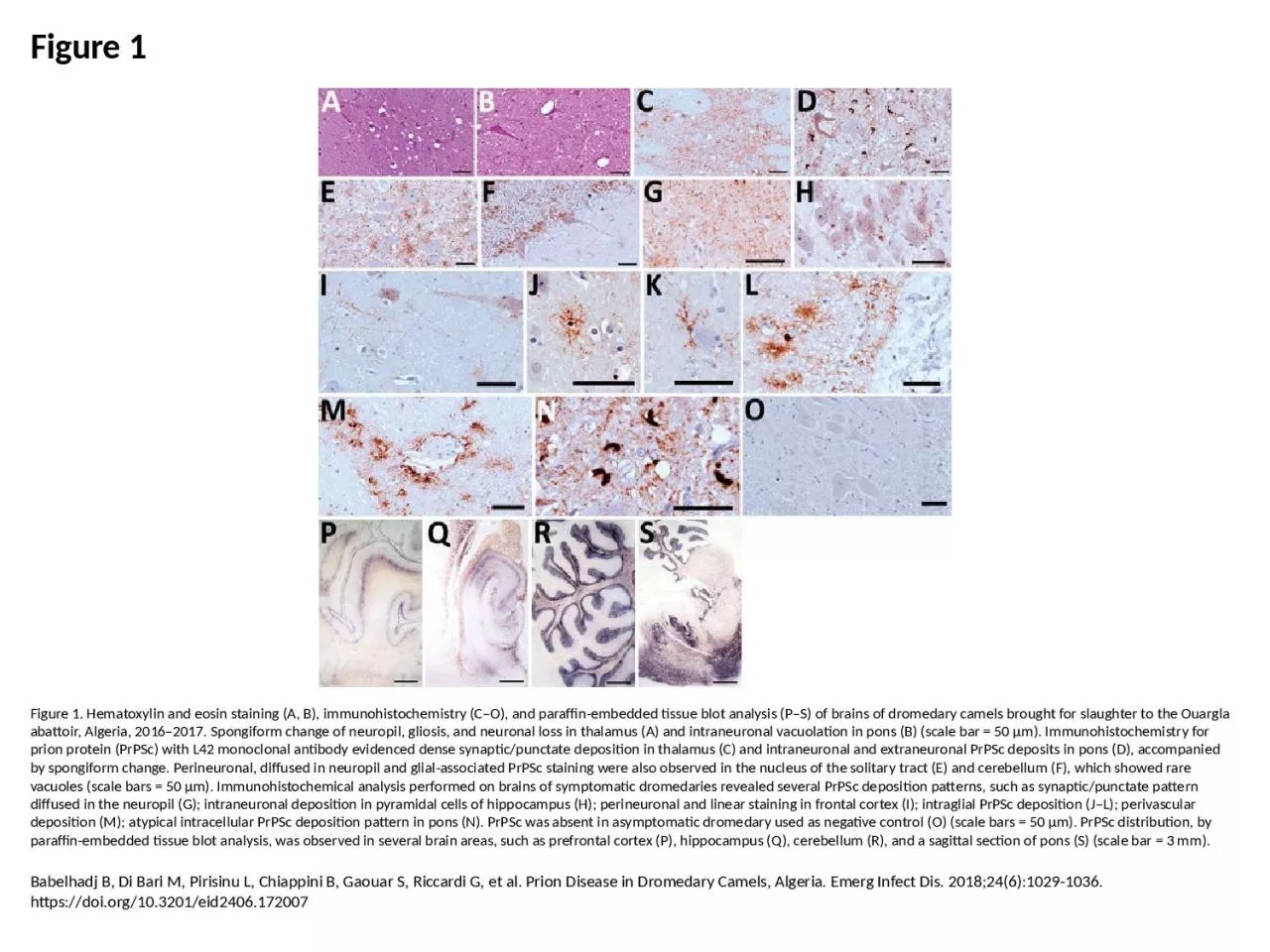
/
Figure 1 Figure 1. Hematoxylin and eosin staining (A, B), immunohistochemistry (C–O), Figure 1 Figure 1. Hematoxylin and eosin staining (A, B), immunohistochemistry (C–O),

Figure 1 Figure 1. Hematoxylin and eosin staining (A, B), immunohistochemistry (C–O), - PowerPoint Presentation

emery
emery . @emery
Follow
67 views
Uploaded On 2023-07-27

Figure 1 Figure 1. Hematoxylin and eosin staining (A, B), immunohistochemistry (C–O), - PPT Presentation

Babelhadj B Di Bari M Pirisinu L Chiappini B Gaouar S Riccardi G et al Prion Disease in Dromedary Camels Algeria Emerg Infect Dis 201824610291036 httpsdoiorg103201eid2406172007 ID: 1012263

deposition prpsc scale pons prpsc deposition pons scale neuropil dromedary staining intraneuronal analysis thalamus cerebellum prion bar spongiform observed

Share:

Link:

Embed:

Download Presentation from below link

Download Presentation The PPT/PDF document "Figure 1 Figure 1. Hematoxylin and eosin..." is the property of its rightful owner. Permission is granted to download and print the materials on this web site for personal, non-commercial use only, and to display it on your personal computer provided you do not modify the materials and that you retain all copyright notices contained in the materials. By downloading content from our website, you accept the terms of this agreement.


Presentation Transcript

1. Figure 1Figure 1. Hematoxylin and eosin staining (A, B), immunohistochemistry (C–O), and paraffin-embedded tissue blot analysis (P–S) of brains of dromedary camels brought for slaughter to the Ouargla abattoir, Algeria, 2016–2017. Spongiform change of neuropil, gliosis, and neuronal loss in thalamus (A) and intraneuronal vacuolation in pons (B) (scale bar = 50 μm). Immunohistochemistry for prion protein (PrPSc) with L42 monoclonal antibody evidenced dense synaptic/punctate deposition in thalamus (C) and intraneuronal and extraneuronal PrPSc deposits in pons (D), accompanied by spongiform change. Perineuronal, diffused in neuropil and glial-associated PrPSc staining were also observed in the nucleus of the solitary tract (E) and cerebellum (F), which showed rare vacuoles (scale bars = 50 μm). Immunohistochemical analysis performed on brains of symptomatic dromedaries revealed several PrPSc deposition patterns, such as synaptic/punctate pattern diffused in the neuropil (G); intraneuronal deposition in pyramidal cells of hippocampus (H); perineuronal and linear staining in frontal cortex (I); intraglial PrPSc deposition (J–L); perivascular deposition (M); atypical intracellular PrPSc deposition pattern in pons (N). PrPSc was absent in asymptomatic dromedary used as negative control (O) (scale bars = 50 μm). PrPSc distribution, by paraffin-embedded tissue blot analysis, was observed in several brain areas, such as prefrontal cortex (P), hippocampus (Q), cerebellum (R), and a sagittal section of pons (S) (scale bar = 3 mm).Babelhadj B, Di Bari M, Pirisinu L, Chiappini B, Gaouar S, Riccardi G, et al. Prion Disease in Dromedary Camels, Algeria. Emerg Infect Dis. 2018;24(6):1029-1036. https://doi.org/10.3201/eid2406.172007