Explore
Featured
Recent
Articles
Topics
Login
Upload
Featured
Recent
Articles
Topics
Login
Upload
Search Results for 'spongiform'
spongiform published presentations and documents on DocSlides.
1 CNS Infection Meninges and Prions
by anderson
2. CNS Infection - Meninges. CNS infections: most ...
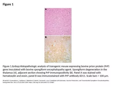
Figure 1 Figure 1. Histopathologic analysis of transgenic mouse expressing bovine prion pr
by rosemary
Piccardo P, Cervenakova L, Vasilyeva I, Yakovleva ...
108A-9 狂犬病 Pathogenesis of rabies virus infection
by williams
. Distribution of animal rabies in the United Stat...
UNIT-3 ‘ Zoonotic disease’
by vivian
(Credit Hours-3+1). Prions. diseases. Transmissib...
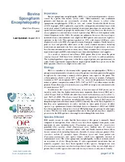
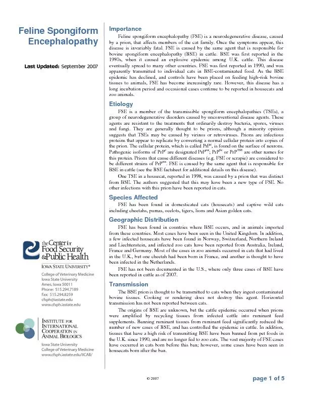
page of Bovine Spongiform Encephalopathy Mad Cow Disease BSE Last Updated May Importance Bovine spongiform encephalopathy BSE is a fatal neurodegenerative disease caused by a prion that mainly af
by calandra-battersby
Other ruminant species cats non human primates an...
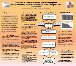
Presence of “Mad Cow Disease” (Bovine Spongiform Enceph
by stefany-barnette
Christina Hill . Department of Biological Sc...
Bovine Spongiform Encephalopathy
by adah
Assist. Prof. Dr. Hussein Al . Naji. Bovine spongi...
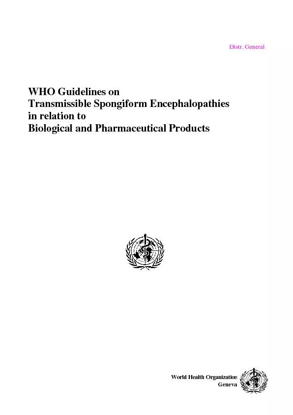
Distr. General WHO Guidelines on Transmissible Spongiform Encephalop
by jane-oiler
World Health Organization Geneva ...
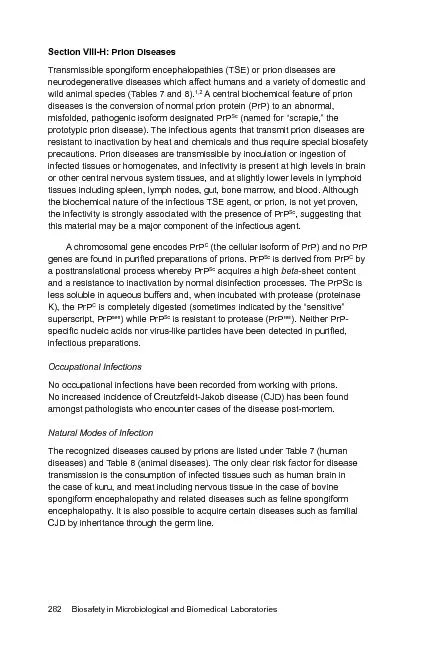
Biosafety in Microbiological and Biomedical Laboratories
by mitsue-stanley
282 Section VIII-H: Transmissible spongiform ence...
Feline Spongiform
by tatyana-admore
FDA Transmissible Spongiform Encephalopathies
by kittie-lecroy
Advisory Committee. 23. rd. Meeting 01 August 20...
The Psychology of a
by stefany-barnette
Prion. Sarah . Themel. This is your brain…. Thi...
Who studies/treats this?
by cheryl-pisano
Ways that the NS can be damaged. What happens aft...
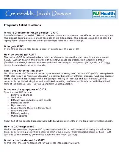
Frequently Asked Questions CreutzfeldtJakob krutsfelt YAHcub dis
by wang
caused by a bacteria, virus or parasite. No. Mos...
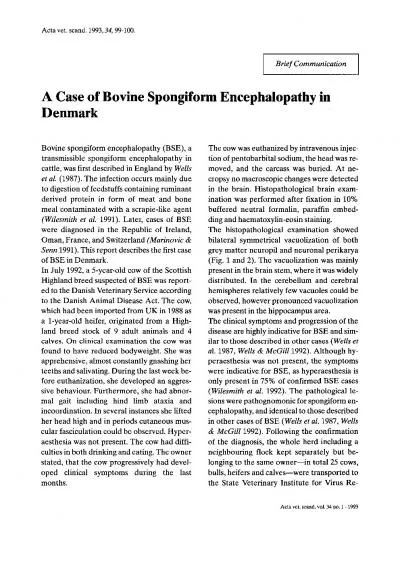
Acta vet scand199334
by dorothy
99-100. Brief Communication A Case of Bovine Spon...
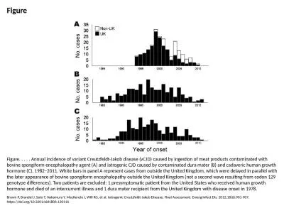
Figure Figure. . . . . Annual incidence of variant Creutzfeldt-Jakob disease (vCJD) caused by inges
by quinn
Brown P, Brandel J, Sato T, Nakamura Y, MacKenzie ...
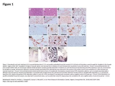
Figure 1 Figure 1. Hematoxylin and eosin staining (A, B), immunohistochemistry (C–O), and paraffi
by emery
Babelhadj B, Di Bari M, Pirisinu L, Chiappini B, G...
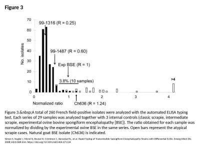
Figure 3 Figure 3. A total of 260 French field-positive isolates were analyzed with the au
by thomas
Simon S, Nugier J, Morel N, Boutal H, Créminon C,...
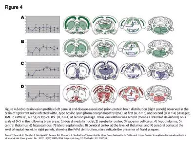
Figure 4 Figure 4. Brain lesion profiles (left panels) and disease-associated prion protei
by heavin
Baron T, Bencsik A, Biacabe A, Morignat E, Bessen ...
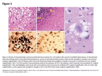
Figure 3 Figure 3. Results of histopathologic and immunohistochemical analyses for a US patient wit
by badra
Maheshwari A, Fischer M, Gambetti P, Parker A, Ram...
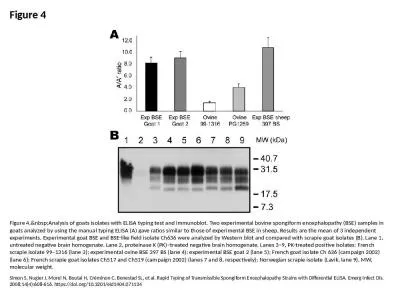
Figure 4 Figure 4. Analysis of goats isolates with ELISA typing test and immunoblot. Two e
by kylie
Simon S, Nugier J, Morel N, Boutal H, Créminon C,...
Load More...