/
Vertebrates and invertebrates Vertebrates and invertebrates

Vertebrates and invertebrates - PowerPoint Presentation

giovanna-bartolotta
giovanna-bartolotta . @giovanna-bartolotta
Follow
700 views
Uploaded On 2016-08-08

Vertebrates and invertebrates - PPT Presentation

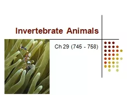
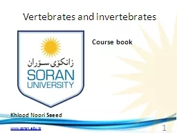
Khlood Noori Saeed Course book 1 SUBJECT OUTLINE 20142015 2 3 Vertebrates and invertebrates Subject title 2 Theory Credit hours 2 Units 2 four stage Stage 120 minute one sessions per week ID: 438136

week cells body cell cells week cell body sponges species system structure vertebrate food water called specialized slits types vertebrates located canals

Share:

Link:

Embed:

Download Presentation from below link

Download Presentation The PPT/PDF document "Vertebrates and invertebrates" is the property of its rightful owner. Permission is granted to download and print the materials on this web site for personal, non-commercial use only, and to display it on your personal computer provided you do not modify the materials and that you retain all copyright notices contained in the materials. By downloading content from our website, you accept the terms of this agreement.


Presentation Transcript

Slide1

Vertebrates and invertebrates

Khlood Noori Saeed

Course book

1Slide2

SUBJECT OUTLINE

2014-20152Slide3

3

Vertebrates and invertebrates

Subject title

2

(

Theory

)

Credit hours 2

Units 2

four stage

Stage

120 minute, one sessions per week

Class schedule

20

weeks

Duration 27 weeks

Assistant Lecturer

Course coordinator

khlood.saeed@soran.edu.iq

Kazem.asiaban@soran.edu.iqSlide4

Description

Teaching students the science of comparative anatomy of vertebrates and invertebrates and the structures of the organs which fond in different living organisms and know how it developed.


2.

Require Background or Experience

Many student know about the human body organs and the functions and their effect.

3

.

Subject Objective

A)Understand the fundamental concepts of comparative anatomy and the scientific vocabulary used in this field.   B) different between vertebrates and invertebrates. . 4. Text book(s) and Readings: Kardong vertebrate comperative anatomy(2).  (3(4 5. Student Materials: 1. Textbook and References 2. Lecture Notes 3. CD and internet resources  6. Collage Facilities 1. Classroom with Whiteboard and projection facilities 2. College library  3data show

4Slide5

Week 1

The vertebrate body

Comparative anatomy:

is the study of structure , of the functional significance of structure , and of the range of variation in structure and function in different species.

Its methods are descriptive and experimental .Comparative anatomy has its roots in philosophy , and its aim is enlightenment.

The benefits of comparative anatomy study:

1-

To attempt study history of different species and the environmental condition under which they rose , flourished , and become extinct.

2-

To help satisfy the curiosity of human mind.

 Vertebrate characteristics [the big four]:Have four definitive structural characteristics:1-The presence of notochord , at least in the embryo.2-The presence of a pharynx with pouches or slits in its wall , at least in the embryo.3-The occurrence of a dorsal , tubular nervous system.4-The presence of vertebral column.5Slide6

Week 2 

Notochord and vertebral column

Notochord

: Is the first skeletal structure to appear in vertebrate embryo

.

It is a rod of living cells.

*Notochord location:

Located immediately ventral to the central nervous system and dorsal to the alimentary canal.

*Notochord extending from:

Extending from the midbrain to the tip of the tail

*Notochord origin: From die roof of the archenteron, or embryonic gut .6Slide7

Week 3

Pharynx:

The pharynx is the region of the alimentary canal exhibiting pharyngeal pouches in the embryo.

The pouches may rupture to the exterior form pharyngeal slits . These slits may remain throughout life , or they may be temporary .

If the slits remain throughout life

, the adult pharynx is the part of the alimentary canal having slits.

If the slits is temporary

, the adult pharynx is part of the alimentary canal connecting the oral cavity and esophagus.

7Slide8

Week 4

Satellite characteristics:

*Skin:

[the integument]

Skin of vertebrates consist of :

1-epidermis

of ectoderm origin .

2-underlying dermis of

mesodermal

origin.8Slide9

Week 5

Metamerism

:

Serial repetition of body structure in the longitudinal axis is known as

metamerism

.

-The successive body segments of earthworms are readily seen externally as well as internally .

-In crayfish the

metamerism is obvious in the caudal region , but in the trunk it is obscured dorsally by the carapace.-In vertebrate metamerism clearly expressed in embryos and is retained in many adult system .No external evidence is seen in vertebrate ??? Because the skin is not metameric .-But if the integument is stripped from the body of fishes , amphibians , and even some reptiles , a series of identical muscle segments is seen .-The serial arrangement of body wall muscles , vertebrae , ribs , spinal nerves , embryonic kidney tubules , segmental arteries and veins is an expression of the fundamental metamerism of vertebrates.9Slide10

Week 6

Digestive organs:

The digestive ( alimentary ) tract exhibits specialized regions for the:

1-acquisition 2-processing 3-temporary storage 4-digestion

5-absorption of food 6-elimination of the unabsorbed residue.


Digestive tract consist of :

1-oral cavity 2-pharynx 3-esophagus 4-stomach

5-intestine ( coiled to increase absorptive area without increasing the body length ) 6-anus or vent

-The tract exhibits a number of ceca or diverticula , including a liver and pancreas.10Slide11

Week 7

Urinogenital

organs:

Urinary and reproductive systems share certain passageways , because urinary and reproductive organs of vertebrate are closely related , kidneys and gonads arise close together in the roof of the

coelom

.

11Slide12

Week 8

Invertebrates

Animals species that do not develop a vertebral column include many animals like insects – worms – clams – crabs – octopus – and other……

The largest animal Phylum or

Superphylum

is also included within invertebrates :

1- Protozoa

2-

Porifera

3-

cnidaria { coelentrata }4- Aschelminthes5- Mollusca6- Annelida7- Echinodermata8- Arthropoda12Slide13

Week 9

Protozoa

General Characters:

1-Unicellular ( or colonial ) and microscopic animals .

2-Most are motile by flagella , cilia or pseudopodia .

3-Most

protozoal

species are aerobic , but some anaerobic species have been found in the human intestine and animal rumen.

4-They occur in all habitats including marine , freshwater ,and terrestrial , including soil.

5-The food inter to the cell ( body of animal ) by mouth of cell or by body wall but the digestive happen in food vacuole in cytoplasm. 6-Osmoregulation in freshwater protozoans is accomplished by contractile vacuoles that pump a hyposomatic urine from the cytoplasm back into the environment.7-Most protozoa reproduce by asexual methods ( binary fission , budding , multiple fission , plasmotomy ) , or sexual reproduction ( syngamy , conjugation ) .8-Some species are parasites of plants and human . 13Slide14

Week 10

Cytoplasm

1-Protoplasm part located outside nuclei.

2-Homogenous , contain many of vacuoles.

3-Reticullar , not

colour

.

4-Divided into :

a-Ectoplasm ( near the cell membrane )

b-Endoplasm ( near the nuclei membrane )

-Ectoplasm : is a gel containing the basal bodies of cilia or flagella , microfilaments .-Endoplasm : or ( sol ) more fluid than ectoplasm and contain organelles such as nuclei , mitochondria and vacuoles and vesicles of various types.14Slide15

Week 11

Locomotor

Organelles

1-Flagella

2-Cilia

3-Pseudopodia

Cilia

Flagella

1-are short hair like appendages 1-are long thread like appendages Extending from the surface of on the surface of living cell. Living cells.2-Rotational, like a motor very 2-Wave –like ,undulating , Fast moving. Sinusoidal , slow movement compared to cilia.3-Many ( hundreds )per cell 3-Few ( less than 10 )per cell4-Eukaryotic cells 4-Eukaryotic and Prokaryotic cells15Slide16

Week 12

Euglena

1-Biflagellated ,

unicell , spindle shape .

2-Most species have photosynthesizing , chloroplast within the body of cell. Chloroplasts contain

pyrenoid

used in the synthesis of

paramylon

, a form of starch energy storage.

3-Have two flagella rooted in basal bodies located in a small reservoir of the front of the cell , one of flagellum is very short while the other is long easily visible with light microscopy.

4-Possess red eyespot ( stigma ) , an organelle composed of carotenoid pigment granules . The red spot itself is not thought to be photosynthetic so that paraflagellar body it filters the sun light located at the base of flagellum.5-Lake a cell wall instead it has pellicle made of protein layer supported by structure of microtubules arranged in strips spiraling around the cell , pellicle give exceptional flexibility and contractility , there is no cellulose.6-Euglenoids are found in many freshwater habitats , and are most abundant in those .7-Small and inconspicuous contractile vacuole is located to one side of the reservoir into which it discharges , it function is osmoregulatory.8-Repreduce a sexually through binary fission a form of cell division , divide longitudinally beginning at the front end of the cell with the duplication of flagellar processes , and stigma .A cleavage forms in the anterior and V-shaped moves toward the posterior , until the two halves are entirely separated , sexual conjugation are rare.16Slide17

Week 13

Amoeba

1-They so tiny need microscope to see them .

2-Living in water , including lakes , ponds , streams , rivers , and puddles .Some can live in the bodies of animals.

3-Move by pseudopodia and help

Amoeba

to eat .

4-Reproduce by binary fission , it means that one

Amoeba

can split in half and make two identical new

Amoeba.5-Cytoplasm divided into two parts:a-endoplasm b-ectoplasm …….both enclosed within a flexible plasma membrane.6-Cell contain single granular nucleus containing DNA.7-Contractile vacuole is used to maintain osmotic equilibrium by excreting excess water from the cell.8-An Amoeba obtain its food by phagocytosis , and particles of organic matter , or by pinocytosis taking in dissolved nutrient through vesicles formed within the cell membrane.17Slide18


Week 14

Phylum :

Porifera

( Sponges )

Sponges are animals of the phylum

porifera

( meaning : pore bearer ). Sponges are a diverse group of sometimes common types with about 5000 species known across the world . Sponges are primarily marine , but around 150 species live in freshwater.

General characters

1-Include a system of pores ( also called ostia ) and canals , through which water passes .2-Sponges are either radially symmetrical or a symmetrical .3-They are supported by a skeleton made up of the protein collagen and spicules , which may be calcareous or siliceous , depending on the group of sponges examined.4-They are multicellular organisms and have an specialized cells that can transform into other types and which often migrate between the main cell layers and mysophyl in the process.5-Sponges do not have nervous , digestive or circulatory systems , instead most rely on maintaining a constant water flow through their bodies to obtain food and O2 and remove wastes.6-All sponges are sessile aquatic animals.7-Many species feed on bacteria and other food particles in the water, some host photosynthesizing microorganism . A few species of sponges that live in food –poor environment have become carnivores that may prey mainly on small crustacea .8-Most species reproduce by sexual reproduction and other by a sexual reproduction.18Slide19

Week 15

Sponge Types :

Sponges have three

different types of body plans:-

1-Asconoid : Sponges are shaped like a simple tube perforated by pores. The open internal part of the tube is called the

spongocoel

, it opening to the outside , the

osculum

.

2-Syconoid : Sponges tend to be larger the asconoids , have a tubular body with a single osculum . The syconoid body wall is thicker and the pores that penetrate it are longer , forming a system of simple canals . These canals are lined by collar cells , the flagella of which move water from the outside , into the spongocoel and out the osculum.3-Leuconoid : These are largest and most complex sponges. These sponges are made up of masses of tissue . Penetrated by numerous canals . Canals lead to numerous small chambers lined with flagellated cells . Water move through the canals , into these chambers , and out via a central canal and osculum.19Slide20


Week 16

Phylum :

Cnidaria

=

Coelenterata

General characteristics :

1-Radial symmetry .

2-Diploblast ( two germ layers during development ectoderm and endoderm ).

3-Adult body with an outer cellular layer ( epidermis or ectodermis )and an inner cellular layer ( gastrodermis ) with anon cellular – layer ( Mesoglea =jelly – like material ) in between .4-Some specialized tissue present .5-Posses unique stinging or adhesive structures ( cnidae ) found in specialized cells (cnidocytes ) most common cnidae called Nematocysts [ that fire like harpoons and used mainly to capture prey also as anchors in some species ].6-Simple nerve net forms a nervous system .7-Alternation of generation with a sexual reproducing polyp stage and sexual reproducing medusa stage variation of this strategy exist throughout the cnidaria .8-Have a ciliated , motile planula larvae .20Slide21


week 17

Hydra

Phylum :

Cnidaria

Class : Hydrozoa

Order :

Anthomedusae

Family : HydridaeGenus : HydraGeneral characteristic of Hydra :1-Small , multicellular , has tubular body up to 10 mm.2-Radial symmetry.3-They can found in most un polluted freshwater , ponds , lakes and streams in the temperature and tropical regions.4-Contain in the foot region basal disc , gland cells in the basal disc secrete a sticky fluid that accounts for its adhesive properties.5-Mouth found in free end of the body surrounded by ( 1-12 ) thin mobile tentacles , each tentacle or cnida ( plural cnidae ) clothed with highly specialized stinging cells ( cnidocytes ) contain specialized structures called ( Nematocysts ).6-The nervous system of hydra is a nerve net , which is structurally simple compared to mammalian nervous system , and there are sensitive nerve cells , located in the body wall and tentacles.7-Reproduction and Excretion occur by diffusion everywhere through the epidermis.8-Hydra undergoes morphallaxis ( tissue regeneration ) when injured or severed.21Slide22


Week 18

Types of cells in

Hydra :

*Epidermis layers :-

1-Epithelio-muscular cells .

2-Sensory cells.

3-Nerve cells.

4-Interstitial cells.

5-Cnidocytes.

6-Germ cells.*Endodermis layers :-1-Epithelio-muscular cells ( digestive cells ).2-Interstitial cells.3-Sensory cells.4-Nerve cells.5-Gland cells. 22Slide23

Week 19

Cnidocyte

:

A

cnidocyte

( also known as a

cnidoblast

or

nematocyte

) is an explosive cell containing one giant

secretory organelle or cnida (plural cnidae ) that defines the phylum : Cnidaria ( corals , sea anemones , hydrae , jelly fish , etc…) .Cnidae are used for prey capture and defense from predators . despite being morphologically simple , lacking a skeleton and usually being sessile , cnidarians prey on fish and crustaceans . A cnidocyte fires a structure that contains the toxin , from a characteristics subcellular organelle called a cnidocyst ( also known as a cnida or nematocyst ) . This is responsible for the sting delivered by a cnidarians .23Slide24

Week 20

Structure and function:

Each

cnidocyte

cell contains an organelle called a

cnida

or

cnidocyst

( e.g. nematocyst )

, which comprises a bulb –shape capsule containing a coild hollow tubule structure attached to it . The immature cnidocyte is referred to as a cnidoblast . The externally oriented side of the cell also has a hair –like trigger called a conidocil .When the trigger is activated , the tubule shaft of the cnidocyst is ejected and in the case of the penetrant nematocyst , the forcefully ejected tubule penetrate the target organism. This discharge takes no more than a few microseconds , and is able to reach accelerations of about 40,000 g .Recent research suggests the process to occur as fast as 700 nanoseconds , thus reaching an acceleration of up to 5,410,000 g .After penetration , the toxic content of the nematocyst is injected into the target organism , allowing the sessile cnidarians to devour it.24