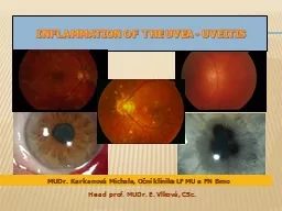
DR VIVEKANAND U What is Uvea Literally Anatomy Iris Ciliary body Choroid UVEITIS Uveitis a term correctly used to describe inflammation of the uveal tract iris ciliary body choroid alone in reality comprises a large group of diverse diseases affecting not only the ID: 1042699
Download Presentation The PPT/PDF document "CLINICAL FEATURES OF IRIDOCYCLITIS" is the property of its rightful owner. Permission is granted to download and print the materials on this web site for personal, non-commercial use only, and to display it on your personal computer provided you do not modify the materials and that you retain all copyright notices contained in the materials. By downloading content from our website, you accept the terms of this agreement.
1. CLINICAL FEATURES OF IRIDOCYCLITIS DR. VIVEKANAND U
2. What is Uvea?Literally
3. AnatomyIrisCiliary bodyChoroid
4. UVEITISUveitis, a term correctly used to describe inflammation of the uveal tract (iris, ciliary body, choroid) alone, in reality comprises a large group of diverse diseases affecting not only the uvea but also the retina, optic nerve and vitreous.
5. Classification IUSG classificationSUN group classification
6. CLASSIFICATIONCLINICALAcute / Chronic / Recurrent ANATOMICALAnterior / Intermediate/ Posterior/ PanuveitisMORPHOLOGICALGranulomatous / Non-granulomatousAETIOLOGICAL
7. Aetiological classification(IUSG)Traumatic Surgical - IOL related uveitisNon surgical ToxicChemicalDrug induced - rifabutinMasquerade syndrome
8. Aetiological classification(IUSG)Infectious Bacterial, fungal, viral, protozoalNon InfectiousKnown systemic association - VKHNot known systemic association - Serpiginous choroiditis
9. Non granulomatous vs GranulomatousOnset - insidiousCourse - chronicInjection - +Pain - +Iris nodules - +KP's - mutton fat Posterior segment - commonly involvedOnset - well definedCourse - acute Injection - +++Pain - +++KP's - fine Iris nodules - absentPosterior segment - rarely involved
10. ANATOMIC CLASSIFICATIONPanuveitis
11. The SUN Working Group Anatomic Classification of Uveitis TypePrimary site of inflammationComponentsAnterior uveitisAnterior chamberIritis, Iridocyclitis, anterior cyclitisIntermediate uveitisVitreousPars planitis, Posterior cyclitis, HyalitisPosterior uveitisRetina or choroidChoroiditis (Focal, multifocal or diffuse)Chorioretinitis,Retinochoroiditis Retinitis,NeuroretinitisPanuveitisAC, Vitreous, Choroid or Retina
12. The SUN Working Group Descriptors of UveitisOnset SuddenInsidious Duration Limited <3 months durationPersistent >3 months duration
13. The SUN Working Group Descriptors of UveitisCourse Acute Episodes characterized by sudden onset & limited durationRecurrent Repeated episodes separated by periods of inactivity without Rx > 3 months in durationChronicPersistent uveitis with relapse in < 3 months after discontinuing treatment
14. Acute non granulomatous anterior uveitis due to collagen diseaseCOMPLETE CLASSIFICATION
15. Symptoms of Anterior uveitis PainRednessPhotophobiaDimness of visionLacrimation
16. Signs of anterior uveitisCONJ - CCCCORNEA - Keratic precipitates FineMutton fatFreshOld
17. KP’SAggregates of inflammatory cells on the endothelial surface of the corneaARLT”S TRIANGLE BASE DOWN TRIANGLE ON THE INF PART OF CORNEACONVENCTION CURRENTS IN AQUEOUS,GRAVITATION OF PARTICLES.
18. TYPE OF KP’Swhite,smoothly roundMutton fat-larger, greasy old-pigmented
19. Anterior chamberFLARE Breakdown of blood aqueous barrier Protein transudation from the iris & ciliary vessels produce opalescence of the aqueous .Slit beam (1mm x 1mm) with maximum intensity is obliquely aimed across the anterior chamber
20. The SUN Working Group Grading Scheme for Anterior Chamber Flare 0 None 1+ Faint 2+Moderate(iris &lens details clear) 3+Marked(iris &lens details hazy) 4+Intense(fibrin or plastic aqueous)Grade Description
21. Anterior chamberCELLSCollection of floating inflammatory cells in the anterior chamberPrimarily lymphocytes & also neutrophilsIndicates active inflammation.
22. Kimura’s grading of AC cells :Trace0-4 cells1+5-10 cells2+11-20 cells3+21-50 cells4+> 50 cells
23. The SUN Working Group Grading Scheme for Anterior Chamber cells 0 <1 0.5+ 1-5 1+ 6-15 2+ 16-25 3+ 26-50 4+ >50GradeCells in Field (1mm by 1mm slit beam)
24. HypopyonCollection of inflammatory cells that settle down in the anterior chamber. Ankylosing spondylitisBehcet’s diseaseInfectionLens induced uveitisMalignancy
25. HyphaemaCollection of RBCs that settle down in the anterior chamber.Viral uveitisTrauma MalignanciesFuchs Heterochromic iridocylitisChronic uveitis with rubeosisAny severe uveitis
26. Iris Posterior synechiaeadhesions between iris & lens capsule.Inflammatory mediators produce fibrin deposits & fibroblast proliferation.Indicates chronic or recurrent inflammation.Most are located at the pupillary border.
27. Festooned pupilMultiple posterior synechiae with the application of mydriatics causes the intervening normal circle of the pupil to dilate.
28. Iris nodules Accumulation of inflammatory cells on the iris surface.Koeppe’s nodule – pupillary border.Busacca’s nodule – iris surface.Berlin’s nodule - angle
29. Seclusio pupillae In severe cases of plastic or recurrent iritis, whole pupillary margin is attached to the lens capsule. Aqueous collects behind the iris and iris bows forwards like a sail – iris bombe. Angle becomes obliterated by adhesion of peripheral iris to posterior surface of the cornea – PAS.IOP raises.
30. Occlusio pupillae The extensive exudation organise across the pupillary area to form a film of opaque fibrous tissue – occlusio pupillae.
31. LENSComplicated cataractVITREOUSOpacities – leucocytes, coagulated fibrin & exudates
32. INTRA OCULAR PRESSURERaised due toACTIVE STAGE Hypertensive iridocyclitis LATER STAGE Secondary glaucoma (pupillary block/PAS)Reduced due to Atrophy of ciliary body Hypotony Phthisis bulbi
33. SEQUELAE OF CHRONIC ANTERIOR UVEITISBand keratopathyIris atrophic patchesMiotic pupilComplicated cataractCyclitic membrane Detachment of ciliary body Pthisis bulbi.
34. Macular edema major cause of severe visual impairmentCataract is common in chronic uveitis and its treatment with corticosteroids.