/
Follow up in Chest Tumors : Value of integrated Follow up in Chest Tumors : Value of integrated

Follow up in Chest Tumors : Value of integrated - PowerPoint Presentation

jaena
jaena . @jaena
Follow
64 views
Uploaded On 2024-01-29

Follow up in Chest Tumors : Value of integrated - PPT Presentation

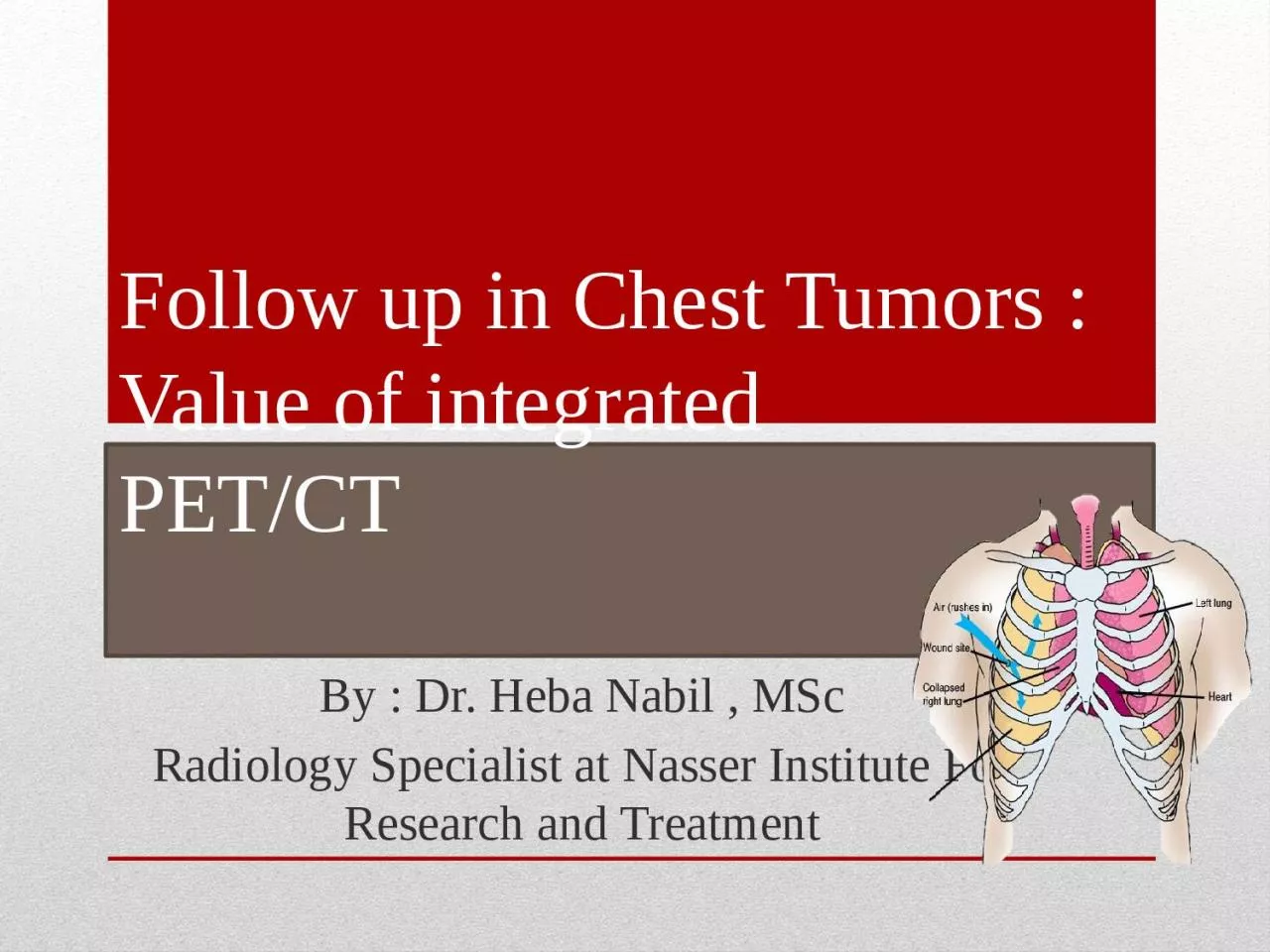
PETCT By Dr Heba Nabil MSc Radiology Specialist at Nasser Institute For Research and Treatment Introduction The management of oncology patients depends on A ccurate local staging of tumor ID: 1041902

patients pet chest treatment pet patients treatment chest images amp study lung patient tumors imaging received staging follow fdg

Share:

Link:

Embed:

Download Presentation from below link

Download Presentation The PPT/PDF document "Follow up in Chest Tumors : Value of int..." is the property of its rightful owner. Permission is granted to download and print the materials on this web site for personal, non-commercial use only, and to display it on your personal computer provided you do not modify the materials and that you retain all copyright notices contained in the materials. By downloading content from our website, you accept the terms of this agreement.


Presentation Transcript

1. Follow up in Chest Tumors : Value of integratedPET/CTBy : Dr. Heba Nabil , MScRadiology Specialist at Nasser Institute For Research and Treatment

2. Introduction

3. The management of oncology patients depends on Accurate local staging of tumor spread Identification of nodal involvement and distant metastases Assessing treatment plans Determining prognosis Evaluating response to treatment.

4. .Indications of PET/CT in chest tumors include Evaluation of the solitary pulmonary nodule Staging of NSCLC, SCLC, mesothelioma and lymphoma Monitoring of treatment response Assessment for recurrent tumor after radio/chemotherapy.

5. .Aim of the work To identify the role of PET-CT in management of chest tumors .

6. Anatomy

7. Lung segments

8.

9. Mediastinum

10.

11. Mediastinal LNs

12.

13. Pathology

14. Chest tumors are subgroubed into

15.

16. Physical Principles

17. PET is a nuclear medicine imaging modality and belongs to the family of emission CT (ECT) which also includes single photon emission CT (SPECT). It is a physiological imaging technique that uses radiopharmaceuticals produced by labeling metabolic markers such as amino acids or glucose with positron-emitting radio nuclides such as fluorine-18. The radio marker is then imaged by coincidence detection of two 511 KeV photons that are produced by annihilation of the emitted positrons.

18. Annihilation reaction: Positrons (β+) released from the nucleus of FDG annihilate with electrons (β-), releasing two coincidence 511-keV photons (γ), which are detected by scintillation crystals (blue rectangles). N neutron, P proton

19. Technique

20.

21. CT TechniqueSingle-phase contrast material-enhanced helical CT is performedAbout 125 ml of a low osmolarity iodinated contrast medium is injected at a rate of 4 ml/sec by using a power injector.For a typical whole body PET/CT study (neck, chest, abdomen, and pelvis), scanning begins at the level of the skull base and extends caudally to the level of the symphysis pubis

22. Typical scout image obtained during an FDG PET/CT study.The blue-purple rectangle represents CT coverage during the study.Each overlapping green rectangle represents PET coverage.Six to seven bed positions are required for PET coverage of the neck, chest, abdomen, and pelvis.

23. PET TechniqueThe total length of CT coverage is an integral number of bed positions scanned during acquisition of PET data.PET is performed on a dedicated PET scanner with a 5-minute emission acquisition per imaging level. The images are acquired in a caudocranial direction from the symphysis pubis to the skull base. Detection of coincidence photons emitted during positron annihilation is the key to PET imaging, whereas accurate co registration of this quantitative/functional information with the CT data is the key to successful PET/CT imaging.

24. PET/CT Fusion & Image Interpretation:Workstations have been available serving to overlay the PET and CT images onto one another, properly putting on consideration the anatomical landmarks for excellent fusion between CT and PET images.

25. The volume of data generated is enormous. PET and CT images are first reconstructed. Then reformatted into coronal and sagittal images to facilitate image interpretation. For each of these sets of PET and CT images, corresponding “fusion” images, combining the two types of data. There are different methods for assessment of radiotracer uptake by normal and pathologic tissues, such as visual inspection and the standardized uptake value (SUV). PET/CT Fusion & Image Interpretation: ( Cont.)

26. Imaging

27. Indication of PET/CT in lung cancerCharacterizing indeterminate pulmonary nodules.Staging and restaging of non-small cell lung cancers, including lymph node metastasis and distant metastasis to adrenal glands or bone.Evaluating for recurrent disease.Planning for radiation therapy.

28. Materials and Methods

29. Patients This is a cross sectional study carried out at Nasser Institute from May 2011 to May 2013 .It included 32 patients. The inclusion criteria for our study were the following:-patients who were suspected to have primary or secondary chest tumors by recent CT, referred for staging before treatment and or follow up after treatment. the exclusion criterion was for uncontrolled diabetic patients The whole study population included 32 patients of variable primary & secondary chest malignancies

30. Patients included performed one or more of the following: 4 patients out of 32 patients had initial PET/CT for staging, intermediate PET/CT and end of treatment PET/CT and follow up, 24 patients out of 32 patients had intermediate PET/CT and end of treatment PET/CT and follow up and 7 patients out of 32 patients had intermediate PET/CT study. The study population was split into two major groups:

31.

32. Number of patientsAge rangeSexPreliminary diagnosis3214-6519 male13 female27 primary chest tumors 8 lung tumors2 pleural tumors3 chest wall tumors7 mediastinal tumors7 lymphoma5 secondary chest tumors

33. Results

34. Aim of the studyFrequencyPercentAssessement before Preparation for BMT26.3 %Assessement of locoregional recurrence/residue or distant metastasis39.4%Assessment of activity of the residual mass13.1%Assessment of treatment response1959.4%Determination of treatment line13.1 %differeniate between residue or irradiation pneumonitis13.1%Follow up515.6%Total32100.0%

35. 28 patients received CTH, 14 received RTH and 12 patients underwent surgery,It is to be noted that in most of patients received mixed lines of treatment that mentioned formerlyCT images were analyzed and we found that

36.

37. Then the fused PET/CT images were analyzed

38. And we reported findings beyond PET/CT resolution in 2 patients out of 32 patients in the form of multiple subcentimetric pulmonary nodules

39.

40.

41. According to these findings 9 patients out of 32 patients were cured &23 patients out of 32 patient were still on treatment

42.

43.

44. Illustrative Cases

45. Case 1

46. Male patient, 49 years old, known to have lung cancer. Received chemotherapy PET/CT scan was requested for assessment of treatment response. PET/CT scan: revealed multiple metabolically active FDG avid wide spread lesions at the RT kidney, lungs (with lymphangitis carcinomatosa) as well as multiple abdominal & mediastinal LNs3 months follow up study: rather stationary course.

47.

48.

49. Case 2

50. Male patient, 57 years old, known to have left lung adenocarcinoma. received chemotherapy & radiotherapy The patient was referred for assessment of the treatment response PET/CT scan: revealed metabolically active FDG avid residual primary lung tumor with hepatic & left suprarenal deposits.9 months follow up study: Regressive course of the MA FDG avid residual left lower lung lobe as well as the distant metastasis

51.

52.

53. Case 3

54. Male patient, 42 years old, known to have left mesothelioma. The patient received chemotherapy& radiotherapy after surgical excision and was referred for assessement of treatment response. PET/CT scan: metabolic active FDG avid local tumoral recurrence at the mediastinal pleura with nodal, hepatic & muscular deposits.

55.

56.

57. Case 4

58. A female patient, 29 years old, known case of HD received chemotherapy was referred for assessement of treatment response PET/CT scan: MA FDG avid multiple nodal lesions above the diaphragm

59.

60.

61. Summary & Conclusion

62. PET/CT imaging is changing the care of patients with lung cancer in several ways:

63. Metabolic and anatomic whole-body staging of patients can be performed in one examination and much reduced scan times, thus, increasing patient comfort.

64. 2. Because of limited patient motion, near ideal fusion of metabolic and anatomic images can be achieved.

65. 3. Anatomic landmarks provided by CT will greatly facilitate the assignment of functional abnormalities to anatomic structures.

66. 4. ‘’ Difficult to image" regions of the body (such as the head and neck and mediastinum) will be evaluated with a high diagnostic accuracy.

67. 5.Fused images can be used to target radiation treatment more accurately and monitor the effects of chemotherapy, surgery, and radiation treatment.

68. By recognizing the relevant radiologic appearances of chest tumors, understanding the appropriateness of staging disease with the TNM classification system, and being familiar with potential imaging pitfalls, radiologists can make an important contribution to treatment and outcome in thoracic cancer patients.

69. Thank You

70.