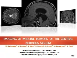
/
IMAGING OF MIDLINE TUMORS OF THE CENTRAL NERVOUS SYSTEM IMAGING OF MIDLINE TUMORS OF THE CENTRAL NERVOUS SYSTEM

IMAGING OF MIDLINE TUMORS OF THE CENTRAL NERVOUS SYSTEM - PowerPoint Presentation

thesoysi
thesoysi . @thesoysi
Follow
364 views
Uploaded On 2020-08-06

IMAGING OF MIDLINE TUMORS OF THE CENTRAL NERVOUS SYSTEM - PPT Presentation

Department of Radiology 1 CHU Hassan II Fez Department of anatomopathology2 CHU Hassan II Fez Department of neuro surgery 3 CHU Hassan II Fez FZ Belhoussine 1 M Boubbou ID: 799983

midline tumors imaging region tumors midline region imaging central nervous system flair mass mri ventricle results axial suprasellar intense

Share:

Link:

Embed:

Download Presentation from below link

Download The PPT/PDF document "IMAGING OF MIDLINE TUMORS OF THE CENTRAL..." is the property of its rightful owner. Permission is granted to download and print the materials on this web site for personal, non-commercial use only, and to display it on your personal computer provided you do not modify the materials and that you retain all copyright notices contained in the materials. By downloading content from our website, you accept the terms of this agreement.


Presentation Transcript

Slide1

IMAGING OF MIDLINE TUMORS OF THE CENTRAL NERVOUS SYSTEM

Department of Radiology 1, CHU Hassan II, Fez.

Department of anatomo-pathology2 CHU Hassan II, Fez.

Department of neuro surgery 3 CHU Hassan II, Fez.

F.Z. Belhoussine1, M. Boubbou1, B. Alami1, K.Moumna2, A.Amarti 2, M.Benzegmout3, S. Tizniti1

NR7

Slide2

IMAGING OF MIDLINE TUMORS OF THE CENTRAL NERVOUS SYSTEM

1

of

25

INTRODUCTION:

The

anatomy

of the

supratentoriel

midline

structures of the

brain

is

complex:Corpus callosum, Third ventricle, Trigone,Pituitary glandPineal gland,… Different types of tumors can arise from these structures including tumors of the trigone and septum, tumors of the falx, third ventricular tumors and pinal region tumors. These tumors share similar features: minimal clinical symptoms despite their occasional large size, mild non-specific intracranial hypertension syndrome, value of MRI for depiction of tumor location, stereotactic biopsy, relative difficulty of surgical management.

Slide3

IMAGING OF MIDLINE TUMORS OF THE CENTRAL NERVOUS SYSTEM

2

of

25

OBJECTIVES:

Illustrate the radiological semiology encephalic tumors of the midline.

Show the value of MRI in the exploration of these tumors

.

Slide4

IMAGING OF MIDLINE TUMORS OF THE CENTRAL NERVOUS SYSTEM

3

of

25

BACKGROUND:

The anatomy of the supra tentoriel midline structures of the brain is complex.

We can distinguish 4 region:

Sellar

and

suprasellar

region.

Corpus callosum region.

Intraventricular

region.

Pineal region.

Falx

region. Brain CT is less sensitive to MRI to contribute for depiction of tumor location, stereotactic biopsy, relative difficulty of surgical management.

Slide5

IMAGING OF MIDLINE TUMORS OF THE CENTRAL NERVOUS SYSTEM

4

of

25

BACKGROUND:

ANATOMY

:

Fig

B:

Sagittal

Tl

MR ; 4: pineal region , 3:

sellar

and

suprasellar

region

34B125AFig A: Coronal Tl MR ; 1: V3 region , 2:corpus callosum region , 5:falx region

Slide6

IMAGING OF MIDLINE TUMORS OF THE CENTRAL NERVOUS SYSTEM

5

of

25

BACKGROUND:

THE MAIN ETIOLOGIES by REGIONS

:

Sellar

/

Suprasellar

region

Microadenoma

.

Macroadenoma

.

Craniopharyngioma.MetastasisMeningiomaIntraventricular ( third ventricle)Colloide cyst ( anterior third ventricle).Astrocytomas.Craniopharyngioma.Hypothalamic astrocytoma.Suprasellar germinoma (involve the anterior third ventricular).Posterior third ventricle: colloid cyst; meningioma; choroide plexus papillomas; metastases.Pineal region:Pinealomas.Germinomas.Teratoma.Pineocytomas

/ pineoblastoma.

Benign cyst

Slide7

IMAGING OF MIDLINE TUMORS OF THE CENTRAL NERVOUS SYSTEM

6

of

25

BACKGROUND:

THE MAIN ETIOLOGIES by REGIONS

:

Trigone

and corpus callosum region

Anaplasic

astrocytomas

.

Glioblastomas

.

Lymphoma

.LipomaMetastases.Fax regionMeningioma.Metastases.Hemangiopericytoma

Slide8

IMAGING OF MIDLINE TUMORS OF THE CENTRAL NERVOUS SYSTEM

7

of

25

MATERIALS AND METHODS :

A retrospective study involving 36 patients collected for department of radiology and

neuro

surgery over 3 years.

Median age is 30 years (2- 77 years).

In all patients conventional magnetic resonance imaging in conjunction with diffusion-weighted imaging (DWI) was performed.

Proton magnetic resonance spectroscopy (MRS) was added in most cases.

MRI examinations were performed in a single session on a machine GE 1.5 Tesla.

Conventional MRI included:

sagittal T1, coronal T2, axial FLAIR, axial T2 *, Diffusion, 3 planes T1 gadolinium and

sequence

spectroscopic. Histological confirmation was performed in all patients.

Slide9

IMAGING OF MIDLINE TUMORS OF THE CENTRAL NERVOUS SYSTEM

8

of

25

RESULTS :

Slide10

IMAGING OF MIDLINE TUMORS OF THE CENTRAL NERVOUS SYSTEM

09

of

25

RESULTS :

Sellar

/

Suprasellar

region

MRI sagittal section (A) and coronal (D) T2 W1 and coronal T1

without

(B) and

with

gadolinium (C)

showing a sellar and suprasellar mass isointense T1, T2 discrete, so is raising homogeneous after contrast.Selected diagnosis: pituitary macroadenoma.Coronal graphic shows pituitary macroadenoma (open arrow). Indentation from diaphragma sella causes "snowman" appearance (curved arrows). Some cystic degeneration & hemorrhage is depicted.Anapath : PITUITARY MACROADENOMASUPRASELLAR MASS ADULT

A

B

C

D

Slide11

IMAGING OF MIDLINE TUMORS OF THE CENTRAL NERVOUS SYSTEM

10

of

25

RESULTS :

Sellar

/

Suprasellar

region

MRI in coronal (A) and sagittal (D) T1, coronal T2 (B) and T1

contrast

Cornale

(c)

objectifying the presence of a large sellar and suprasellar process with a heterogeneous isointense T1 (A and D), discrete hyperintense T2 (B), with enhancement of heterogeneously after contrast (C). this process presents an important extension latérosellaire responsible hydrocephalus upstream.Sagittal graphic shows a predominantly cystic, partially solid, suprasellar mass with focal rim calcifications. Note small intrasellar component and fluid-fluid level.Anapath : CRANIOPHARYNGIOMASUPRASELLAR MASS CHILDAB

C

D

Slide12

IMAGING OF MIDLINE TUMORS OF THE CENTRAL NERVOUS SYSTEM

11

of

25

RESULTS :

Sellar

/

Suprasellar

region

CRANIOPHARYNGIOMA

Benign

dysontogenetic

epithelial tumor derived from

Rathke pouch epithelium.Two types: Adamantinomatous and papillary. Location:Surgical division of craniopharyngioma into three groups: Sellar/ Pre chiasmatic / Retrochiasmatic. Best diagnostic clue:CT Finding: Partially Ca++, partially solid, cystic suprasellar mass in a child.MR Finding: - High signal intensity suprasellar mass on pre-contrast T1WI- Tl C+: Solid portions enhance heterogeneously, cyst walls enhance strongly.

Slide13

IMAGING OF MIDLINE TUMORS OF THE CENTRAL NERVOUS SYSTEM

12

of

25

RESULTS :

Sellar

/

Suprasellar

region

CRANIOPHARYNGIOMA

Pathology

:

Most common pediatric intracranial tumor of

non-glial

origin.Approximately 54% of all pediatric sellar/chiasmatic region tumors are CPs Clinical Issues:Clinical profile: Pediatric patient with morning headache, visual defect, short stature.Age: Bimodal age distribution (peak 5-15 Years papillary CP > SOy).64-96% overall 10 year survival.Diagnostic Checklist:Use NECT to detect Ca++ if MR diagnosis is in question.

Slide14

IMAGING OF MIDLINE TUMORS OF THE CENTRAL NERVOUS SYSTEM

13

of

25

RESULTS :

Sellar

/

Suprasellar

region

Anapath

:MENINGIOMA

Fig

A,B, C and D show a

sphenoid

ridge mass slightly intense with cortex in axial and sagittal T1W1(fig A, D) and axial T2W1(Fig C)witch enhance homogeneously & intensely (fig B)ABDC

Slide15

IMAGING OF MIDLINE TUMORS OF THE CENTRAL NERVOUS SYSTEM

14

of

25

RESULTS :

Intraventricular

(3

rd

ventricle

)

A

D

C

31 years old. Headaches with visual disturbances neglected for a year. MRI: round lesion of the anterior third ventricle, although limited in homogeneous

hyperintense T1 and T2, clogging the holes of Monro, causing a biventricular hydrocephalus active upstream (FLAIR hyper intense peri-ventricular).Coronal T1 Flair W1 T2Anapath: COLLOID CYSTAxial graphic shows a classic CC at the foramen of Monro causing mild/moderate obstructivehydrocephalus. Note fornices and choroid plexus are elevated, stretched over the cyst (arrows).

Slide16

IMAGING OF MIDLINE TUMORS OF THE CENTRAL NERVOUS SYSTEM

15

of

25

RESULTS :

Intraventricular

(3

rd

ventricle

)

Anapath

:PILOCYTIC

ASTROCYTOMA

26

years old, Headache, nausea and vomiting, MR find a mass in the third ventricle, hypo intense of Gray matter in T1W1, hyper intense to CSF in Flair and ringlike enhancement of cyst. ADCAxial FSPGR 3DCoronal T1W1 Gado+Coronal T1 Flair W1

Slide17

IMAGING OF MIDLINE TUMORS OF THE CENTRAL NERVOUS SYSTEM

16

of

25

RESULTS :

Intraventricular

(3

rd

ventricle

)

A

D

C

38 years old. Headaches with visual disturbances and vomiting. MRI:

Intraventricular Well delineated, lobulated mass in third ventricle iso- intense In T1W1, hyper intense in T2W1, strongly and homogeneously enhanced.Coronal T1 Flair W1 T2Anapath: PAPILLOMA • Typically pediatric tumors, lateral ventricle• In adults, 4th ventricle, third ventricle in 10%• Enhancing papillary mass, hydrocephalus commonSAG t1

COR T1 GADO +

COR T2 flair

SAG T1 GADO +

Slide18

IMAGING OF MIDLINE TUMORS OF THE CENTRAL NERVOUS SYSTEM

17

of

25

RESULTS :

Pineal

region

A

D

C

14 years old. Headaches with visual disturbances and vomiting. MRI: pineal region mass

arround

the posterior third ventricle with calcifications in CT,

i

so- intense In T1W1, hyper intense in T2W1, Slightly hyperintense flair , causing a triventricular hydrocephalus active upstream and homogeneously enhanced.T2Anapath: Germinoma•Engulfs" calcified pineal gland• Intensely enhancing pineal mass, often homogeneous• Often CSF spread at diagnosis• Hyperdense on CT• Typically young male patientsSAG t1SAG T1 GADO +Ca+Axial CT

SAG T1

SAG T1 Gado+

Axial T2 flair

COR T2

Slide19

IMAGING OF MIDLINE TUMORS OF THE CENTRAL NERVOUS SYSTEM

18

of

25

RESULTS :

Pineal

region

A

D

C

31 years old.

Fainting, convulsive crisis last year. MRI: Round cyst hypo intense T1, hyper intense T2 without calcifications

.

T2

PINEAL CYST •Round, smooth cystic mass• Typically < 1 cm, may be up to 2 cm• Variable calcification and cyst fluid• No or minimal rim enhancement, compressed enhancing gland often seen posteriorly• May be indistinguishable from PC on imagingSAG t1SAG T1 GADO +COR T1SAG T1AXIAL T2

Slide20

IMAGING OF MIDLINE TUMORS OF THE CENTRAL NERVOUS SYSTEM

19

of

25

RESULTS :

Pineal

region

A

D

C

37 years old. Headache.

MRI: lobular mass

occupating

trijimenal region slightly hyperintense to CSF on T1W1, doesn’t completely null on flair causing triventricular hydrocephalus active upstream, hyper intense on T2 , without enhancement.T2 Anapath: EPIDERMOID CYSTSAG t1SAG T1 GADO +v4COR T1COR flair T2

Axial T2

SAGT1 gado +

• Most

epidermoid

cysts resemble CSF, not fat

• No

dermal

appendages

• 4-9x more common than

dermoid

• Off-

midline

>

midline

: 40-50% in CPA, 10-15%

para

sellar

/middle

fossa

, 10%

diploic

• MRI:

Isointense

to CSF

except

restricts

on diffusion

Slide21

IMAGING OF MIDLINE TUMORS OF THE CENTRAL NERVOUS SYSTEM

20

of

25

RESULTS :

Pineal

region

C

14 years old. Headache.

.MRI: lobular mass

occupating

trijimenal

region

slightly hyperintense to CSF on T1W1 flair, causing triventricular hydrocephalus active upstream, hyper intense on T2 flair, without enhancement.Coronal T1 Flair W1 Anapath: ASTROCYTOMA LOW GRADECor T2 flairCor T1 gado+ SAG T1 Axial SPGR

Slide22

IMAGING OF MIDLINE TUMORS OF THE CENTRAL NERVOUS SYSTEM

21

of

25

RESULTS :

Trigone

and corpus

callosum

region

A

D

C

55 years old. Headache and memory troubles.

MRI: enhancing lesion involving the entire corpus

callosum showing minimal mass effect on the ventricle. Contrast enhanced axial T1W1.Coronal T1 Flair W1 T2 Anapath: LYMPHOMASAG t1SAG T1 GADO +v4COR T1COR flair T2Axial T2•Periventricular enhancing mass•Often crosses corpus callosum•

Typically iso-intense/hypointense

on T2WI• Necrosis common in AIDS related lymphoma.

Intravascular lymphoma may appear diffusely infiltrating.

Axial flair T1

Axial T2

Axial T1 Gado +

Slide23

IMAGING OF MIDLINE TUMORS OF THE CENTRAL NERVOUS SYSTEM

22

of

25

RESULTS :

Trigone

and corpus

callosum

region

A

D

C

55 years old. Headache and memory troubles.

MRI:

Heterogenous mass involving the entire corpus callosum . It’s hypo intense on T1W1 and shows heteregenous enhancement lesion involving the entire corpus callosum showing minimal mass effect on the ventricle. Contrast enhanced axial T1W1.T2 Anapath: CORPUS CALLOSUM GLIOMA SAG t1SAG T1 GADO +COR T1Axial T2

Coronal T1 Flair W1

Sagittal T1

TDM axial C+

Slide24

IMAGING OF MIDLINE TUMORS OF THE CENTRAL NERVOUS SYSTEM

23

of

25

CONCLUSION :

A

SAG t1

COR T1

Tumors of the midline of the brain are complex and diverse.

The magnetic resonance imaging is of major interest, allowing a particularly informative topographic analysis, a diagnostic aid, bringing sometimes a tissue characterization and support the choice of treatment, specifying the path and the target in stereotactic biopsies.

Slide25

IMAGING OF MIDLINE TUMORS OF THE CENTRAL NERVOUS SYSTEM

24

of

25

BIBLIOGRAPHY :

A

SAG t1

COR T1

1-

A.Osborn

,

S.Blaser

,

K.Salzman

. Diagnostic Imaging :Brain.

2- Tumeurs cranio encéphalique de la ligne médiane. C.Delmaire; JY.Gauvrit; EL Hajj; G.Soto.Ares; JR2006, 87: 764- 78.3- Tous en selle sur l’hypophyse. R.Richard, C. Vandendries, F. Benoudiba, N. Hocine, M. Adoui, G. Nasser, D. Ducreux. SFR 2011.4- Imagerie des tumeurs de la région pinéale : à propos de 26 cas S. Belkacem, M. Fikri, J. Faik Ouahab, N. Ech-Cherif El Kettani, MR. El Hassani, M. Jiddane . SFR2011 .5- Aide au diagnostic des lésions kystiques intracrâniennes .Y Alaoui Lamrani, M Maâroufi, I Kamaoui, N Hammas, H Ammor, L Benjelloun, M Boubbou, N Sqalli Houssaini, A Amarti, S Tizniti. SFR 2011.

Slide26

IMAGING OF MIDLINE TUMORS OF THE CENTRAL NERVOUS SYSTEM

25

of

25

A

SAG t1

COR T1

THANKS