/
Single Cell Transcriptomics of the TAZ-deficient Murine Corneal Endothelium Single Cell Transcriptomics of the TAZ-deficient Murine Corneal Endothelium

Single Cell Transcriptomics of the TAZ-deficient Murine Corneal Endothelium - PowerPoint Presentation

jocelyn
jocelyn . @jocelyn
Follow
0 views
Uploaded On 2024-03-13

Single Cell Transcriptomics of the TAZ-deficient Murine Corneal Endothelium - PPT Presentation

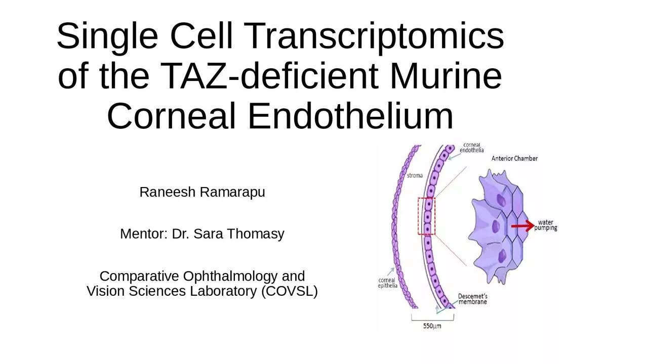
Raneesh Ramarapu Mentor Dr Sara Thomasy Comparative Ophthalmology and Vision Sciences Laboratory COVSL Corneal Endothelium Hexagonal single cell layer Nonproliferative in most species Active cells that constantly pump out water maintain corneal transparency ID: 1047097

stress taz cell corneal taz stress corneal cell endothelial upregulates fuchs downregulated dystrophy fecd functional ko2moko wt2mo genes analysis

Share:

Link:

Embed:

Download Presentation from below link

Download Presentation The PPT/PDF document "Single Cell Transcriptomics of the TAZ-d..." is the property of its rightful owner. Permission is granted to download and print the materials on this web site for personal, non-commercial use only, and to display it on your personal computer provided you do not modify the materials and that you retain all copyright notices contained in the materials. By downloading content from our website, you accept the terms of this agreement.


Presentation Transcript

1. Single Cell Transcriptomics of the TAZ-deficient Murine Corneal EndotheliumRaneesh RamarapuMentor: Dr. Sara ThomasyComparative Ophthalmology and Vision Sciences Laboratory (COVSL)

2. Corneal EndotheliumHexagonal single cell layer Non-proliferative in most species Active cells that constantly pump out water – maintain corneal transparency Ref [1]

3. The Pathology of Interest: Fuchs Endothelial Corneal Dystrophy (FECD)Ref [2]

4. The Pathology of Interest: Fuchs Endothelial Corneal Dystrophy (FECD)Polygenic Disease >300 million human patients globally under the age of 30 [3]Ref [2]

5. The Pathology of Interest: Fuchs Endothelial Corneal Dystrophy (FECD)Polygenic Disease >300 million human patients globally under the age of 30 [3] Characterized by [4,5]Accelerated Loss of CEn Altered Descemet Membrane (DM) stiffness Aberrant ECM accumulation Guttae Ref [3]

6. The Pathology of Interest: Fuchs Endothelial Corneal Dystrophy (FECD)Only treatment is corneal transplant Low accessibility Need for alternative non-surgical therapeutics! Ref [3]

7. Our Model of FECD: TAZ-deficient miceCOVSL modelled late-onset FECD using Wwtr1 deficient mice [6]

8. Our Model of FECD: TAZ-deficient miceCOVSL modelled late-onset FECD using Wwtr1 deficient mice [6]Reduced CEn densityabnormal CEn morphologySofter DMThinner corneas Altered expression and localization of Na/K-ATPase and ZO-1

9. Wwtr1 (TAZ)Encodes transcriptional co-activator with PDZ-binding motif (TAZ)Multifunctional Gene [7-13]Mechanotransducer of DM stiffness Ref [7]

10. Wwtr1 (TAZ)Encodes transcriptional co-activator with PDZ-binding motif (TAZ)Multifunctional Gene [7-13]Mechanotransducer of DM stiffness Central orchestrator of multiple pathways HippoWnt TGF-BetaNF-kB

11. Goal of the project Define the transcriptomic landscape of TAZ deficiency

12. Goal of the project Define the transcriptomic landscape of TAZ deficiencyWe have single cell transcriptomic data of 3 genotypes of 3 age groups: Wildtype (WT): 2-, 6-, 11-month-old Wwtr1 Heterozygous knockout (TAZ HET): 2-, 6-, 11-month-old Wwtr1 Homozygous knockout (TAZ KO): 2-, 6-, 11-month-old

13. Single Cell RNA-sequencingHigh dimensionality data analysis Sequence all the RNA from individual cells Allows us to assess heterogenous tissues such as the cornea

14. ScRNA-seq Analysis in a nutshellAllows us to engage with and interpret multidimensional complex data by

15. ScRNA-seq Analysis in a nut shell Allows us to engage with and interpret multidimensional complex data by Dimensionality reductionRef [14]

16. ScRNA-seq Analysis in a nut shell Allows us to engage with and interpret multidimensional complex data by Dimensionality reductionBut how do we define cell types?Ref [14]

17. ScRNA-seq Analysis in a nut shell Allows us to engage with and interpret multidimensional complex data by Dimensionality reductionBut how do we define cell types?K-means ClusteringRef [14]Ref [15]

18. ScRNA-seq Analysis in a nut shell Allows us to engage with and interpret multidimensional complex data by Dimensionality reductionBut how do we define cell types?K-means ClusteringWe now have statistically defined cell types that can be further analyzed

19. WT, TAZ HET and TAZ KO CEn cells clustered separately from the CEp and LimbusThese were validated with previously described markers

20. WT 2MO versus KO 2MO CEnKOWT

21. Gene OntologyCompiles the 1000s of upregulated genes between WT and KO Compares it to a database of genes categorized and labelled by function and networks

22. Gene Ontology

23. Gene OntologyBased on comparisons to multiple databases and Electron microscopy work, we focused onEndoplasmic reticulum stress Mitochondrial function

24. WT2MO v KO2MOKO downregulated functional markersSlc4a11 – anion exchangerSlc4a4 – Na/bicarbonate transporterAqp1/5 – Aquaporins

25. WT2MO v KO2MOKO downregulated functional markersInterestingly it downregulates type I collagens responsible for DM stiffness

26. WT2MO v KO2MOKO downregulated functional markersInterestingly it downregulates type I collagens responsible for DM stiffnessAlthough fibroblastic marker fibronectin is upregulated

27. WT2MO v KO2MOKO downregulated functional markersKO upregulates ER Stress transcriptsHspa5 is integral to the ER stress response [16]

28. WT2MO v KO2MOKO downregulated functional markersKO upregulates ER Stress transcriptsCalnexin regulates mitochondrial function with ER stress, calcium movement and ER stress associated cell death [17,18]

29. WT2MO v KO2MOKO downregulated functional markersKO upregulates ER Stress transcriptsKO upregulates calcium binding genesThese genes are upregulated with ER stress and calcium binding [19, 20]

30. WT2MO v KO2MOKO downregulated functional markersKO upregulates ER Stress transcriptsKO upregulates calcium binding geneKO upregulates genes associated with stress metabolism

31. WT2MO v KO2MOKO downregulated functional markersKO upregulates ER Stress transcriptsKO upregulates calcium binding geneKO upregulates genes associated with stress metabolism Slc2a1 – glucose transporter Pkm – PhosphofructokinaseFgfr1 – associated with metabolic changes in stress [21]Ldha – lactate dehydrogenase

32. WT2MO v KO2MOKO downregulated functional markersKO upregulates ER Stress transcriptsKO upregulates calcium binding geneKO upregulates genes associated with stress metabolism KO upregulates mitochondrial genes

33. Putting it togetherImproper TAZ signaling negatively impacts the CEn healing response due to cytoplasmic retentionThis coupled with TAZ’s key role in orchestrating multiple pathways likely causes the trifecta of ER stress, change in metabolism and mitochondrial upregulation

34. WT11MO vs KO11MOKOWT

35. WT11MO vs KO11MOKO11MO has only downregulated genes except RIK gene (unknown function, maybe associated with mitophagy/mitochondrial function)GO indicates KO11MO are likely low functioning WT11MO cells (downregulation of protein synthesis and regulation of apoptosis)

36. ConclusionsWe have a greater understanding of the mechanisms driving CEn dysfunction in FECD in the TAZ model With age, TAZ dysfunction causes a reduction in cell function (at least at the transcriptomic level)

37. Future directionValidate these transcriptomic differences through qPCR and localization through fluorescent in situ hybridizationFunctionally validate the pathways of interest using immunohistochemistry

38. AcknowledgementsI’d like to thank Drs. Sara Thomasy and Brian Leonard for their incredible support both in and outside the lab I would like to thank the members of the wet lab portion (Sangwan Park, Nayeli Echeverria, Michelle Ferneding, Sophie Le and Monica Ardon) for procuring this data I would also like to thank Dr. Vijay Raghunathan for his critical comments that improved these projectsFinancial support was provided by the Students Training in Advanced Research (STAR) Program through the NIH T35 OD010956-22 grant

39. ReferencesMeir, Y.-J. J., Chen, H.-C., Chen, C.-C. & Ma, H.-K. D. Revisiting Existing Evidence of Corneal Endothelial Progenitors and Their Potential Therapeutic Applications in Corneal Endothelial Dysfunction. Adv. Ther. 37, 1034–1048 (2020)Aiello F, Gallo Afflitto G, Ceccarelli F, Cesareo M, Nucci C. Global Prevalence of Fuchs Endothelial Corneal Dystrophy (FECD) in Adult Population: A Systematic Review and Meta-Analysis. J Ophthalmol. 2022;2022: 3091695.Xia D, Zhang S, Nielsen E, et al. The ultrastructures and mechanical properties of the Descement's membrane in Fuchs endothelial corneal dystrophy. Sci Rep. 2016; 6: 23096. Matthaei M, Hribek A, Clahsen T, Bachmann B, Cursiefen C, Jun AS. Fuchs Endothelial Corneal Dystrophy: Clinical, Genetic, Pathophysiologic, and Therapeutic Aspects. Annu Rev Vis Sci. 2019;5: 151–175.Cui, Z. et al. Pathological molecular mechanism of symptomatic late-onset Fuchs endothelial corneal dystrophy by bioinformatic analysis. PLoS One 13, e0197750 (2018)Park S, Leonard B, Kim S, Mccorkell ME, Murphy CJ, Raghunathan V, et al. Evaluation of function-related corneal endothelial markers in TAZ deficient mice over time. Invest Ophthalmol Vis Sci. 2021;62: 840–840.Varelas X, Sakuma R, Samavarchi-Tehrani P, Peerani R, Rao BM, Dembowy J, et al. TAZ controls Smad nucleocytoplasmic shuttling and regulates human embryonic stem-cell self-renewal. Nat Cell Biol. 2008;10: 837–848. Dupont S, Morsut L, Aragona M, Enzo E, Giulitti S, Cordenonsi M, et al. Role of YAP/TAZ in mechanotransduction. Nature. 2011;474: 179–183. Okumura N, Nakamura T, Kay EP, Nakahara M, Kinoshita S, Koizumi N. R-spondin1 regulates cell proliferation of corneal endothelial cells via the Wnt3a/β-catenin pathway. Invest Ophthalmol Vis Sci. 2014;55: 6861–6869. Morgan JT, Murphy CJ, Russell P. What do mechanotransduction, Hippo, Wnt, and TGFβ have in common? YAP and TAZ as key orchestrating molecules in ocular health and disease. Exp Eye Res. 2013;115: 1–12. Okumura N, Hashimoto K, Kitahara M, Okuda H, Ueda E, Watanabe K, et al. Activation of TGF-β signaling induces cell death via the unfolded protein response in Fuchs endothelial corneal dystrophy. Sci Rep. 2017;7: 6801. Okumura N, Minamiyama R, Ho LT, Kay EP, Kawasaki S, Tourtas T, et al. Involvement of ZEB1 and Snail1 in excessive production of extracellular matrix in Fuchs endothelial corneal dystrophy. Lab Invest. 2015;95: 1291–1304. Losick VP, Jun AS, Spradling AC. Wound-Induced Polyploidization: Regulation by Hippo and JNK Signaling and Conservation in Mammals. PLoS One. 2016;11: e0151251https://www.jkobject.com/blog/umap-explanation/https://towardsdatascience.com/k-means-a-complete-introduction-1702af9cd8cCai, Yu, Jyothi Arikkath, Lu Yang, Ming-Lei Guo, Palsamy Periyasamy, and Shilpa Buch. 2016. “Interplay of Endoplasmic Reticulum Stress and Autophagy in Neurodegenerative Disorders.” Autophagy 12 (2): 225–44.Guérin, R., Arseneault, G., Dumont, S. & Rokeach, L. A. Calnexin is involved in apoptosis induced by endoplasmic reticulum stress in the fission yeast. Mol. Biol. Cell 19, 4404–4420 (2008)Gutiérrez, T. et al. The ER chaperone calnexin controls mitochondrial positioning and respiration. Sci. Signal. 13, (2020)Ren, Bingyu, Yanmei Huang, Chen Zou, Yingying Wu, Yuru Huang, Jiazuan Ni, and Jing Tian. 2019. “Transcriptional Regulation of Selenoprotein F by Heat Shock Factor 1 during Selenium Supplementation and Stress Response.” Cells 8 (5). https://doi.org/10.3390/cells8050479.Shin, Young-Hee, Hana Cho, Bo Young Choi, Jonghoon Kim, Jaeyoung Ha, Sang Won Suh, and Seung Bum Park. 2021. “Phenotypic Discovery of Neuroprotective Agents by Regulation of Tau Proteostasis via Stress-Responsive Activation of PERK Signaling.” Angewandte Chemie 60 (4): 1831–38.Nies VJ, Sancar G, Liu W, van Zutphen T, Struik D, Yu RT, Atkins AR, Evans RM, Jonker JW, Downes MR. Fibroblast Growth Factor Signaling in Metabolic Regulation. Front Endocrinol (Lausanne). 2016 Jan 19;6:193. doi: 10.3389/fendo.2015.00193. PMID: 26834701; PMCID: PMC4718082.