/
First week of  Development First week of  Development

First week of Development - PowerPoint Presentation

jovita
jovita . @jovita
Follow
0 views
Uploaded On 2024-03-13

First week of Development - PPT Presentation

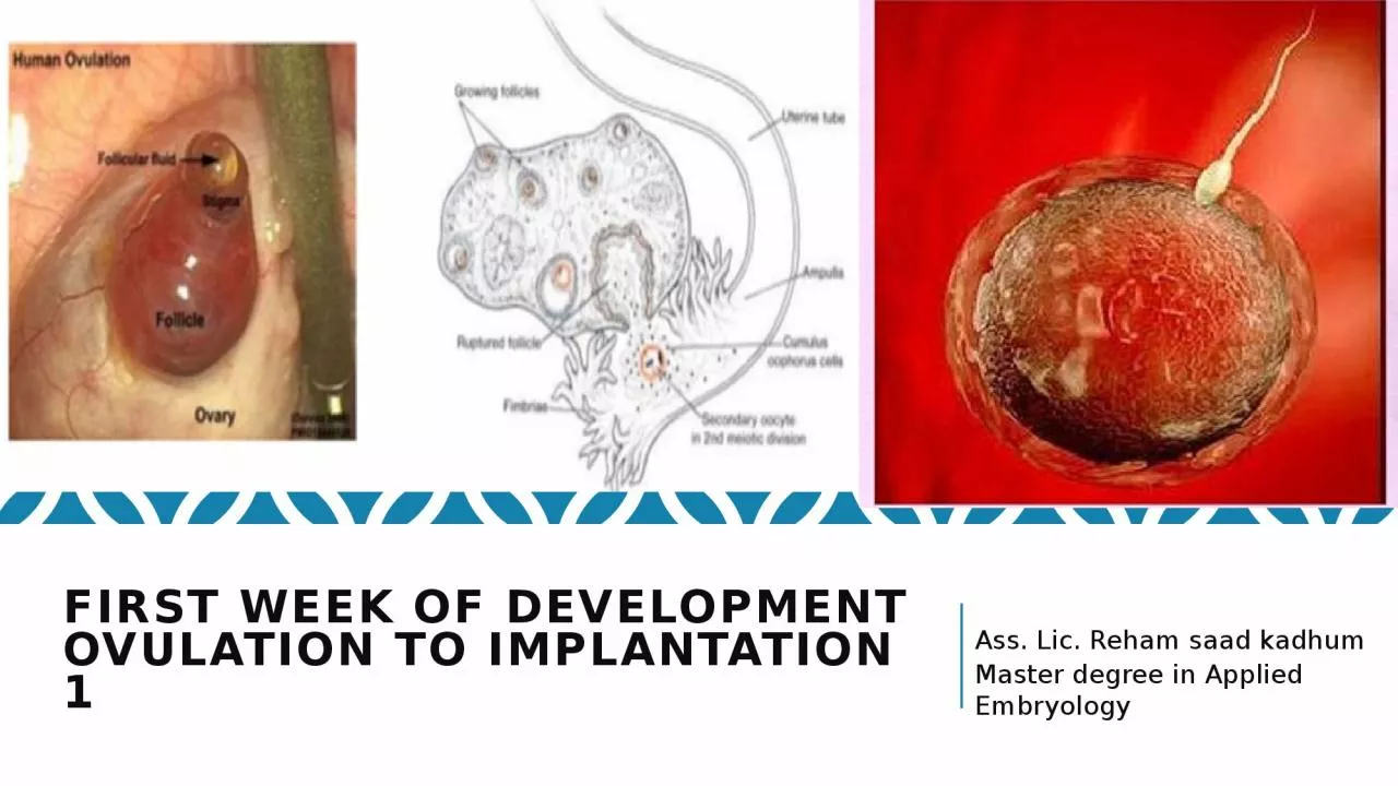
Ovulation to implantation 1 Ass Lic Reham saad kadhum Master degree in Applied Embryology Objectives Initially period the first week Development Ovulation Fertilization ID: 1048235

ovulation oocyte corpus cells oocyte ovulation cells corpus fertilization meiosis ovary uterine follicle progesterone luteum membrane secondary acrosomal follicular

Share:

Link:

Embed:

Download Presentation from below link

Download Presentation The PPT/PDF document "First week of Development" is the property of its rightful owner. Permission is granted to download and print the materials on this web site for personal, non-commercial use only, and to display it on your personal computer provided you do not modify the materials and that you retain all copyright notices contained in the materials. By downloading content from our website, you accept the terms of this agreement.


Presentation Transcript

1. First week of DevelopmentOvulation to implantation 1 Ass. Lic. Reham saad kadhum Master degree in Applied Embryology

2. Objectives

3. Initially period - the first week Development:Ovulation.Fertilization.Cleavage Blastocyst formation.

4. Introduction The primary oocyte maturates in the mature follicle and stops in the diplotene stage to complete meiosis I resulting in: Secondary oocyte. First polar body (containing little cytoplasm). The secondary oocyte begins meiosis II, and then the mature follicle raptured and thus ovulation the secondary oocyte outside the ovary as it is in metaphase of meiosis II. If fertilization occurs; the secondary oocyte completed meiosis II resulting in:Tertiary mature oocyte.Secondary polar body.

5. OVARIAN CYCLE Its sexual monthly cycle controlled by hypothalamus , begin at puberty Gonadotropin- releasing hormone (GnRH), produced by the hypothalamus, acts on cells of the anterior lobe (adenohypophysis) of the pituitary gland, which in turn secrete gonadotropins. These hormones, follicle-stimulating hormone (FSH) and luteinizing hormone (LH), stimulate and control cyclic changes in the ovary.FSH :Stimulate the development of ovarian follicles and produce estrogen by follicular cells. LH:Stimulate ovulation.Stimulate follicular cells and corpus luteum to produce progesterone.

6.

7. Estrogen action : The uterine endometrium enters the follicular or proliferative phase.■ Thinning of the cervical mucus occurs to allow passage of sperm.■ The anterior lobe of the pituitary gland is stimulated to secrete LH. At mid cycle, there is an LH surge that:■ Elevates concentrations of maturation- promoting factor, causing oocytes to complete meiosis I and initiate meiosis II■ Stimulates production of progesterone by follicular stromal cells (luteinization)■ Causes follicular rupture and ovulation

8.

9. Ovulation 1. vesicular follicle grows rapidly to a diameter of 25 mm to become a mature vesicular (graafian) follicle. In the days preceding the ovulation under influence of FCH 2. LH surge cause the oocyte complete meiosis I and enter meiosis II but oocyte is arrested in metaphase approximately 3 hours before ovulation the surface of the ovary begins to bulge locally, and at the apex, an avascular spot, the stigma, appears High level of LH : lead toa. increase activity of collagenase enzyme , which lead to digestion of collagen fibers surrounding the follicle.b. Increase level of prostaglandins, which lead to local muscular contraction in the walls of the ovary.Those contractions extrude the oocyte, which together with its surrounding granulosa cells from the región of the cumulus oophorus breaks free (ovulation) and floats out of the ovary

10. Oocyte transport : due to 1. Fimbriae , starts to sweep over the surface of the ovary shortly before ovulation . 2. Rhythmic contraction of uterine tube, this leads to pushing the ova toward the uterus in about 4 days.3. Movement of the cilia on the uterine tube epithelial lining. Note In humans, the fertilized oocyte reaches the uterine lumen in approximately 3 to 4 days.

11.

12. Corpus LuteumAfter ovulation, granulosa cells remaining in the wall of the ruptured follicle, together with cells from the theca interna, are vascularized by surrounding vessels. Under the influence of LH, these cells develop a yellowish pigment and change into lutein cells, which form the corpus luteum and secrete estrogens and progesterone Progesterone, together with some estrogen, causes the uterine mucosa to enter the progestational or secretory stage in preparation for implantation of the embryo.

13. The corpus luteum continúes to grow and forms the corpus luteum of pregnancy (corpus luteum graviditatis). By the end of the third month, this structure may be one third to one half of the total size of the ovary. Yellowish luteal cells continué to secrete progesterone until the end of the fourth month; thereafter, they regress slowly as secretion of progesterone by the trophoblastic component of the placenta becomes adequate for maintenance of pregnancy. Removal of the corpus luteum of pregnancy before the fourth month usually leads to abortion.

14. Corpus Albicansis the fibrous tissue resulting after generation of the corpus luteum at the 9thday after ovulation if no fertilization occur. It formation accompanied by decreased progesterone secretion resulting in menstrual bleeding. Corpus atreticum: is the connective tissue in the ovary that replaces the degenerating follicles in each ovarian cycle (5-15 follicles grow only once reach maturity).

15. FertilizationDefinition:A process by which the spermatozoon and oocyte unit and give rise to a new organism , the Zygot.Site of fertilization:Spermatozoa ascend from vagina enter the cervix and uterine tube with the help of muscular contraction of the uterus and uterine tube, until they reach the Ampullary region of the uterine tube.Sperm can survive few days inside the female , and the oocyte die 12-24 hours after ovulation.

16. Sperm changes in prepation for fertilization:Spermatozoa are not able to fertilize the oocyte immediately upon arrival in the female genital. Following changes must take place.1.Capacitation:Removal of glycoprotein coat from the plasma membrane that covers the acrosomal region, it takes about 7 hours.2.Acrosomal reaction:fusion between the plasma membrane and outer acrosomal membrane, lead to release of acrosomal contents (acrosin, hyaluronidase, and trypsin like substance,) needed to penetrate corona radiata and zona pellucida

17. Phases of fertilization:1.Penetration of corona radiata:out of 300-500 sperms reach the site of fertilization only one fertilize the ovum the rest help it to penetrate the corona radiata. 2. Penetration of zona pellucida:by the aid of enzymes released from acrosomal membrane.3. Fusion of oocyte-sperm cell membranes:The head and tail of the spermatozoon enter the cytoplasm of the oocyte , but the plasma membrane is left behind on the oocyte surface.

18. Oocyte responds to the spermatozoon entrance1.Cortical and zona reactions:Zona pellucida changes its structure and composition and remove species-specific receptors , and this prevent polyspermy .2. Resumption of second meiotic division .3. Metabolic activation of the egg.

19. Results of fertilization1. Restoration of diploid number .2. Determination of sex.3. Initiation of cleavage. With out fertilization Oocyte degenerate.

20. Clinical Correlate Infertility:Incidence : about 15 %.Causes :1.Insufficient number of sperms ( normally 100 million /ml), less than 20 million it may be the cause)2. Obstruction of tubes.3. immunity to sperms.4. no ovulation, and others.

21.