/
Ovarian Follicles Ovarian Follicles

Ovarian Follicles - PowerPoint Presentation

lindy-dunigan
lindy-dunigan . @lindy-dunigan
Follow
466 views
Uploaded On 2016-09-09

Ovarian Follicles - PPT Presentation

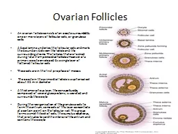
An ovarian follicle consists of an oocyte surrounded by one or more layers of follicular cells or granulosa cells A basal lamina underlies the follicular cells and marks the boundary between the follicle and the surrounding ID: 463019

corpus cells endometrium menstrual cells corpus menstrual endometrium phase oocyte follicular granulosa cycle follicle layer follicles luteum ovarian ovulation

Share:

Link:

Embed:

Download Presentation from below link

Download Presentation The PPT/PDF document "Ovarian Follicles" is the property of its rightful owner. Permission is granted to download and print the materials on this web site for personal, non-commercial use only, and to display it on your personal computer provided you do not modify the materials and that you retain all copyright notices contained in the materials. By downloading content from our website, you accept the terms of this agreement.


Presentation Transcript

Slide1

Ovarian Follicles

An ovarian follicle consists of an oocyte surrounded by one or more layers of follicular cells, or granulosa cellsA basal lamina underlies the follicular cells and marks the boundary between the follicle and the surrounding stroma. The follicles that are formed during fetal life—primordial follicles—consist of a primary oocyte enveloped by a single layer of flattened follicular cells These cells are in the first prophase of meiosis The oocyte in the primordial follicle is a spherical cell about 25 m in diameterA thick amorphous layer, the zona pellucida, composed of several glycoproteins, is secreted and surrounds the oocyte During the reorganization of the granulosa cells to form the antrum, some cells of this layer concentrate at a certain point on the follicular wall. This group forms a small hillock of cells, the cumulus oophorus, that protrudes toward the interior of the antrum and contains the oocyte Slide2

A group of

granulosa cells concentrates around the oocyte and forms the corona radiata. These granulosa cells accompany the oocyte when it leaves the ovary.As the follicles grow Liquid (liquor folliculi) begins to accumulate between the follicular cells. The small spaces that contain this fluid coalesce, and the granulosa cells reorganize themselves to form a larger cavity, the antrum follicles are then called secondary or antral folliclesFollicular fluid contains components of the plasma and products secreted by follicular cells. Glycosaminoglycans, several proteins (including steroid-binding proteins), and high concentrations of steroids (progesterone, androgens, and estrogens) are present.Slide3

While modifications are taking place in the

oocyte and granulosa layer, the fibroblasts of the stroma immediately around the follicle differentiate to form the theca folliculiThis layer subsequently differentiates into the theca interna and the theca externa The cells of the theca interna, when completely differentiated, acquire the ultrastructural characteristics of cells that produce steroids. These cells are known to synthesize a steroid hormone—androstenedione—that is transported to the granulosa layer.The cells of the granulosa, under the influence of follicle-stimulating hormone, synthesize an enzyme, aromatase, that transforms androstenedione into estrogenSlide4

Follicular Atresia

Most ovarian follicles undergo atresia, in which follicular cells and oocytes die and are disposed of by phagocytic cellsFollicles at any stage of development (primordial, primary, preantral, and antral) may undergo atresiaThis process is characterized by cessation of mitosis in the granulosa cells, detachment of granulosa cells from the basal lamina, and death of the oocyte and granulosa cells. Although follicular atresia takes place from before birth until a few years after menopause, there are times at which it is particularly intenseAtresia is greatly accentuated just after birth, when the effect of maternal hormones ceases, and during puberty and pregnancy, when marked qualitative and quantitative hormonal modifications take place.Slide5

Interstitial Cells

Although granulosa cells and the oocytes undergo degeneration during follicular atresia, the theca interna cells frequently persist in isolation or in small groups throughout the cortical stroma and are called interstitial cellsPresent from childhood through menopause, interstitial cells are active steroid secretors, stimulated by LHSlide6

Ovarian Cycle

At puberty, the female begins to undergo regular monthly cycles.These sexual cycles are controlled by the hypothalamus. Gonadotropin-releasing hormone (GnRH) produced by the hypothalamus acts on cells of the anterior pituitary gland, which in turn secrete GonadotropinsThese hormones, follicle-stimulating hormone (FSH) and luteinizing hormone (LH), stimulate and control cyclic changes in the ovary.Slide7

At the beginning of each ovarian cycle, 15 to 20

primary (preantral) stage follicles are stimulated to grow under the influence of FSH.Thus, FSH rescues 15 to 20 of these cells from a pool of continuously forming primary folliclesUnder normal conditions, only one of these follicles reaches full maturity, and only one oocyte is discharged; the others degenerate and become atretic.In the next cycle, another group of primary follicles is recruited, and again, only one follicle reaches maturityConsequently, most follicles degenerate without ever reaching full maturity.Slide8

When a follicle becomes

atretic, the oocyte and surrounding follicular cells degenerate and are replaced by connective tissue, forming a corpus atreticum.FSH also stimulates maturation of follicular (granulosa) cells surrounding the oocyte.proliferation of these cells is mediated by growth differentiation a member of the transforming growth factor-β (TGF-β) family.Slide9

In cooperation,

granulosa and thecal cells produce estrogens that(a) cause the uterine endometrium to enter the follicular or proliferative phase;b) cause thinning of the cervical mucus to allow passage of sperm(c) stimulate the pituitary gland to secrete LH.Slide10

At mid-cycle, there is an

LH surge thata) elevates concentrations of maturation-promoting factor, causing oocytes to complete meiosis I and initiate meiosis II;b) stimulates production of progesterone by follicular stromal cells (luteinization);(c) causes follicular rupture and ovulation.Slide11

OVULATION

In the days immediately preceding ovulation, under the influence of FSH and LH, the secondary follicle grows rapidly to a diameter of 25 mm.Coincident with final development of the secondary follicle, there is an abrupt increase in LH that causes the primary oocyte to complete meiosis I and the follicle to enter the preovulatory stageMeiosis II is also initiated, but the oocyte is arrested in metaphase approximately 3 hours before ovulation.Slide12

In the meantime, the

surface of the ovary begins to bulge locally, and at the apex, an avascular spot, the stigma, appears.The high concentration of LH increases collagenase activity, resulting in digestion of collagen fibers surrounding the follicleSlide13

Prostaglandin levels

also increase in response to the LH surge and cause local muscular contractions in the ovarian wallThose contractions extrude the oocyte, which together with its surrounding granulosa cells from the region of the cumulus oophorus, breaks free (ovulation) and floats out of the ovarySome of the cumulus oophorus cells then rearrange themselves around the zona pellucida to form the corona radiataSlide14

CORPUS LUTEUM

After ovulation, granulosa cells remaining in the wall of the ruptured follicle, together with cells from the theca interna, are vascularized by surrounding vessels.Under the influence of LH, these cells develop a yellowish pigment and change into lutean cells, which form the corpus luteum and secrete the hormone progesteroneProgesterone, together with estrogenic hormones, causes the uterine mucosa to enter the progestational or secretory stage in preparation for implantation of the embryo.Slide15

CORPUS ALBICANS

If fertilization does not occur, the corpus luteum reaches maximum development approximately 9 days after ovulation.It can easily be recognized as a yellowish projection on the surface of the ovarythe corpus luteum shrinks because of degeneration of lutean cells and forms a mass of fibrotic scar tissue, the corpus albicans.Slide16

Simultaneously, progesterone production

decreases, precipitating menstrual bleeding.If the oocyte is fertilized, degeneration of the corpus luteum is prevented by human chorionic gonadotropin (hCG)a hormone secreted by the syncytiotrophoblast of the developing embryo.The corpus luteum continues to grow and forms the corpus luteum of pregnancy (corpus luteum graviditatis).By the end of the third month, this structure may be one-third to one-half of the total size of the ovary.Slide17

Yellowish

luteal cells continue to secrete progesterone until the end of the fourth month;they regress slowly as secretion of progesterone by the trophoblastic component of the placentaRemoval of the corpus luteum of pregnancy before the fourth month usually leads to abortion.Slide18

Uterus at Time of ImplantationSlide19

The wall of the uterus consists of three layers:

(a) endometrium or mucosa lining the inside wall;(b) myometrium, a thick layer of smooth muscle;(c) perimetrium, the peritoneal covering lining the outside wallSlide20

Endometrium

The endometrium consists of epithelium and a lamina propria containing simple tubular glands that sometimes branch in their deeper portionsIts covering epithelial cells are a mixture of ciliated and secretory simple columnar cellsThe endometrial layer can be subdivided into two zones:(1) The basalis is the deepest one, adjacent to the myometrium; it contains lamina propria and the closed tips of the uterine glands2) The functionalis contains the remainder of the lamina propria and the glands, as well as the surface epitheliumWhereas the functionalis undergoes profound changes during the menstrual cycles, the basalis remains mostly unchanged.Arcuate arteries are circumferentially oriented in the middle layers of the myometrium. From these vessels, two sets of arteries arise to supply blood to the endometrium: straight arteries, which supply the basalis, and spiral arteries, which bring blood to the functionalis.Slide21

At the time of implantation, the mucosa of the uterus is in the

secretory phaseduring which time uterine glands and arteries become coiled and the tissue becomes succulent.As a result, three distinct layers can be recognized in the endometrium:a superficial compact layer, an intermediate spongy layer, and a thin basal layerNormally, the human blastocyst implants in the endometrium along the anterior or posterior wall of the body of the uteruswhere it becomes embedded between the openings of the glandsSlide22

The Menstrual Cycle

Estrogens and progesterone control the organs of the female reproductive system.The proliferation and the differentiation of epithelial cells and the associated connective tissues depend on these hormonesAfter menopause, the diminished synthesis of these hormones causes a general involution of the reproductive organs.After puberty, the ovarian hormones, under the stimulus of the anterior lobe of the pituitary, cause the endometrium to undergo cyclic structural modifications during the menstrual cycleThe duration of the menstrual cycle is variable but averages 28 days.Slide23

Menstrual cycles usually start between 12 and 15 years of age and continue until about age 45–50 years

the female is fertile only during the years when she is having menstrual cyclesthe beginning of the menstrual cycle is taken as the day when menstrual bleeding appears. The menstrual discharge consists of degenerating endometrium mixed with blood from the ruptured blood vesselsThe menstrual phase lasts 3–4 days on averageThe next phases of the menstrual cycle are called the proliferative and secretory (or luteal) phases.The duration of the proliferative phase is variable, 10 days on average The secretory phase begins at ovulation and lasts about 14 days.Slide24

The Proliferative, Follicular, or Estrogenic Phase

After the menstrual phase, the uterine mucosa is relatively thin (about 0.5 mm). The beginning of the proliferative phase coincides with the rapid growth of a small group of ovarian follicles that, when the cycle began, was probably at the transition from preantral to antral follicles.Estrogens act on the endometrium, inducing cell proliferation and reconstituting the endometrium lost during menstruation. (Estrogen also acts on other parts of the reproductive system, eg, inducing the production of cilia by epithelial cells of the oviduct.)During the proliferative phase, the endometrium is covered by a simple columnar epithelium. The glands, formed by simple columnar epithelial cells, are straight tubules with narrow lumens At the end of the proliferative phase, the endometrium is 2–3 mm thick.Slide25

The Secretory

, or Luteal, PhaseThe secretory phase starts after ovulation and results from the action of progesterone secreted by the corpus luteumActing on glands already developed by the action of estrogen, progesterone further stimulates the gland cells.The epithelial cells begin to accumulate glycogen below their nucleiglycoprotein secretory products dilate the lumens of the glandsOne important feature of this phase is that the glands become highly coiled In this phase, the endometrium reaches its maximum thickness (5 mm) as a result of the accumulation of secretions and of edema in the stromaMitoses are rare during the secretory phase.Slide26

If fertilization has taken place, the embryo has been transported to the uterus and attaches to the uterine epithelium during the

secretory stage, around 7 or 8 days after ovulationIt is thought that the secretion of the glands is the major source of embryonic nutrition before embryo implantation.Slide27

The Menstrual Phase

When fertilization of the oocyte and embryo implantation do not occur and the corpus luteum ceases functioning, the consequent rapid decrease of blood levels of progesterone and estrogens causes menstruationSeveral factors are involved in the shedding of the endometrium, such as cycles of contraction and relaxation of the spiral arteries, activation (by lack of progesterone) of locally produced matrix metalloproteinases, and local release of prostaglandins, cytokines, and nitric oxideThese factors lead to breakdown of blood vessel walls and basement membranes as well as collagen of the endometrial lamina propriaAt the end of the menstrual phase, the endometrium is usually reduced to a thin layer of lamina propria, the blind ends of uterine glands (both of which present in the basalis layer)Slide28

During the following 3 or 4 days, the compact and spongy layers are expelled from the uterus, and the basal layer is the only part of the

endometrium that is retainedThis layer, which is supplied by its own arteries, the basal arteries,functions as the regenerative layer in the rebuilding of glands and arteries in the proliferative phaseSlide29

Summary of Events of the Menstrual

CycleStage of Cycle:   Proliferative:Main actions of pituitary hormones  Follicle-stimulating hormone stimulates rapid growth of ovarian follicles.Main events in the ovary  Growth of ovarian follicles; dominant follicle reaches preovulatory stage.Dominant ovarian hormone  Estrogens, produced by the growing follicles, act on vagina, tubes, and uterus.Main events in the endometrium  Growth of the mucosa after menstruation.Slide30

Summary of Events of the Menstrual

CycleStage of Cycle:  Secretory or Luteal Main actions of pituitary hormones  Peak of luteinizing hormone at the beginning of the secretory stage, secreted by stimulation of estrogen, induces ovulation and development of the corpus luteum.Main events in the ovary  Ovulation. Development of the corpus luteum. Degeneration of the corpus luteum.Dominant ovarian hormone Progesterone, produced by the corpus luteum, acts mainly on the uterus.Main events in the endometrium  Further growth of the mucosa, coiling of glands, secretion.Slide31

Summary of Events of the Menstrual

CycleStage of Cycle:  MenstrualDominant ovarian hormone  Progesterone production ceases.Main events in the endometrium  Shedding of part of the mucosa about 14 days after ovulation.