/
Korean J Gastroenterol Vol 59 No 1 5357httpdxdoiorg104166k Korean J Gastroenterol Vol 59 No 1 5357httpdxdoiorg104166k

Korean J Gastroenterol Vol 59 No 1 5357httpdxdoiorg104166k - PDF document

layla
layla . @layla
Follow
342 views
Uploaded On 2022-08-21

Korean J Gastroenterol Vol 59 No 1 5357httpdxdoiorg104166k - PPT Presentation

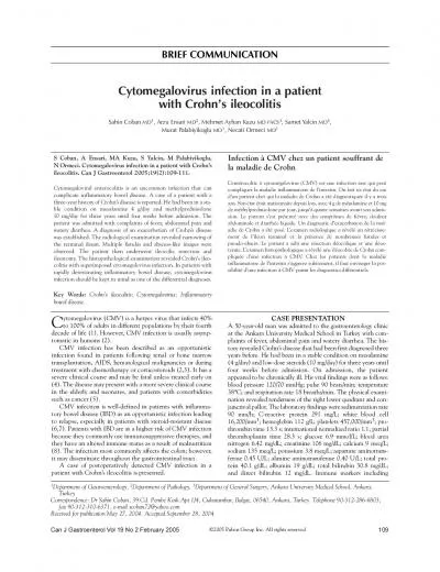
장성열 등 간암으로 오인된 CastOeman 병The Korean Journal of Gastroenterology Fig 1 Liver CT findings A CT image on hepatic arterial phase showed a 21 cm diameter enhancing mass in t ID: 939272

disease castleman liver mass castleman disease mass liver showed vascular multicentric report node lymph cancer lobe case phase hepatic

Share:

Link:

Embed:

Download Presentation from below link

Download Pdf The PPT/PDF document "Korean J Gastroenterol Vol 59 No 1 5357h..." is the property of its rightful owner. Permission is granted to download and print the materials on this web site for personal, non-commercial use only, and to display it on your personal computer provided you do not modify the materials and that you retain all copyright notices contained in the materials. By downloading content from our website, you accept the terms of this agreement.


Presentation Transcript

Korean J Gastroenterol Vol. 59 No. 1, 53-57http://dx.doi.org/10.4166/kjg.2012.59.1.53CASE REPORT 장성열 등. 간암으로 오인된 CastOeman 병The Korean Journal of Gastroenterology Fig. 1. Liver CT findings. (A) CT image on hepatic arterial phase showed a 2.1 cm diameter enhancing mass in the caudate lobe of the liver (arrow). (B) CT image on delayed phase showed that the nodule became isodense, compared with the liver parenchyma. Fig. 2. Liver MRI findings. (A) Contrast-enhanced (gadolinium ethoxybenzyl diethylenetriamine pentaacetic acid) T1-weighted MRI on heparterial phase showed a 2.2 cm diameter, exophytic enhancing nodular lesion in the caudate lobe of the liver. (B, C) Contrast-enhanced T1-weighted MRI on delayed phase (at 3 minutes and 20 minutes) showed no contrast uptake in the hepatic nodule. Fig. 3. Angiography findings. Celiac angiography showed a round extrahepatic hypervascular mass supplied by the common hepaticartery (arrow).ten with a sense of fever and chills. She denied alcohol intake and smoking. She had undergone tonsillectomy 11 months ago. On admission, her vital signs were: blood pressure, 117/ 66 mmHg; heart rate, 93 beats per minute; respiratory rate, 18 breaths per minute; and body temperature, 36.6C. On physical examination,

she presented right upper quadrant tenderness, but no rebound tenderness. Hepatosplenome-galy, ascites, and lymph node enlargement were not observed.Laboratory blood analysis was performed and the follow-ing results were obtained: white blood cells, 8,000/mm; he-moglobin, 12.4 g/dL; hematocrit, 36.6%; platelets, 242,000/ ; AST, 20 IU/L; ALT, 26 IU/L; total bilirubin, 0.38 mg/dL; ALP, 228 IU/L; and GGT, 25 IU/L. Viral markers of HBV showed a positive HBsAg, a negative HBeAg, a positive antibody to HBeAg, and a serum HBV DNA level of 4,123 IU/mL. Tumor marker results were as follows (with reference ranges in pa-rentheses): AFP, 1.46 ng/mL (0-7 ng/mL); carbohydrate CA 19-9, 12.66 U/mL (0.1-34 U/mL); and proteins induced by vi-tamin K antagonists (PIVKA-II), 16 mAU/mL (0.1-40 mAU/ mL). CT and MRI revealed a 2.2 cm sized, exophytic hyper-vascular mass in the caudate lobe of the liver (Figs. 1, 2). Celiac angiography was performed because HCC was sus-pected. The celiac angiogram demonstrated a hypervascular mass with rapid wash-out supplied by the common hepatic artery; the mass was extrahepatic in location (Fig. 3). With these radiologic studies, we suspected an extrahepatic hy-pervascular mass rather than exophytic HCC. The patient underwent laparoscopic resection

of the extra- Jang SY, et al. A Case of Castleman’s Disease Mimicking a Hepatocellular CarcinomaVol. 59 No. 1, January 2012lymphoma: cyclophosphamide, vincristine, doxorubicin and prednisone (CHOP) or dexamethasone (CVAD) can be used to multicentric Castleman’s disease.4,5Castleman’s disease is a rare lymphoid disorder that oc-curs predominantly in the mediastinum. Here, we report an unusual case of Castleman’s disease located near the cau-date lobe of the liver with review of the literature. A hyper-vascular mass in a patient with chronic hepatitis B was pre-sumed to be a HCC. However, pathological examination re-vealed a Castleman’s disease mass of the HV type. On the ba-sis of this result, Castleman’s disease should be considered as a rare differential diagnosis in patients with a hyper-vascular perihepatic mass, even if the patients are infected with HBV.1.Castleman B, Iverson L, Menendes VP. Localized mediastinal lymph-node hyperplasia resembling thymoma. Cancer 1956; 9:822-830.2.Keller AR, Hochholzer L, Castleman B. Hyaline-vascular and plasma-cell types of giant lymph node hyperplasia of the me-diastinum and other locations. Cancer 1972;29:670-683.3.Gaba AR, Stein RS, Sweet DL, Variakojis D. Multicentric giant lymph node hyperplasia. Am J Clin Pathol 197

8;69:86-90.4.Casper C. The aetiology and management of Castleman dis-ease at 50 years: translating pathophysiology to patient care. Br J Haematol 2005;129:3-17.5.van Rhee F, Stone K, Szmania S, Barlogie B, Singh Z. Castleman disease in the 21st century: an update on diagnosis, assess-ment, and therapy. Clin Adv Hematol Oncol 2010;8:486-498.6.Bowne WB, Lewis JJ, Filippa DA, et al. The management of uni-centric and multicentric Castleman's disease: a report of 16 cases and a review of the literature. Cancer 1999;85:706-717.7.Bucher P, Chassot G, Zufferey G, Ris F, Huber O, Morel P. Surgical management of abdominal and retroperitoneal Castleman's disease. World J Surg Oncol 2005;3:33.8.Madan R, Chen JH, Trotman-Dickenson B, Jacobson F, Hunsak-er A. The spectrum of Castleman's disease: Mimics, radiologic pathologic correlation and role of imaging in patient management. Eur J Radiol 2012;81:123-131.9.Rahmouni A, Golli M, Mathieu D, Anglade MC, Charlotte F, Vasile N. Castleman disease mimicking liver tumor: CT and MR features. J Comput Assist Tomogr 1992;16:699-703.10.Farkas E, Tóth B, Besznyák I. Retroperitoneal Castleman tumor. Orv Hetil 1993;134:413-416.11.Peck D, Lum PA. Castleman disease in the porta hepatis: bipha-sic helical computed tomography. Can Assoc Radi

ol J 1996; 47:410-412.12.Cirillo RL Jr, Vitellas KM, Deyoung BR, Bennett WF. Castleman disease mimicking a hepatic neoplasm. Clin Imaging 1998; 22:124-129.13.Uzunlar AK, Ozate M, Yaldiz M, Büyükbayram H, Ozaydin M. Castleman's disease in the porta hepatis. Eur Radiol 2000;10: 1913-1915.14.Al-Salamah SM, Khan IA, Khalid K, Al-Shakweer WA. Castle-man disease presenting as obstructive jaundice. Saudi Med J 2005;26:111-113.15.Sato N, Kondo S, Saito K, et al. Hyaline vascular-type Castle-man's disease in the hepatoduodenal ligament: report of a case. Surg Today 2006;36:647-650.16.Karami H, Sahebpour AA, Ghasemi M, et al. Hyaline vas-cular-type Castleman's disease in the hilum of liver: a case report. Cases J 2010;3:74.17.User IR, Akçören Z, Karnak I. Castleman disease: an unusual diagnosis of a portal mass in an 8-year-old boy. J Pediatr Surg 2011;46:e9-e11.18.Waterston A, Bower M. Fifty years of multicentric Castleman's disease. Acta Oncol 2004;43:698-704.19.Chung YE, Kim MJ, Park YN, et al. Varying appearances of chol-angiocarcinoma: radiologic-pathoelation. Radiogra-phics 2009;29:683-700.20.Chronowski GM, Ha CS, Wilder RB, Cabanillas F, Manning J, Cox JD. Treatment of unicentric and multicentric Castleman dis-ease and the role of radiotherapy. Cancer 2001;92:670-