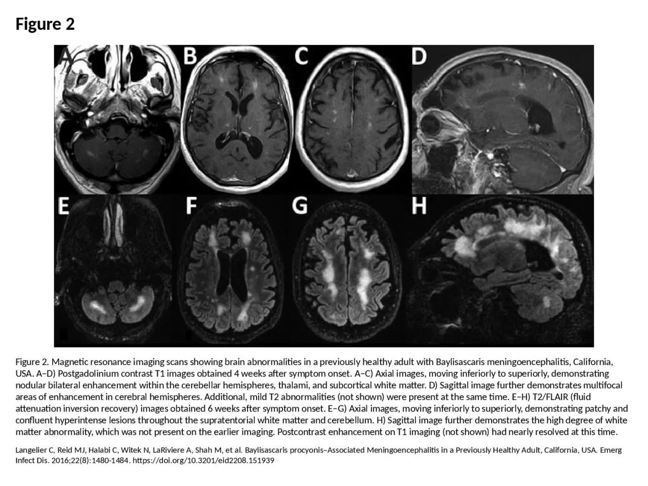
/
Figure 2 Figure 2. Magnetic resonance imaging scans showing brain abnormalities in a previously Figure 2 Figure 2. Magnetic resonance imaging scans showing brain abnormalities in a previously

Figure 2 Figure 2. Magnetic resonance imaging scans showing brain abnormalities in a previously - PowerPoint Presentation

lucy
lucy . @lucy
Follow
64 views
Uploaded On 2024-01-13

Figure 2 Figure 2. Magnetic resonance imaging scans showing brain abnormalities in a previously - PPT Presentation

Langelier C Reid MJ Halabi C Witek N LaRiviere A Shah M et al Baylisascaris procyonisAssociated Meningoencephalitis in a Previously Healthy Adult California USA Emerg Infect Dis 201622814801484 httpsdoiorg103201eid2208151939 ID: 1039737

matter images enhancement white images matter white enhancement imaging sagittal image hemispheres shown symptom present time demonstrating superiorly inferiorly

Share:

Link:

Embed:

Download Presentation from below link

Download Presentation The PPT/PDF document "Figure 2 Figure 2. Magnetic resonance im..." is the property of its rightful owner. Permission is granted to download and print the materials on this web site for personal, non-commercial use only, and to display it on your personal computer provided you do not modify the materials and that you retain all copyright notices contained in the materials. By downloading content from our website, you accept the terms of this agreement.


Presentation Transcript

1. Figure 2Figure 2. Magnetic resonance imaging scans showing brain abnormalities in a previously healthy adult with Baylisascaris meningoencephalitis, California, USA. A–D) Postgadolinium contrast T1 images obtained 4 weeks after symptom onset. A–C) Axial images, moving inferiorly to superiorly, demonstrating nodular bilateral enhancement within the cerebellar hemispheres, thalami, and subcortical white matter. D) Sagittal image further demonstrates multifocal areas of enhancement in cerebral hemispheres. Additional, mild T2 abnormalities (not shown) were present at the same time. E–H) T2/FLAIR (fluid attenuation inversion recovery) images obtained 6 weeks after symptom onset. E–G) Axial images, moving inferiorly to superiorly, demonstrating patchy and confluent hyperintense lesions throughout the supratentorial white matter and cerebellum. H) Sagittal image further demonstrates the high degree of white matter abnormality, which was not present on the earlier imaging. Postcontrast enhancement on T1 imaging (not shown) had nearly resolved at this time.Langelier C, Reid MJ, Halabi C, Witek N, LaRiviere A, Shah M, et al. Baylisascaris procyonis–Associated Meningoencephalitis in a Previously Healthy Adult, California, USA. Emerg Infect Dis. 2016;22(8):1480-1484. https://doi.org/10.3201/eid2208.151939