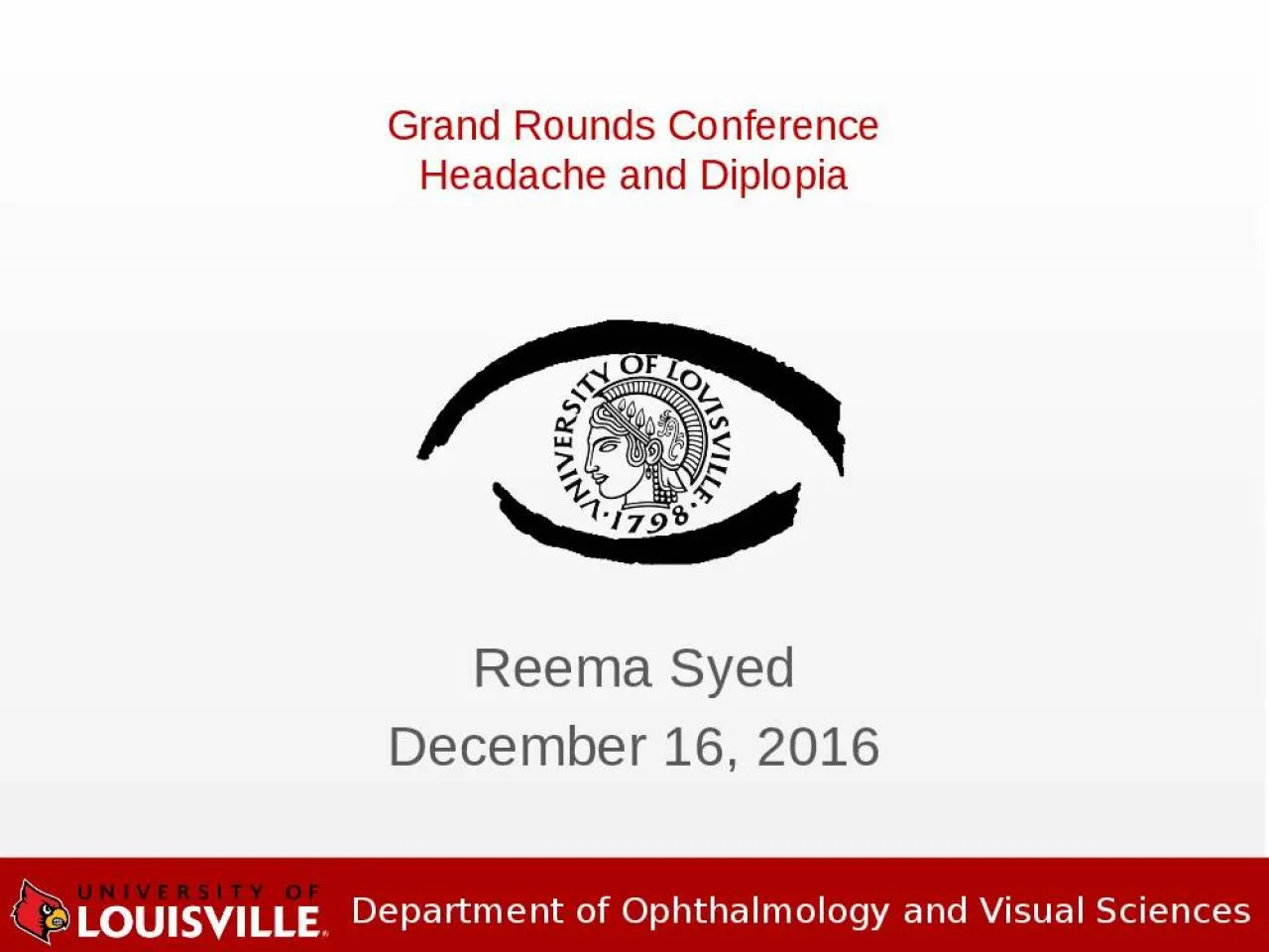
/
Reema  Syed December 16, 2016 Reema  Syed December 16, 2016

Reema Syed December 16, 2016 - PowerPoint Presentation

maisie
maisie . @maisie
Follow
343 views
Uploaded On 2022-02-24

Reema Syed December 16, 2016 - PPT Presentation

Grand Rounds Conference Headache and Diplopia CC Leftsided headache for 2 weeks Double vision for 2 days HPI 24 yr o female with worsening achy left frontal and retroorbital pain x 2 wks ID: 909674

fibrous left dysplasia bone left fibrous bone dysplasia orbital rectus mmhg frontal pain history vision primary follow recession optic

Share:

Link:

Embed:

Download Presentation from below link

Download Presentation The PPT/PDF document "Reema Syed December 16, 2016" is the property of its rightful owner. Permission is granted to download and print the materials on this web site for personal, non-commercial use only, and to display it on your personal computer provided you do not modify the materials and that you retain all copyright notices contained in the materials. By downloading content from our website, you accept the terms of this agreement.


Presentation Transcript

Slide1

Reema SyedDecember 16, 2016

Grand Rounds Conference

Headache and Diplopia

Slide2

CC

Left-sided headache for 2 weeks

Double vision for 2 daysHPI24 yr/o female with worsening achy left frontal and retro-orbital pain x 2 wksNew onset binocular oblique diplopia that resolves with covering either eye.Pertinent negatives: trauma, nausea/vomiting, pain with eye movements

Subjective

Slide3

History

Past Medical History

: Fibrous dysplasia of frontal and ethmoid sinuses Medications: oral contraceptives, Hydrocodone/AcetaminophenAllergies

: Penicillin

Family History

: non-contributory

ROS

: negative

Slide4

Exam

OD OSBCVA: 20/20 20/20-2Pupils:

tr

RAPD OS

IOP (mmHg):

12 10

EOM: Color vision: 6/6 5/6 Anterior Segment: WNL WNL

0

0

0

0

0

-2

-2

-3

-1

-3

-3

0

Slide5

Primary gaze:

Esotropia

16 PD, Left

hypertropia

12 PD

Differential IOP: 10 mmHg primary gaze, 24 mmHg

upgaze

, 20 mmHg abduction

Slide6

Dilated Fundus Exam

OD: WNL

OS: grade 2 disc edema

Slide7

Assessment

24 year female with left retro-orbital pain, mechanical strabismus with possible left optic neuropathy OS

DDx:Fibrous dysplasia with growth or secondary malignant transformation Plan:MRI brain and orbits

Slide8

Patient presented 2 days later with loss of vision to CF@ 2ft OS, 2+ RAPD, increased optic disc edemaStarted on Prednisone 60 mg

qday

Follow-up

Slide9

Normal

brain parenchyma

Groundglass appearance of frontal bones, frontal sinuses and left ethmoid air cells, compatible with fibrous dysplasia. T1 with Gadolinium: enhancing mass encroaching on and crowding left orbital apexFollow-up

Slide10

Combined approach with neurosurgery

Craniotomy with left orbital decompression

Debulking of fibrous dysplasia Removal of cystic structure along optic canalOrbital roof left unrepaired to allow for decompressionFollow-up

Slide11

Pathology

Fibrous dysplasia with secondary aneurysmal bone cyst

Slide12

Staged strabismus surgery:

Left

inferior rectus recession (6mm) Developed >50 PD left hypotropia due to scarring and 8PD Esotropia Left superior rectus resection (8mm) + medial rectus recession (4 mm on adjustable suture)

Follow-up

Slide13

6 month follow-up

BCVA 20/25 OS

Residual left

hypotropia

, left upper lid ptosis

Planned right superior rectus recession

Slide14

Fibrous Dysplasia

B

enign disorder of bone - normal bone is replaced by fibrous tissue with islands of immature woven bone.2.5% of primary bone tumors in the first 3 decades of lifeMost commonly affects long bones of the extremities or the craniofacial skeleton.

Slide15

Three forms:

monostotic

(single skeletal site): 75%polyostotic (multiple sites): 20-25%McCune Albright Syndrome (polyostotic fibrous dysplasia with endocrine dysfunction and café au

lait

spots):

3%

Fibrous Dysplasia

Slide16

Activating somatic mutation

of

a gene on chromosome 20, encoding stimulatory G protein. Triggers an arrest of typical bone maturation.Functionally impaired osteoblasts that produce spicules of poorly organized bone.Pathophysiology

Slide17

Pain

, swelling and

disfigurementHeadache, proptosis, nasal obstruction, extraocular muscle palsies, trigeminal neuralgia and epiphora.Most common neurologic complications: visual impairment and hearing loss

Signs and Symptoms

Slide18

Generally considered a benign, pediatric disease

that becomes

dormant by adulthoodMalignant transformation (incidence of 0.4% to 6.7%): osteosarcoma, fibrosarcoma and chondrosarcomaBenign but locally aggressive aneurysmal bone cysts (rare case reports) Prognosis

Slide19

Thank you