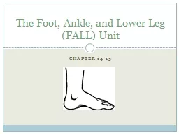
httpnhlsicom20130213erikkarlssonoutindefinitelywithlaceratedachillestendonscthpt2a10amperefsihp Problem Bony Anatomy of the Lower Leg Ligaments of the Lateral Ankle Bony Anatomy of the Ankle Joint ID: 908900
Download Presentation The PPT/PDF document "The Ankle and Lower Leg http://www.youtu..." is the property of its rightful owner. Permission is granted to download and print the materials on this web site for personal, non-commercial use only, and to display it on your personal computer provided you do not modify the materials and that you retain all copyright notices contained in the materials. By downloading content from our website, you accept the terms of this agreement.
Slide1
The Ankle and Lower Leg
Slide2http://www.youtube.com/watch?v=T5qCI0T4Fhs
http://nhl.si.com/2013/02/13/erik-karlsson-out-indefinitely-with-lacerated-achilles-tendon/?sct=hp_t2_a10&eref=sihp
Slide3Problem?
Slide4Bony Anatomy of the Lower Leg
Slide5Ligaments of the Lateral Ankle
Slide6Bony Anatomy of the Ankle Joint
Slide7Ligaments of the Medial Ankle
Slide8Muscles of the Ankle and Lower Leg
Slide9Syndesmosis of the Lower Leg
Slide10Achilles Tendon Stretching
A tight heel cord may limit
dorsiflexion
and may predispose athletes to ankle injury
Should routinely stretch before and after practice
Stretching should be performed with knee extended and flexed 15-30 degrees
Strength Training
Static and dynamic joint stability is important in preventing injury
Develop a balance in strength throughout the range
Preventing Injury in the Lower Leg and Ankle
Slide11Slide12Neuromuscular Control Training
Can be enhanced by training in controlled activities on uneven surfaces or a balance board
Shoes
Can be an important factor in reducing injury
Shoes should not be used in activities they were not made for
Preventive Taping and
Orthoses
Tape can provide some prophylactic protection
However, improperly applied tape can disrupt normal biomechanical function and cause injury
Lace-up braces have even been found to be effective in controlling ankle motion
Slide13History
Past history
Mechanism of injury
When does it hurt?
Type of, quality of, duration of pain?
Sounds or feelings?
How long were you disabled?
Swelling?
Previous treatments?
Assessing the Lower Leg and Ankle
Slide14Observations
Postural deviations?
Genu
valgum
or
varum
?
Difficulty with walking?
Deformities, asymmetries or swelling?
Color and texture of skin, heat, redness?
Obvious pain?
Is range of motion normal?
Palpation
Begin with bony landmarks and progress to soft tissue
Attempt to locate areas of deformity, swelling and localized tenderness
Slide15Special Test - Lower Leg
Percussion/bump testUsed when fracture is suspected
Percussion test is a blow to the tibia, fibula or heel to create vibratory force that resonates w/in fracture causing pain
Compression test
Compression test involves compression of tibia and fibula either above or below site of concern
Slide16Ligament Tests
Anterior drawer test
Used to determine damage to anterior
talofibular
ligament primarily and other lateral ligament secondarily
A positive test occurs when foot slides forward and/or makes a clunking sound as it reaches the end point
Talar
tilt test
Performed to determine extent of inversion or
eversion
injuries
With foot at 90 degrees
calcaneus
is inverted and excessive motion indicates injury to
calcaneofibular
ligament and possibly the anterior and posterior
talofibular
ligaments
If the
calcaneus
is
everted
, the deltoid ligament is tested
Slide17Anterior Drawer Test
Talar Tilt Test
Bump Test
Compression Test
Slide18Functional Tests
While weight bearing the following should be performed
Walk on toes (plantar flexion)
Walk on heels (
dorsiflexion
)
Hops on injured ankle
Start and stop running
Change direction rapidly
Run figure eights
High Knees
Butt Kicks
Carioka
Ankle Injuries: Sprains
Single most common injury in athletics caused by sudden inversion or
eversion
moments
Inversion Sprains
Most common and result in injury to the lateral ligaments
Anterior
talofibular
ligament is injured with inversion, plantar flexion and internal rotation
Occasionally the force is great enough for an avulsion fracture to occur w/ the lateral
malleolus
Recognition and Management of Injuries to the Ankle
Slide20Severity of a sprain- graded (1-3)
With inversion sprains the foot is forcefully inverted or occurs when the foot comes into contact w/ uneven surfaces
Slide21Slide22Eversion Ankle Sprains
-(Represent 5-10% of all ankle sprains)
Etiology
Bony protection and ligament strength decreases likelihood of injury
Eversion force resulting in damage to deltoid and possibly fx of the fibula
Deltoid can also be impinged and contused with inversion sprains
Slide23Syndesmotic
Sprain (high ankle sprain)Etiology/ MOIInjury to the distal tibiofemoral
joint (anterior/posterior
tibiofibular
ligament)
Injured w/ increased external rotation or
dorsiflexion
Injured in conjunction w/ medial and lateral ligaments
May require extensive period of time in order to return to play
Slide24Slide25Graded Ankle Sprains
Signs of Injury
Grade 1
Mild pain and disability; weight bearing is minimally impaired; point tenderness over ligaments and no laxity
Grade 2
Feel or hear pop or snap; moderate pain w/ difficulty bearing weight; tenderness and edema
Positive
talar
tilt and anterior drawer tests
Possible tearing of the anterior
talofibular
and
calcaneofibular
ligaments
Grade 3
Severe pain, swelling,
hemarthrosis
, discoloration
Unable to bear weight
Positive
talar
tilt and anterior drawer
Instability due to complete
ligamentous
rupture
Slide26Care
Must manage pain and swelling
Apply horseshoe-shaped foam pad for focal compression
Apply wet compression wrap to facilitate passage of cold from ice packs surrounding ankle
Apply ice for 20 minutes and repeat every hour for 24 hours
Continue to apply ice over the course of the next 3 days
Keep foot elevated as much as possible
Avoid weight bearing for at least 24 hours
Begin weight bearing as soon as tolerated
Return to participation should be gradual and dictated by healing process
Slide27Ankle Fractures/Dislocations
Cause of Injury
Number of mechanisms – often similar to those seen in ankle sprains
Signs of Injury
Swelling and pain may be extreme with possible deformity
Care
Splint and refer to physician for X-ray and examination
RICE to control hemorrhaging and swelling
Once swelling is reduced, a walking cast or brace may be applied, w/ immobilization lasting 6-8 weeks
Rehabilitation is similar to that of ankle sprains once range of motion is normal
Slide28Slide29Tibial
and Fibular Fractures
Cause of Injury
Result of direct blow or indirect trauma
Fibular fractures seen with
tibial
fractures or as the result of direct trauma
Signs of Injury
Pain, swelling, soft tissue insult
Leg will appear hard and swollen (
Volkman’s
contracture)
Deformity – may be open or closed
Care
Immediate treatment should include splinting to immobilize and ice, followed by medical referral
Restricted weight bearing for weeks/months depending on severity
Slide30Slide31Stress Fracture of Tibia or Fibula
Cause of Injury
Common overuse condition, particularly in those with structural and biomechanical insufficiencies
Result of repetitive loading during training and
conditioning
Signs of Injury
Pain with activity
Pain more intense after exercise than before
Point tenderness; difficult to discern bone and soft tissue pain
Bone scan results (stress fracture vs.
periostitis
)
Slide32Care
Eliminate offending activityDiscontinue stress inducing activity 14 daysUse crutch for walking
Weight bearing may return when pain subsides
After pain free for 2 weeks athlete can gradually return to activity
Biomechanics must be addressed
Slide33Medial
Tibial Stress Syndrome (Shin Splints)
Cause of Injury
Pain in anterior portion of shin
Stress fractures, muscle strains, chronic anterior compartment syndrome,
periosteum
irritation
Caused by repetitive
microtrauma
Weak muscles, improper footwear, training errors,
varus
foot, tight heel cord, hypermobile or pronated feet and even forefoot supination can contribute to MTSS
May also involve, stress fractures or
exertional
compartment syndrome
Slide34Shin Splints (continued
)
Signs of Injury
Diffuse pain about
disto
-medial aspect of
lower
leg
As condition worsens ambulation may be painful, morning pain and stiffness may also increase
Can progress to stress fracture if not treated
Care
Physician referral for X-rays and bone scan
Activity modification
Correction of abnormal biomechanics
Ice massage to reduce pain and inflammation
Flexibility program for
gastroc
-soleus complex
Arch taping and orthotics
Slide35Shin Contusion
Cause of Injury
Direct blow to lower leg (impacting
periosteum
anteriorly)
Signs of Injury
Intense pain, rapidly forming hematoma w/ jelly like consistency
Increased warmth
Care
RICE, NSAID’s and analgesics as needed
Maintaining compression for hematoma (which may need to aspirated)
Fit with doughnut pad and
orthoplast
shell for protection
Slide36Compartment Syndrome
Cause of Injury
Rare acute traumatic syndrome due to direct blow or excessive exercise
May be classified as acute, acute
exertional
or chronic
Signs of Injury
Excessive swelling compresses muscles, blood supply and nerves
Deep aching pain and tightness is experienced
Weakness with foot and toe extension and occasionally numbness in dorsal region of foot
Slide37Pulselessness
Pallor (pale color)ParesthesiaPressureParalysis
5 P’s of Compartment Syndrome
Slide38Care
If severe acute or chronic case, may present as medical emergency that requires surgery to reduce pressure or release fascia
RICE, NSAID’s and analgesics as needed
Avoid use of compression wrap = increased pressure
Surgical release is generally used in recurrent conditions
May require 2-4 month recovery (post surgery)
Conservative management requires activity modification, icing and stretching
Surgery is required if conservative management fails
Slide39Slide40Achilles Tendonitis
Cause of Injury
Inflammatory condition involving tendon, sheath or
paratenon
Tendon is overloaded due to extensive stress
Presents with gradual onset and worsens with continued use
Decreased flexibility exacerbates condition
Signs of Injury
Generalized pain and stiffness, localized proximal to calcaneal insertion, warmth and painful with palpation, as well as thickened
May progress to morning stiffness
Slide41Care
Resistant to quick resolution due to slow healing nature of tendonMust reduce stress on tendon, address structural faults (orthotics, mechanics, flexibility)
Aggressive stretching and use of heel lift may be beneficial
Use of anti-inflammatory medications is suggested
Slide42Achilles Tendon Rupture
Slide43Achilles Tendon Rupture
CauseOccurs w/ sudden stop and go; forceful plantar flexion w/ knee moving into full extensionCommonly seen in athletes
>
30 years old
Generally has history of chronic inflammation
Signs of Injury
Sudden snap (kick in the leg) w/ immediate pain which rapidly subsides
Point tenderness, swelling, discoloration; decreased ROM
Obvious indentation and positive Thompson test
Slide44Care
Usual management involves surgical repair for serious injuries Non-operative treatment consists of RICE, NSAID’s, analgesics, and a non-weight bearing cast for 6 weeks to allow for proper tendon healing
Must work to regain normal range of motion followed by gradual and progressive strengthening program
Slide45Achilles Tendon Repair