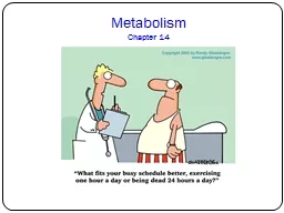
/
Lecture7 METABOLISM OF NUCLEIC ACIDS (BCH 304) Lecture7 METABOLISM OF NUCLEIC ACIDS (BCH 304)

Lecture7 METABOLISM OF NUCLEIC ACIDS (BCH 304) - PowerPoint Presentation

molly
molly . @molly
Follow
65 views
Uploaded On 2023-11-17

Lecture7 METABOLISM OF NUCLEIC ACIDS (BCH 304) - PPT Presentation

Topic 3 DNA REPAIR SO ANADOZIE PhD DNA DAMAGE DNA is the repository of genetic information in each living cell the cellular DNA must be replicated duplicated maintained and pass down to the daughter cells ac ID: 1032649

base dna repair damage dna base damage repair mutation excision mutations strand change types caused altered form result bases

Share:

Link:

Embed:

Download Presentation from below link

Download Presentation The PPT/PDF document "Lecture7 METABOLISM OF NUCLEIC ACIDS (BC..." is the property of its rightful owner. Permission is granted to download and print the materials on this web site for personal, non-commercial use only, and to display it on your personal computer provided you do not modify the materials and that you retain all copyright notices contained in the materials. By downloading content from our website, you accept the terms of this agreement.


Presentation Transcript

1. Lecture7METABOLISM OF NUCLEIC ACIDS (BCH 304)Topic 3DNA REPAIR S.O ANADOZIE (Ph.D.)

2. DNA DAMAGE DNA is the repository of genetic information in each living cell, the cellular DNA must be replicated (duplicated), maintained, and pass down to the daughter cells accuracy. During this process error are made in the DNA and therefore needs repair because its integrity and stability are essential to life. Some chemicals resulting from the environment such as radiation and oxidative damage causes mutation in the DNA and possibly diseases if not repaired. Examples includes:Skin cancer, caused by excessive exposure to UV radiation in the form of sunlight.

3. DNA DAMAGE Tobacco smoking which can lead to mutations in lung cells and subsequently cancer of the lung.DNA is also subject to oxidative damage from by-products of metabolism, such as free radicals.A "proofreading" enzyme normally recognizes and corrects many of these errors, some mutations survive this process. Human DNA undergoes lasting, uncorrected errors range from 1 x 10⁻⁴ to 1 x 10⁻⁶ mutations per gamete for a given gene. A rate of 1 x 10⁻⁶ means one mutation is expected to occur at a specific locus per one million gametes.

4. DNA DAMAGE (CTD)Mutation rates in other organisms are often much lower.Majority of mutations that occurs in the DNA do not necessary, encode proteins, thus, will not have serious effect on the organism.However, not true in all cases. Since mutations do occur in the coding regions of DNA. There are situations in which the change in a single base pair in the human genome can cause a serious disease e.g. sickle cell anaemia.The DNA damage done by physical chemical and environmental agents can be classified into four groups with different types.

5. Types of DNA damage (CTD) In spontaneous deamination cytosine gets deaminated to form uracil, while adenine forms hypoxanthine.Spontaneous depurination also occur due to cleavage of glycosyl bonds (that connect purine to the backbone). 2,000 - 10,000 purines are lost per mammalian cells in 24h

6. The depurinated sites are called abasic sites. Because they were detected in purines, they are called apurinic (AP) sites. The AP sites is usually used for any base lacking DNA.Production of reactive oxygen species (ROS) is often associated with alteration of bases e.g. formation and oxidative damage to DNA increases the advancement of age.Ultraviolet radiations result in the formation of covalent links between adjacent pyrimidines along the DNA strand to form pyrimidine dimersDNA chain breaks can be caused by ionizing radiations (e.g. X-rays).Types of DNA damage (CTD)

7. MUTATIONSThe genetic macromolecule DNA is highly stable with regards to its base composition and sequence. However, DNA is not totally exempt from gradual change.Mutation refers to change in the DNA structure of a gene.Mutagens are substances that causes mutationsThe changes that occur in DNA mutation are reflected in replication, transcription and translation.

8. Types of mutationsThere are basically two major types of mutations; point mutations and frameshift mutations.Point mutation: The replacement of one base pair by another.They are of two sub-types;Transition: A purine or pyrimidine is replaced by another CGAG transition CGGG GCTC GCCC

9. Types of mutationsTransversion: The replacement of purine with a pyrimidine or vice versa. CGAG transversion CGTG GCTC GCACFrameshift: This occurs when one or more base pairs are inserted or deleted from the DNA, respectively causing altered reading frame of the mRNA which can be insertion or deletion. CGAG insertion GCATG; deletion CGC GCTAG GCG

10. Consequences of point mutationsThe change in a single base sequence in point mutation may cause one of the following;1. Silent mutation: The codon (of mRNA) containing the changed base may code for the same amino acids e.g UCA codes for serine and change in the third base (UCU) still codes for serine. - This is due to the degeneracy of the genetic code. Therefore, there are no detectable effects in silent mutation.

11. Consequences of point mutations2. Missense mutation: The change base may code for a different amino acids. E.g. UCA codes for serine and ACA codes foe threonine.- Missense amino acid may be acceptable, partially acceptable or unacceptable with regard to the function of a protein molecules. - Sickle cell anaemia is a typical example of missense mutation.3. Nonsense mutation: The codon with altered base may become a termination (nonsense) codon. E.g. Change in the second base of serine codon (UCA) may change to UAA. The altered codon acts as a stop signal and results in termination of the protein synthesis.

12. Consequences of frame shift mutationsInsertion and deletion of a base in a gene results in an altered reading frame of the mRNA.The mRNA (containing codons) does not recognize that a base is missing or a new base added.Translation continues as there are no punctuations in the reading codons’Protein synthesized in this form will have several altered amino acids and/or prematurely terminated protein.Mutations in cancer occur as a result of alterations in DNA structure.

13. REPAIR OF DNA

14. DNA repairDamage to DNA caused by replication errors or mutations may have serious consequences. DNA repair processes exist in both prokaryotic and eukaryotic organisms, and many of the proteins involved have been highly conserved throughout evolution. The cell possesses an inbuilt system to repair the damage DNA. This can be achieved via:Base excision-repairNucleotide excision-repairMismatch repairDouble-stranded break repair

15. Base excision-repairBase excision repair (BER) handles the spontaneous DNA damage caused by free radicals and other reactive species generated by metabolism.The bases cytosine, guanine and adenine can undergo spontaneous depurination to respectively form uracil, hypoxanthine and xanthine.The altered bases do not exist in the normal DNA and therefore need to be removed. This is carried out by base excision repair.A defective DNA in which cytosine is deaminated to uracil is acted upon by the enzyme uracil DNA glycosylase. This result in the removal of the defective base uracil.The enzyme endonuclease cuts the backbones of DNA strand near the defect and removes a few bases.

16. Base excision-repairDNA polymerase and DNA ligase repair the gap created by DNA damage.

17. Nucleotides excision repairDNA damage resulting from ultra violent light, ionizing radiation and environmental factors result in the modification of certain bases, strand breaks and cross-linkages.Nucleotide excision-repair is ideally suited for such large scale defects in DNA.DNA double helix is unwound after the identification of defect in DNA to expose the damaged part.An excision nuclease (exinucleases) cut the DNA on the other size a (up stream and downstream) of the damaged DNA.DNA polymerase fills the gap caused by nucleotide excision and get ligated by DNA ligase.An example of disease caused by nucleotide excision is the xeroderma pigmentosum (XP).

18. Nucleotides excision repair

19. Mismatch repairDefects in the copying of daughter DNA. E.g. copying of cytosine instead of thymine to incorporate opposite to adenineCorrect a single mismatch base pair e.g. C to A instead of T to A.Template strand of DNA exists in a methylated form while the newly formed strand is not methylated.The enzymes GATC endonucleases cuts the strand at the adjacent methylated GATC sequence, followed by an endonucleases to remove the defective strand. A new DNA strand is then synthesized to replaced the damaged one.Example of mismatch disease is the hereditary nonopolyposis colon cancer.

20. Mismatch repairs

21. Double-strand breaks (DSBs) in DNA are dangerous and are caused by ionizing radiation, including gamma rays and X-rays. They result in genetic recombination leading to chromosomal rearrangements, in which pieces of one chromosome become attached to another chromosome and finally cell death.They can be repaired by homologous recombination or non-homologous end joining.Homologous recombination (HRR) occurs in yeasts while non-homologous (NHEJ) and joining dominates in mammals.In NHEJ, an enzyme called DNA ligase IV uses overhanging pieces of DNA adjacent to the break to join and fill in the ends. Additional errors can be introduced during this process. In HRR, the homologous chromosome itself is used as a template for repair.Double-stranded break repair

22. REVIEW AND QUESTIONSStay safe and please engage in further readingAll the best