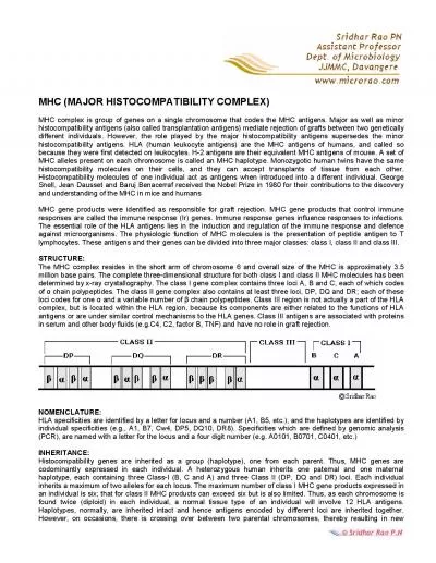
/
MHC STRUCTURE ANALYSIS OF THE MAJOR HISTOCOMPATIBILITY COMPLEX MHC STRUCTURE ANALYSIS OF THE MAJOR HISTOCOMPATIBILITY COMPLEX

MHC STRUCTURE ANALYSIS OF THE MAJOR HISTOCOMPATIBILITY COMPLEX - PowerPoint Presentation

molly
molly . @molly
Follow
342 views
Uploaded On 2022-06-15

MHC STRUCTURE ANALYSIS OF THE MAJOR HISTOCOMPATIBILITY COMPLEX - PPT Presentation

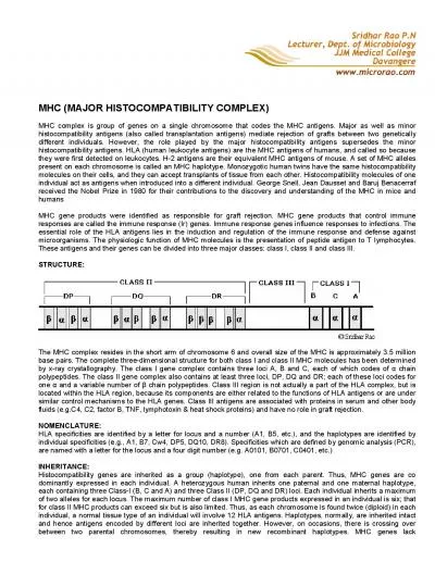
SUMMARY 1 Introduction 2 MHC class II structure 3 Labile regions 4 NonClassical HLA 5 The peptide binding site 6 CD4 HLA interaction 7 Conclusions INTRODUCTION MHCI ID: 919427

hla mhc cd4 classical mhc hla classical cd4 binding residues structure class peptide helix domain mbp sequence proteins molecules

Share:

Link:

Embed:

Download Presentation from below link

Download Presentation The PPT/PDF document "MHC STRUCTURE ANALYSIS OF THE MAJOR HIST..." is the property of its rightful owner. Permission is granted to download and print the materials on this web site for personal, non-commercial use only, and to display it on your personal computer provided you do not modify the materials and that you retain all copyright notices contained in the materials. By downloading content from our website, you accept the terms of this agreement.


Presentation Transcript

Slide1

MHC

STRUCTURE ANALYSIS OF THE MAJOR HISTOCOMPATIBILITY COMPLEX

Slide2

SUMMARY

1|

Introduction

2|

MHC class II structure3| Labile regions4| Non-Classical HLA5| The peptide binding site6| CD4 – HLA interaction7| Conclusions

Slide3

INTRODUCTION

Slide4

MHC-I

Expressed

in

all

nucleated cellsInteracts with CD8 T cells and NKPresents inner antigensInvolved in the control of damaged cells

MHC-II

Expressed

only in APCsInteracts with CD4 T cellsPresents outern patogenic antigensInvolved in activating adaptative immune response thru T-helper cells

T-CD8 and T-CD4 cells are not able to recognize antigens without binding to MHC proteins

MHC-I & MHC-II

Slide5

MHC

-II TYPES

Classical

MHC-II

antigen presentation DRThree types DQ

DP

Non-

classical

MHC-II indirectly involved in antigen presentationDM: promoter for antigenic

peptide load in classical MHC-II proteinsDO: interacts with DM, modulating it’s function and

the set of antigens

bound

to

the MHC-II

Slide6

GENE STRUCTURE

Slide7

Folding

in

the

RER.

Binding to the unvariant chainModifications at golgi’s systemTransportation in vesiclesUnvariant chain is degradated to CLIP

Fusion

with

the lysosome. Antigenic peptide load.Transportion to the membrane FOLDING AND PROCESSING

Slide8

PROTEIN FAMILY (SCOP)

Slide9

MHC CLASS II STRUCTURE

Slide10

STRUCTURE

Slide11

DOMAINS: PEPTIDE BINDING GROOVE

Slide12

DOMAINS: IG-LIKE

Slide13

MHC-I

MHC-II

COMPARISON OF MHC-I & MHC-II STRUCTURES

Slide14

COMPARISON OF MHC-I & MHC-II STRUCTURES

MHC-I

MHC-II

In MHC-II

we find:Open ends

Longer

peptides

(13-18 residues)Anchor residues all over the cleftPeptides at constant elevation

Slide15

MHC appears 460-540 million years ago in a jawed vertebrate common antecessor

Genes for MHC class I and class II appear since the common antecessor

MHC-II PHYLOGENY

Slide16

LABILE REGIONS

Slide17

|

Structural

superimpositions| 4 superimpositions:Mixing all classical HLA-IIOnly DROnly DQOnly DP| Alignment of the

obtained

structures

HLA-II STRUCTURES COMPARISON

Slide18

3

HLA-II

DR

3

HLA-II DQ2 HLA-II DPHLA-II SUPERIMPOSITIONSc = 8.80RMSD = 0.86

Slide19

HLA-II

DR

HLA-II

DQ

HLA-II DP

Sc = 9.35

RMSD = 0.81

Sc = 9.14

RMSD = 0.49Sc = 9.70RMSD = 0.84HLA-II SUPERIMPOSITION

Slide20

VARIABLE REGIONS

β2

Ig-like

domain

Helical

kink on the β1 domain3

10 helix on the α chain

Slide21

10 º

IG-LIKE

Β

2 DOMAIN

Slide22

A-B

loop

IG-LIKE

Β

2 DOMAIN

Slide23

Sequence

alignment (A-B loop)

IG-LIKE Β2 DOMAIN

A-B

loop

Slide24

Β

1-HELICAL KINK

From

β62 to β70Involved in conformational changes

Higher

desviation

at βAsp-66

Slide25

HLA-II

DR

proteins

show a

higher desviation at

β

Asp

66

Β1-HELICAL KINKHLA-II DRHLA-II DQHLA-II DP

Slide26

The

β

1-helical

kink changes it’s conformation in response to interactions with other proteins1H15: cristal contact1ZGL:

TCR

Β

1-HELICAL KINK

Slide27

Sequence

alignment

Β1-HELICAL KINK

Slide28

3

10

HELIX

3

residues/turnH bonds every i and i + 3 residuesUnestable structure: RARE

Slide29

HLA-II

DQ

proteins

show a

higher desviation at

the

3

10

helix HLA-II DRHLA-II DQHLA-II DP310 HELIX

Slide30

Glicines

3PL6

1UVQ

310 HELIX

3

10

helix unwinds and rotates 20 º for allowing interaction with MHC-DMGlycines

on positions α52 and α53 increase structural lability of the 310 helix Increased affinity for HLA-DM

Slide31

Sequence

a

lignment

310 HELIX

Slide32

Deletions

on

α52 reduce affinity

for HLA-II DM DQA*02DQA*04

HLA-DM

deficient

interaction

Autoinmune disease asociation:Celiac diseaseDiabetis mellitus 1Multiple sclerosis

Insertion of a Gly on position α53 restores DQA*02 – MHC-DM interactions310 HELIX

Slide33

NON-classical

HLA-ii

Slide34

HLA-II

DR

HLA-II

DM

HLA-II DONON-CLASSICAL HLA-II

Slide35

Superimposition

with

classical HLA-II

3C5J (DR)2BC4 (DM)4I0P (DO)NON-CLASSICAL HLA-II

Slide36

Sequence

is

not

conserved

NON-CLASSICAL HLA-II

Slide37

Non-

clasical

HLA-II

proteins seem to be remote paralogs

of classical MHC-II proteinsDifferent sequenceDifferent functionSame structureNON-CLASSICAL HLA-II

Slide38

THE PEPTIDE BINDING SITE

Slide39

BINDING GROOVE

α

1

and

β1 domains contributing approximately equal halves 2 a-helices flanking an 8-stranded β-sheet floor

MCH

class II is open ended

Variable length allowed

Peptide protrusion Non anchoring of C and N terminal

Slide40

BINDING POCKETS

Constituted

by a set of residues of

α

1 and β1 domainsPrincipal pockets bind P1, P4, P6 and P9

Additional contribution of P3, P7 and P10

HLA-DR2a binding MBP 84-104

P1: Phe92P4: Ile95P6: Thr97P9: Thr100

Slide41

BINDING SPECIFICITY - HLA-DR2a vs. HLA-DR2b

Polymorphic

MHC class II proteins

Different architecture, charge and shape of binding pockets

Different binding specificity

HLA-DR2 haplotype includes two

isotypes

co-expression of two DR β chains: DRB1*1501 and DRB5*0101Same α-subunit DRA*0101HLA-DR2a (DRA*0101,DRB5*0101)HLA-DR2b (DRA*0101, DRB1*1501)

Both isotypes can present Myelin Basic Protein (MBP)

Slide42

P1 POCKET

Phe24α, Ile31α, Phe32α, Trp43α, Ala52α, Phe54α, Asn82β, Val85β,

xxx86β

and Phe89βHLA-DR2aGly86β – preference for bulky aromatic residues such as Phe92 of MBP HLA-DR2bVal86β - preference for smaller aliphatic residues such as Val89 of MBP

Slide43

P1 POCKET

Phe24α, Ile31α, Phe32α, Trp43α, Ala52α, Phe54α, Asn82β, Val85β,

xxx86β

and Phe89βHLA-DR2aGly86β – preference for bulky aromatic residues such as Phe92 of MBP HLA-DR2bVal86β - preference for smaller aliphatic residues such as Val89 of MBP

Slide44

P4 POCKET

HLA-DR2a

Arg71β – preference for relative small aliphatic residues

such as

Ile95 of MBP HLA-DR2bAla71β - preference for large hydrophobic residues such as Phe92 of MBP (which is P1 anchor in HLA-DR2a)Gln9α, Asn62α, Tyr13β, Tyr26β, xxx71β, Ala74β, and Tyr78β

Slide45

P4 POCKET

HLA-DR2a

Arg71β – preference for aliphatic residues

such as

Ile95 of MBP Gln9α, Asn62α, Tyr13β, Tyr26β, xxx71β, Ala74β, and Tyr78βHLA-DR2bAla71β - preference for large hydrophobic residues such as Phe92 of MBP (which is P1 anchor in HLA-DR2a)

Slide46

PEPTIDE SHIFT

3-position shift between MBP 84-102

(green and blue) and

MBP

85-99 (orange and purple)Superimposition of HLA-DR2a/MBP84-102 and HLA-DR2b/

MBP

85-99

Pocket 1

Phe92Val98Pocket 4Ile95Phe92Pocket 6Thr97Asn94

Pocket 9Thr100Thr97

Slide47

BOND NETWORK

Hydrogen

bond network highly

conserved

Stereotyped mode of binding across the spectrum of peptide-MCHII interactionsMBP residues form a total of 12 hydrogen bonds with seven residues of the HLA-DR2 complex

Slide48

BOND NETWORK

Hydrogen

bond network highly

conserved

Stereotyped mode of binding across the spectrum of peptide-MCHII interactionsContact interactions between MBP residues

and HLA-DR2

complex

Slide49

DEIFHVD

v

v

v

v

v

Β

sheet

a

helix

3

10

helix

Slide50

Β

sheet

a

helix

Slide51

HLA-DR – CD4 INTERACTION

Slide52

Hypervariable

chains

Coreceptor

CD45

Slide53

STRUCTURE

K

D

General cell

surface molecules1-100µMCTLA-4 and CD800.2 µMHuman CD8 binding to mouse MHC I10µMHuman CD8 binding to HLA I200µMHuman CD4 binding to mouse MHC II200µMHuman CD4 binding to HLA II>2mM

.

DISSOCIATION CONSTANTS

T-cells as a scan with high speed and sensitivity

Slide54

CD4 CORRECEPTOR

F

our

Ig-like domainsD1 binds MHC II (N-term)D2 is important in folding

Slide55

T45W >> Q40Y, P48L > K35P, F43L > G47S > K46R, S60R, D63R > S42G > L44T

EXPERIMENT

CD4-DM

T45W

and Q40Y Kd 8.8 µM

11

interaction

sites with β2 and α2 domainsMutagenesis testMost important: Threonine 45 Tryptophan Second mutation: Glutamine 40 Tyrosine

CD4 INTERACTION SITES

Slide56

Close

similarity

Sc = 8.93

RMS = 0.95SUPERIMPOSITION OF CD4 WT – CD4 DM

Slide57

Particular

residues

mutated

Gln TyrThr TrpSUPERIMPOSITION OF CD4 WT – CD4 DM

Slide58

11

residues

contact

14 HLA-DR1 residues (hydrophobic interactions)Region 1 – beta

sheet

(

β

2 contact)Region 2 – alpha helix (α2 contact)

Slide59

REASONS FOR THE INTERACTION BETWEEN CD4-HLADR1

Slide60

Lys46 – Ser144

CD4

close

to DR1

HIDROGEN BONDS

Slide61

HIDROGEN BONDS

Slide62

Pro48 – Val142

Leu44 – Ser144

VAN DER WAALS INTERACTIONS (REGION 1)

Slide63

Lys35 –

Glu

162

Phe43 – Thr145/Ile148/Leu158

VAN DER WAALS INTERACTIONS (REGION 1)

Slide64

PHE 43 HYDROPHOBIC POCKET

Slide65

PHE 43 HYDROPHOBIC POCKET

Slide66

Arg59 – Glu88

Asp63 – Lys176

VAN DER WAALS INTERACTIONS (REGION 2)

Slide67

Sc =

9.39

RMS = 0.70

SUPERIMPOSITION FREE-HLA vs HLA-CD4DM

Slide68

REASONS FOR INCREASED AFFINITY

Slide69

CD4 DEEP GROOVE

In the wild-type CD4– HLA-DR1 complex, there exists a deep groove on the CD4 side

Mutation

1

 Threonine 45 Triptophan

Slide70

Trp

45

side

chain

makes hydrophobic contacts with β2 Val 143

Slide71

GLN40 TYR

Second

mutation

: Glutamine 40 Tyrosine

Slide72

GLN40 TYR

Second

mutation

: Glutamine 40 Tyrosine

Slide73

ALPHA CHAIN ALIGNMENTS

Slide74

BETA CHAIN ALIGNMENTS

Slide75

BETA CHAIN ALIGNMENTS

Slide76

CONCLUSIONS

Slide77

Despite the fold is conserved sequence variability in MHC II protein is in the peptide binding site.

The beta 2

Ig

-like domain is one of the most labile regions in MHC II. It makes sense for it’s function.The first helical kink

at the alpha helix from beta 1 domain is involved in conformational changes related to antigenic peptide binding and TCR interactions.The 3 10 helix flexibility is necessary for correct interaction with MHC II DM moleculesNon-classical MHC-II seem to be remote paralogs of classical ones.All HLA-CD4 contacting residues are conserved across all HLA typesCD4 engages in the same way HLA-DP, DQ and DRIncreased affinity confers no survival advantage probably to avoid autoimmunityCONCLUSIONS

Slide78

BIBLIOGRAPHY

Slide79

Painter

CA,

Stern

LJ. Conformational variation

in structures of classical and nonclassical MHCII proteins and functional implications. Immunol Rev. 2012; 250(1): 144-157.Kulski JK, Shiina T, Anzai T, Kohara S, Inoko H. Comparative genomic analysis of the MHC: the evolution of class

I

duplication

blocks, diversity and complexity from shark to man. Immunol Rev. 2002; 190: 95–122.Otha Y, Okamura K, McKinney EC, Bartl S, Hashimoto K, Flajnik MF. Primitive synteny of vertebrate major histocompatibility complex class I and class II genes. PNAS. 2000; 97(9): 4712-4717.

Owen JA, Punt J, Stanford SA. Kuby inmunología. 7ª Ed. Mexico: McGraw Hill; 2014.Li Y, Li H, Martin R, Mariuzza R. Structural basis for the binding of an immunodominant peptide

from Myelin Basic Protein in diferent

registers

by

two HLA-DR2 proteins. JMB. 2000; 304: 177-188

Smith K, Pyrdol J, Gauthier L. Crystal structure

of HLA-DR1 (DRA*0101, DRB1*1501) complexed with a peptide

from

human

Myelin

Basic

Protein

. J. Exp.

Med

. 1998; 188: 1511-1520

Davis

JS,

Ikemizu

S,

Evans

JE,

Fugger

L,

Bakker

RT, Van der

Merwe

PA.

The

nature

of molecular

recognition

by

T cells. Nat

immunol

. 2003; 4: 217-24

Cole

DK,

Pumphrey

NJ,

Boulter

JM, Sami M, Bell JI,

Gostick

E,

Price

DA,

Gao

GF,

Sewell

AK,

Jakobsen

BK.

Human

TCR-

binding

affinity

is

governed

by

MHC

class

restriction

. J

Immunol

. 2007; 178(9):

5727-34

Janeway

CA Jr.

The

T cell receptor as a

multicomponent

signalling

machine

: CD4/CD8 coreceptors

and

CD45 in T cell

activation

. Annu

Rev

Immunol

.

1992;10:645-74

Wang

JH,

Meijers

R,

Xiong

Y,

Liu

JH,

Sakihama

T,

Zhang

R,

Joachimiak

A,

Reinherz

EL. Crystal

structure

of

the

human

CD4 N-terminal

two-domain

fragmentcomplexed

to a

class

II MHC

molecule

.

Proc

Natl

Acad

Sci

U S A. 2001;98(19):10799-804

Wang

XX, Li Y,

Yin

Y, Mo M,

Wang

Q,

Gao

W,

Wang

L,

Mariuzza

RA.

Affinity

maturation

of

human

CD4

by

yeast

surface

display

and

crystal

structure

of a CD4-HLA-DR1 complex.

Proc

Natl

Acad

Sci

U S A. 2011;108(38):15960-5

Slide80

PEM questions

Slide81

The

MHC:

a

) Has four extracellular domains b

) Is called H-2 in human c) The stability doesn’t depends on the structure bounded to it d) Type I has only one alpha domain e) Class I is very different from MHC class IIAbout the MHC proteins is true that:a) MHC molecules are only present in mammalsb) Polimorphic variation tends to accumulate on the Ig-like domain.c) MHC class II non classical molecules show low sequence identity with classical MHC molecules.d) Humans have an enormous set of MHC class II proteinse) Most of the MHC cristalized structures contain the transmembrane domain

Which is the correct answer about the β2 domain on the MHC-II molecules?

a

) Some

MHC structures miss this domainb) This domain shows an angle of 50º of rotationc) It’s involved in antigen presentationd) It’s a beta + alpha folde) The most variable structure in the region is the A-B loop.Which of the next proteins is known to interact with the helical kink from the β1 domain of the MHC-II?a)CD4b)CD8c)TCRd)KIRe)TLR-4

Slide82

The non-classical types of MHC class II have:

a) Different

structure, different sequence and different function than the classical MHC-II.

b) Different structure, same sequence and same function than the classical MHC-II.

c) Different structure, different sequence and same function than the classical MHC-II.d) Same structure, same sequence and same function than the classical MHC-II.e) Same structure, different sequence and different function than the classical MHC-II.The peptide binding groove of the MHC class II molecules:a) Not allow peptides with variable length.b) Binds the presented peptide with quite low specificity.c) Have a high conserved sequence within all the types of MHC-II classical and non-classical.d) Is composed by only residues of the alpha domain.e) Binds the presented peptide with covalent bonds.The TCR

molecules

:

Have

three main components: the hypervariable chains, the coreceptor and an tyrosine phosphatase The TCR doesn’t needs any help in order to maximize the immune response The TCR uses ten molecules each time to do the signal transduction Are not from immune response Always needs a co-receptor in order to make the signal transduction Which is the animal, that lives nowadays, with the simplest MHC gene structure?a)Sharksb) Jelly-fishc) Dogsd) Bony fishese) Snails

Slide83

The

coreceptor

CD4: Binds only MHC I molecules

Binds only MHC II molecules Doesn’t participate in immune response Has the same dissociation constant than CD8 Binds with a really high affinity with the MHC In order to interact CD4 and HLA: There are only hydrogen bonds making the interaction Only two residues of each structure are the contacting residues The interactions are in the alpha 1 and beta 1 domains of HLA Both structures make disulphide bonds between them

There

are two regions of CD4 in contact with HLA

Slide84

THANK YOU FOR YOUR ATTENTION!

Slide85

PROTEIN FAMILY (CATH)

Slide86

DISULPHIDE

BONDS

Slide87

Chain

α

Chain

βConservationVariationPolimorphic changes tend to

appear

on

the

peptide binding sitePOLIMORFIC VARIATION