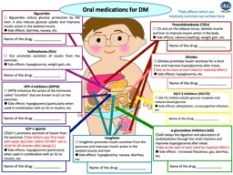
/
Insulin Resistance in Liver Diseases Insulin Resistance in Liver Diseases

Insulin Resistance in Liver Diseases - PowerPoint Presentation

pagi
pagi . @pagi
Follow
65 views
Uploaded On 2023-11-18

Insulin Resistance in Liver Diseases - PPT Presentation

Lt Col Prof Dr Shakeel Ahmed Mirza MBBS MRCP UK FRCP London Classified Medical Specialist Gastroenterologist MH Rwp Hepatologist amp Endocrinologist Prof of Medicine AFPGMI Layout ID: 1032961

liver insulin fatty nash insulin liver nash fatty resistance risk disease syndrome amp patients normal glucose alcohol diabetes bmi

Share:

Link:

Embed:

Download Presentation from below link

Download Presentation The PPT/PDF document "Insulin Resistance in Liver Diseases" is the property of its rightful owner. Permission is granted to download and print the materials on this web site for personal, non-commercial use only, and to display it on your personal computer provided you do not modify the materials and that you retain all copyright notices contained in the materials. By downloading content from our website, you accept the terms of this agreement.


Presentation Transcript

1.

2. Insulin Resistance in Liver DiseasesLt Col Prof Dr Shakeel Ahmed MirzaMBBS, MRCP ( UK ), FRCP ( London )Classified Medical SpecialistGastroenterologist MH Rwp Hepatologist & EndocrinologistProf of MedicineAFPGMI

3. LayoutWhat is Insulin Resistance ?What is mechanism of Insulin Resistance ?Causes & its Relationship to ObesityAtherosclerosisHypertriglyceridemiaEpidemiologyDiagnosis Management

4. Introduction The condition in which normal amounts of insulin are inadequate to produce a normal insulin response from fat, muscle and liver cells.

5. Insulin ResistanceHyperinsulinemic individuals are at risk for developing Diabetes, Dyslipidemia, Hypertension & ultimately Cardiovascular diseasePatients with Metabolic Syndrome are 3.5 times as likely to die from Cardiovascular disease compared to normal people

6. Insulin ResistanceHyperinsulinemia increases serum free fatty acid levels.Chronic hyperinsulinemia promotes de novo hepatic lipogenesisActivate profibrotic cytokines

7. Mechanism It includes Genetic or Primary target cell defects Autoantibodies to insulin & Accelerated insulin degradation

8. CAUSES of IRObesityPCODLipodystrophic states -.Werner syndrome Rabson-Mendenhall syndrome Pineal hypertrophic syndrome Immune insulin resistanceLow titer immunoglobulin (Ig) G anti-insulin antibodies are present in most patients receiving insulin. Cushing syndrome and Acromegaly

9. What Happens When Insulin Reaches Cells ?9Insulin binding to IR will cause autophosphorylation and phosphorylation of IRS at Tyrosine, then IRS will be activated and bind to following components to activate the signal cascade.Glucose will be transport from blood into cells.Phosphorylation at Serine will block the IRS function.

10. GLUT4 Is The Transporter Of Glucose In Muscle And Adipose Tissue10GLUT4 will be relocated from the cytoplasm to membrane Foster et al, J. Biol. Chem., 2001Blue: DNARed: GLUT4Green: Transferon

11. What Will Happen If GLUT4 Doesn’t move right? Insulin Resistance: An impaired biological response to insulin-Resistance to insulin-stimulated glucose uptake-Increased lipolysis/FFAs11McFarlane SI, et al. J Clin Endocrinol Metab. 2001

12. Mechanisms Relating Insulin Resistance and DyslipidemiaFat CellsLiverInsulinIRXFFA

13. Mechanisms Relating Insulin Resistance and DyslipidemiaFat CellsLiverInsulinIRX TG Apo B VLDLVLDLFFA

14. (hepaticlipase)Mechanisms Relating Insulin Resistance and DyslipidemiaFat CellsLiverKidneyInsulinIRX(CETP)CE TG Apo B VLDLVLDLHDLTGApo A-1FFA

15. (hepaticlipase)Mechanisms Relating Insulin Resistance and DyslipidemiaFat CellsLiverKidneyInsulinIRX(CETP)CE TG Apo B VLDL(CETP)VLDLHDL(lipoprotein or hepatic lipase)SDLDLLDLTGApo A-1TGCEFFA

16. DEVELOPMENT OF NAFLD

17. Relation Between Insulin Resistance and HypertriglyceridemiaOlefsky JM et al. Am J Med. 1974;57:551-560.* Total area under 3-hour response curve (mean of 2 tests).625500400300200100100200300400500600Insulin Response to Oral Glucose*Plasma TG (mg/dL)r = 0.73P < 0.0001

18. NAFLD Fatty liver disease can range from fatty liver alone (steatosis)fatty liver associated with inflammation (steatohepatitis). This condition can occur with the use of alcohol (alcohol-related fatty liver) or in the absence of alcohol (nonalcoholic fatty liver disease [NAFLD]).

19. What is NASH ?? Nonalcoholic steatohepatitis (NASH) is the term used to describe the distinct clinical entity in which patients lack a history of significant alcohol consumption but have liver biopsy findings indistinguishable from alcoholic steatohepatitis

20. NAFLD EpidemiologyMost common cause for elevated liver function tests in the United States due to the ongoing obesity epidemic in the United States.Associated with the use of alcohol. This may occur with as little as 10 oz of alcohol ingested per week. Identical lesions also can be caused by other diseases or toxins.

21. EpidemiologyAccording to National Cholesterol Education Program Adult Treatment Panel III, insulin resistance syndrome is affecting about 24% of US adults aged greater than 20 years. The syndrome is more common in older people and in Mexican Americans, and will increase in prevalence as populations age and become more obese

22. EpidemiologyIt consists of the co-occurrence of metabolic risk factors for type 2 diabetes and cardiovascular disease , including Overall obesityCentral obesityDyslipidemia Hyperglycemia &Hypertension.

23. Obesity 60% of adults have BMI>25 (85th%)22% of adults have BMI>30 (95th%)In 1990, there was no state with over 15% adults with BMI>3025% of children have BMI over 85th%Lee, Arch Peds Ad Med, 162(7):682 July 2008

24. Abdominal obesity is linked to an increased risk of coronary heart diseaseWaist circumference has been shown to be independently associated with increased age-adjusted risk of CHD, even after adjusting for BMI and other cardiovascular risk factors0.00.51.01.52.02.53.0<69.8 69.8<74.2 74.2<79.2 79.2<86.3 86.3<139.71.272.06 2.312.44p for trend = 0.007Relative riskQuintiles of waist circumference (cm)Rexrode KM et al, 1998CHD: coronary heart disease; BMI: body mass index

25. Abdominal obesity increases the risk of developing type 2 diabetes<7171–75.976–8181.1–8686.1–9191.1–96.3>96.324201612840Relative riskWaist circumference (cm)Carey VJ et al, 1997

26.

27. HOW IS IT DIAGNOSED?

28. CLINICAL PRESENTATIONVariable clinical presentation Typically asymptomatic, but may have hepatomegaly and abdominal discomfortLiver enzymes may be normal in >75% of cases, making them insensitive in detecting NAFLDWhen increased, usually only modestly and limited to aminotransferasesALT upper limits of normal: <30 in Male…<20 in Female …..

29. HISTORYHistory of weight fluctuations and if obese, for how long Document features of the metabolic syndrome (truncal obesity, HTN, low HDL, high TG, glucose intolerance)Current and past medicationsFamily history of liver disease, diabetes,obesity

30. CLINICAL FEATURESFatigueMalaiseA Dull Ache in upper right abdomen, a possible sign of an enlarged liverAdvance stage (e.g. cirrhosis)Lack of appetiteWeight lossNauseaEasy BrusingWeaknessFatigueBleedingItching on Hands and FeetEncephalopathyLiver Failure

31. BIOCHEMICAL MARKERSSerum ALT levelsSerum Gamma-glutamyltransferase (GGT)Non-esterified fatty acids(NEFAs)GlucoseInsulin resistanceAdiponectin levelsOthers

32. WorkupNo laboratory studies can help definitively establish a diagnosis of fatty liver or NASH.AminotransferasesThe only abnormality may be an elevated aspartate aminotransferase (AST) or ALT level. These levels may be elevated as much as 10-fold. However, the AST and ALT levels may be normal in some patients with fatty liver or NASHAlkaline phosphataseThis level can be elevated in some patients with NASH.Usually, it is less than 2- to 3-times normal.

33. LipidsHyperlipidemia may be present.Increased triglycerides are common in children and in patients with metabolic syndrome.Iron studiesElevations in serum ferritin, iron, and/or decreased transferrin saturation may occur in patients with NASH.Although iron overload occurs in a small proportion of patients with NASH, these patients have more severe disease. Viral serological markers: Before a diagnosis of NASH can be made, viral markers should be tested and viral infection excluded.

34. Autoimmune markers, such as antinuclear antibody (ANA) and anti–smooth muscle antibody (ASMA), are often slightly elevated in NASH.Positive antibodies are associated with more severe fibrosis levels.In the appropriate clinical setting, serum protein electrophoresis (SPEP) and anti–liver-kidney antibody may lead to a diagnosis of autoimmune liver disease.Fasting insulin and glucose levels will alert the clinician to potential glucose intolerance and may lead to more effective therapies

35. OTHER NONINVASIVE TESTS Other noninvasive commercial tests for fibrosis (eg, FIBROSpect, FibroSURE, FibroScan) have not yet been proven in the Western population for NASH; however, large studies are ongoing.

36. Radiographic StudiesUS :Liver more echogenic than the R kidneySpleen same density as the L kidneyCT :Hypodense Liver density less than the spleenMRI :Hypodense on the T2 image

37. US : NAFLD / NASHLiver more echogenic than the R kidneySpleen same density as the L kidney

38. DIAGNOSISDiagnosis of NAFLD can often be made by imaging stuAdies, including U/S, CT or MRI – detects presence of fat

39. DIAGNOSIS (cont.)MR spectroscopy accurately measures hepatic triglyceride contentHas advantage over U/S, CT and MRI as it is quantitative rather than qualitative

40. CT Scan: NASH/NAFL Hypodense Liver density less than the spleen

41. MRI: NAFL / NASHHypodense on the T2 image

42. Hey, But Should We Do a Biopsy?

43. Microvesicular SteatosisCentral placed nucleus surround by numerous dropletsAcute condition with the impairment of fatty acid -oxidationPoor prognosis, frequently death results

44. Microvesicular Fat

45. HISTOLOGYHistologic evidence of steatohepatitis may disappear with progression to cirrhosis Thus, significant proportion of cryptogenic cirrhosis is likely related to unrecognized NASH

46. Microvesicular SteatosisAcute fatty liver pregnancyJamaican vomiting sicknessDrugsAspirinValproic acidTetracylineMDMA (ecstasy)KetoprofenAmiodaroneStavudineHeat strokeReye’s syndromeMetabolicUrea cycle disordersMitochodrial cytopathiesWolman’s diseaseCholesterol ester storage diseaseNavajo neuropathy

47. Macrovesicular SteatosisNucleus displaced by single fat vacuoleAssociations:ObesityDiabetesAlcoholMedicationsCachexia, rapid wt lossHepatitis C (genotype 3)

48. Macrovesicular Fat (NAFL)

49. HISTOLOGYHistologic diagnosis of NAFL requires presence of ≥ 5% steatosisIndistinguishable from alcoholic fatty liver

50. Steatohepatitis (NASH)Insulin resistance syndromeLipid abnormalitiesHyperlipidemiaHypolipproteinemiaRapid wt lossShort bowel syndromeGastric bypassJejunal-ileal bypassLipodystrophyTPN (choline deficiency)Wilson’sDrug induced

51. NASH involves presence of steatosis with evidence of inflammation and hepatocyte injury:Ballooning Mallory bodiesHISTOLOGY

52. NASH & Mallory’s Hyaline

53. NASH & Perisinusoidal Fibrosis

54. DIAGNOSTIC APPROACHRF/Metabolic SyndromeFatty Liver onImagingElevated Transamminases R/O other Liver diseaseImage liverMeasure AST/ALT,Assess EtOH useImage liverExcessiveEtOHLimitedEtOHAFLDNAFLDBiopsyNegPosNegReproduced from AGA Guidelines

55. Management Approach

56. Screening/Public Health ApproachPublic EducationScreening for at risk individuals:Blood Sugar/ HbA1cLipidsBlood pressureTobacco useBody habitusFamily history

57. Life-Style Modification: Is it Important?ExerciseImproves CV fitness, weight control, sensitivity to insulin, reduces incidence of diabetesWeight lossImproves lipids, insulin sensitivity, BP levels, reduces incidence of diabetesGoals: Brisk walking - 30 min./day 10% reduction in body wt.

58. Smoking Cessation / Avoidance:A risk factor for development in children and adultsBoth passive and active exposure harmfulA major risk factor for:insulin resistance and metabolic syndromemacrovascular disease (PVD, MI, Stroke)microvascular complications of diabetespulmonary disease, etc.

59. IR in Liver MedicationsAntioxidantsVitamin EVitamin CMetforminThiazolidinedionesRosiglitazonePioglitazoneOrlistatHepatoprotective AgentsUrsodeoxycholic AcidAlternative TherapiesMilk ThistleAlpha – Lipoic AcidOmega – 3 Fatty Acids

60. Diabetes Control - How Important?Goals: FBS - premeal <110, postmeal <180. HbA1c <7%For every 1% rise in Hb A1c there is an 18% rise in risk of cardiovascular events & a 28% increase in peripheral arterial diseaseEvidence is accumulating to show that tight blood sugar control in both Type 1 and Type 2 diabetes reduces risk of CVD

61. BP Control - How Important?Goal: BP.<130/80Hypertension:ACE inhibitors, ARBsOthers - thiazides, calcium channel blockers, beta blockers, alpha blockersCentral acting Alfa agonist : MoxolidinDylipidemia:Statins, Fibrates, NiacinPlatelet inhibitors:ASA, clopidogrel

62. Lipid Control - How Important?Goals: HDL >40 mg% (>1.1 mmol /l)LDL <100 mg/dL (<3.0 mmol /l)TG <150 mg% (<1.7 mmol /l)Placebo Statin Year of follow-up% patients01234561020300Risk reduction=24%(p<0.0001)Heart Protection Study Collaborative Group, 200219.8% of statin-treatedpatients had a majorcardiovascular event by 5 years

63. The END!Thank You! Oh, sorry, not the end, just the beginning …..63